
10.1: Introduction to lipids
- Page ID
- 14976


Introduction to Lipids
(Thanks to Rebecca Roston for providing a cohesive organizational framework and image templates)
Lipids are organic molecule molecules that are soluble in organic solvents, such as chloroform/methanol, but sparingly soluble in aqueous solutions. These solubility properties arise since lipids are mostly hydrophobic. One type, triglycerides, is used for energy storage since they are highly reduced and get oxidized to release energy. Their hydrophobic nature allows them to pack efficiently through self-association in an aqueous environment. Triglycerides are also used for insulation since they conduct heat poorly, which is good if you live in a cold climate but bad if you wish to dissipate heat in a hot one. Triglycerides also offer padding and mechanical protection from shocks (think walruses). Another type of lipid forms membrane bilayers, which separate cellular contents from the outside environment, or separate intracellular compartments (organelles) from the cytoplasm. Some lipids are released from cells to signal other cells to change to specific stimuli in a process called cell signaling. From a more molecular perspective, lipids can act as cofactors for enzymes, pigments, antioxidants, and water repellents. As we saw with proteins, lipid structure mediates their function. So let's probe their structures.
Lipids can be split into structural classes in a variety of ways. An earlier classification divided them into those that release fatty acids on based-catalyzed hydrolysis to form soaps in a saponification reaction and those that don't. A much better and broader classification is based on if the lipids contain fatty acids or isoprenoids, as shown in Figure \(\PageIndex{1}\) below.

Figure \(\PageIndex{1}\): Fatty acids and isoprenoid lipids
The nonpolar chains of the fatty acid are drawn in the figure above in the lowest energy zig-zag fashion as we saw when we discussed the main chain conformation of proteins (Chapter 4.1). In that chapter, we started with the exploration of a long 12 C chain carboxylic acid, dodecanoic acid. In the lowest energy conformation, the dihedral angles are all + 1800 to minimize torsional strain in the molecule. Rotation around one C-C bond can produce a gauche form, which introduces a kink into the chain as shown in Figure \(\PageIndex{2}\).

Fatty Acids
Fatty acids can be free or covalently linked by ester or amide link to a base molecule like triglycerides or membrane lipids. The key principle that we learned with our study of proteins, that structure determines function, also applies to lipids. The figure below shows three different types of molecules, a free fatty acid, a wax with an esterified fatty acid, and a glycolipid with a fatty acid connected by an amide link in another type of lipid (glycosphingolipid). Each has different properties leading to different functions. Waxes for instance are very nonpolar and water-insoluble. They are amorphous solids at room temperature but, depending on their structure, can easily melt to form high-viscosity liquids. They are used as a coating on the surfaces of leaves to help prevent water loss. The glycolipids (glyco- as it contains a monosaccharide group) are constituents of membrane bilayers. Figure \(\PageIndex{3}\) shows the general structures of fatty acid-containing waxes and other lipids.

Fatty acids vary in length, usually contain an even number of carbon atoms, and can be saturated (have no double bonds in the acyl chain), or unsaturated (with either one -monounsaturated - or multiple - polyunsaturated - cis double bond(s)). The double bonds are NOT conjugated as they are separated by a methylene (-CH2-) spacer. Fatty acids can be named in many ways.
- symbolic name: given as x:y Δ a,b,c where x is the number of carbon atoms in the chain, y is the number of double bonds, and a, b, and c are the positions of the start of the double bonds counting from C1 - the carboxyl carbon. Double bonds are usually cis (Z).
- systematic name using IUPAC nomenclature. The systematic name gives the number of carbon atoms in the chain (e.g. hexadecanoic acid for 16:0). If the fatty acid is unsaturated, the base name reflects the number of double bonds (e.g. octadecenoic acid for 18:1 Δ 9 and octadecatrienoic acid for 18:3Δ 9,12,15).
- common name: (e.g. oleic acid, 18:1Δ9), which is found in high concentration in olive oil)
They can be named most easily with a symbolic name. Figure \(\PageIndex{4}\) shows examples of fatty acids and their symbolic names.

There is an alternative to the symbolic representation of fatty acids, in which the carbon atoms are numbered from the distal end (the n or ω end) of the acyl chain (the opposite end from the alpha carbon). Hence 18:3 Δ 9,12,15 could be written as 18:3 (ω -3) or 18:3 (n -3), where the terminal C is numbered one and the first double bond starts at C3.
The most common saturated fatty acids found in biochemistry textbooks are listed in the table below. Note how the melting point increases with the length of the hydrocarbon chain. This arises from increasing noncovalent induced dipole-induced dipole attractions between the long chains. Heat must be added to lessen these attractions to allow melting. Table \(\PageIndex{1}\) below shows examples of fatty acids and their symbolic names.
| Symbolic | common name | systematic name | structure | mp(C) |
|---|---|---|---|---|
| 12:0 | Lauric acid | dodecanoic acid | CH3(CH2)10COOH | 44.2 |
| 14:0 | Myristic acid | tetradecanoic acid | CH3(CH2)12COOH | 52 |
| 16:0 | Palmitic acid | Hexadecanoic acid | CH3(CH2)14COOH | 63.1 |
| 18:0 | Stearic acid | Octadecanoic acid | CH3(CH2)16COOH | 69.6 |
| 20:0 | Arachidic aicd | Eicosanoic acid | CH3(CH2)18COOH | 75.4 |
Table \(\PageIndex{1}\): Examples of fatty acids and their symbolic names
Table \(\PageIndex{2}\) below shows common unsaturated fatty acids. Arachidonic acid is an (ω -6) fatty acid while docosahexaenoic acid is an (ω -3) fatty acid. Note the decreasing melting point for the 18:X series with an increasing number of double bonds.
| Symbol | common name | systematic name | structure | mp(C) |
|---|---|---|---|---|
| 16:1Δ9 | Palmitoleic acid | Hexadecenoic acid | CH3(CH2)5CH=CH-(CH2)7COOH | -0.5 |
| 18:1Δ9 | Oleic acid | 9-Octadecenoic acid | CH3(CH2)7CH=CH-(CH2)7COOH | 13.4 |
| 18:2Δ9,12 | Linoleic acid | 9,12 -Octadecadienoic acid | CH3(CH2)4(CH=CHCH2)2(CH2)6COOH | -9 |
| 18:3Δ9,12,15 | α-Linolenic acid | 9,12,15 -Octadecatrienoic acid | CH3CH2(CH=CHCH2)3(CH2)6COOH | -17 |
| 20:4Δ5,8,11,14 | arachidonic acid | 5,8,11,14- Eicosatetraenoic acid | CH3(CH2)4(CH=CHCH2)4(CH2)2COOH | -49 |
| 20:5Δ5,8,11,14,17 | EPA | 5,8,11,14,17-Eicosapentaenoic- acid | CH3CH2(CH=CHCH2)5(CH2)2COOH | -54 |
| 22:6Δ4,7,10,13,16,19 | DHA | Docosohexaenoic acid | 22:6w3 |
Table \(\PageIndex{2}\): Common unsaturated fatty acids
Let's consider how the presence of double bonds in fatty acids influences their melting points. Figure \(\PageIndex{5}\) shows common variants of fatty acids each with 18 carbon atoms. Compare the Lewis structure and spacefill models below. What a difference a cis double bond makes!
The double bonds in fatty acids are cis (Z), which introduces a "permanent" kink into the chain, similar to the"temporary" kink in the saturated dodecanoic acid carboxylic acid with a single gauche bond (Figure 2). The kink is permanent since there is no rotation around the double bond unless it is broken (which can happen through photoisomerization reactions). The more double bonds, the greater the kinking. The more kinks, the less chance for van der Waals contacts between the acyl chains, and reduces induced-dipole-induced dipole interactions between the chains, leading to lowered melting points.

Now in your mind, replace the cis double bond in oleic acid with a trans. You should now see a long "zig-zag" shaped molecule with no kinks. Trans fatty acids are rare in biology but are produced in the industrial partial hydrogenation of fats, which is done to decrease the number of double bonds and make the fats more solid-like and tastier while decreasing rancidity. These trans fatty acids would pack closer together and affect the structure and function of the lipid in a given environment (such as a membrane bilayer). Increased consumption of trans fatty acids is associated with an increased risk of cardiovascular disease.
Table \(\PageIndex{3}\): shows the percentages of fatty acids in different oils/fats
| FAT | <16:0 | 16:1 | 18:0 | 18:1 | 18:2 | 18:3 | 20:0 | 22:1 | 22:2 | . |
|---|---|---|---|---|---|---|---|---|---|---|
| Coconut | 87 | . | 3 | 7 | 2 | . | . | . | . | . |
| Canola | 3 | . | 11 | 13 | 10 | . | 7 | 50 | 2 | |
| Olive Oil | 11 | . | 4 | 71 | 11 | 1 | . | . | . | . |
| Butter-fat | 50 | 4 | 12 | 26 | 4 | 1 | 2 | . | . | . |
Table \(\PageIndex{3}\): Percentages of fatty acids in different oils/fats
The fatty acid composition differs in different organisms:
- animals have 5-7% of fatty acids with 20-22 carbons, while fish have 25-30%
- animals have <1% of their fatty acids with 5-6 double bonds, while plants have 5-6% and fish 15-30%
As there are essential amino acids that cannot be synthesized by humans, there are also essential fatty acids that must be supplied by the diet. There are only two, one each in the n-6 and n-3 classes:
- n-6 class: α-linoleic acid (18:2 n-6, or 18:2Δ9,12) is a biosynthetic precursor of arachidonic acid (20:4 n-6 or 20:4Δ5,8,11,14)
- n-3 class: linolenic acid (18:3 n-3, or 18:3Δ9,12,15) is a biosynthetic precursor of eicosapentaenoic acid (EPA, 20:5 n-3 or 20:5Δ5,8,11,14,17) and to a much smaller extent, docosahexaenoic acid (DHA, 22:6 n-3 or 22:6Δ4,7,10,13,16,19).
These two fatty acids are essential since mammals cannot introduce double bonds in fatty acids beyond carbon 9. These essential precursor fatty acids are substrates for intracellular enzymes such as elongases and desaturases (to produce 20:4 n-6, 20:5 n-3, and 22:6 n-3 fatty acids), and beta-oxidation type enzymes in the endoplasmic reticulum and another organelle, the peroxisome. The peroxisome is involved in the oxidative metabolism of straight-chain and branched fatty acids, peroxide metabolism, and cholesterol/bile salt synthesis. Animals fed diets high in plant 18:2(n-6) fats accumulate 20:4(n-6) fatty acids in their tissues while those fed diets high in plant 18:3(n-3) accumulate 22:6(n-3). Animals fed diets high in fish oils accumulate 20:5 (EPA) and 22:6 (DHA) at the expense of 20:4(n-6).
Many studies support the claim that diets high in fish that contain abundant ω-3 fatty acids, in particular EPA and DHA, reduce inflammation and cardiovascular disease. ω-3 fatty acids are abundant in high-oil fish (salmon, tuna, sardines), and lower in cod, flounder, snapper, shark, and tilapia.
Some suggest that contrary to images of early hominids as hunters and scavengers of meat, human brain development might have required the consumption of fish which is highly enriched in arachidonic and docosahexaenoic acids. A large percentage of the brain consists of lipids, which are highly enriched in these two fatty acids. These fatty acids are necessary for the proper development of the human brain and in adults. Deficiencies in these might contribute to ADHD, dementia, and dyslexia. These fatty acids are essential in the diet and probably could not have been derived in high enough amounts from the eating of the brains of other animals. The mechanism for the protective effects of n-3 fatty acids in health will be explored later in the course when we discuss prostaglandins synthesis and signal transduction.
Aggregates of Fatty Acids in Aqueous Solution: Micelles
Structure determines both properties and function. It should be obvious that free, unesterified fatty acids are very (but not completely) insoluble in water. When added to water, they saturate the solution at a very low concentration and then phase separate out into aggregates called micelles. The structure of a micelle formed from dodecylsulfate, a common detergent with a sulfate instead of a carboxylate head group, is shown below. All of the nonpolar Cs and Hs of the long alkyl chains are "buried" and are not exposed to water, whereas the sulfate head groups are solvent exposed.
Figure \(\PageIndex{6}\) shows an interactive iCn3D model of image
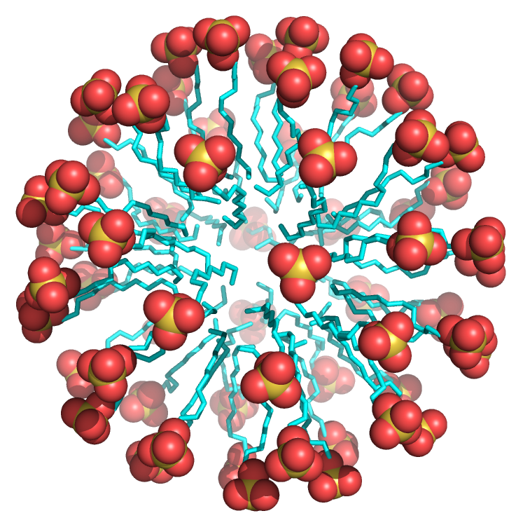
 Figure \(\PageIndex{6}\): Sodium dodecylsulfate micelle (Copyright; author via source).
Click the image for a popup or use this external link: https://structure.ncbi.nlm.nih.gov/i...KECTiGaPPScED7
Fatty acids (carboxylates and sulfates) are amphiphilic, with a larger polar/charged head group that tapers down to a hydrophobic tail, forming a cone-like structure. This cone structure allows the packing of many of these single chains amphiphiles into a micelle, as shown in Figure \(\PageIndex{7}\).

Free fatty acids are transported in the cell and the blood not in micelles but by fatty acid-binding proteins. The most abundant protein in the blood, albumin, binds and transports fatty acids. Figure \(\PageIndex{8}\) shows an interactive iCn3D model of hexadecanoic acid bound to human albumin (1E7H). The fatty acids are shown in colored spacefill rendering.
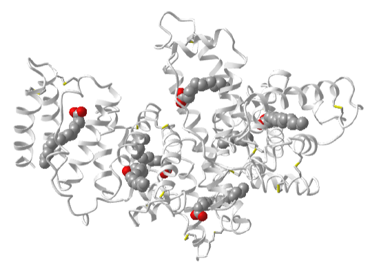
Figure \(\PageIndex{8}\): Hexadecanoic acid bound to human albumin (1E7H). (Copyright; author via source).
Click the image for a popup or use this external link: https://structure.ncbi.nlm.nih.gov/i...CGUdMj11vNevF6
Waxes
You are familiar with ear wax and also the waxy surface of plants. Ear wax contains long-chain fatty acids (saturated and unsaturated) and alcohols derived from them. (They also contain isoprenoid derivatives like squalene and cholesterol). The waxy cuticle surface layers of plants contain very-long-chain fatty acids ( C20–C34) and their derivatives including alkanes, aldehydes, primary and secondary alcohols, ketones, and esters, (They also contain isoprenoid derivatives as well). We will consider waxes as very long-chain fatty acids and their derivatives, as shown in Figure \(\PageIndex{9}\).

These molecules are extremely nonpolar and as such makes great barriers preventing water loss through leaves and water penetration into the ear. This group of molecules clearly shows that the properties (insolubility, high melting point) and function (hydrophobic barrier/protection) arises from structure (very long chain carbon molecules, few electronegative atoms, and lack of C=C double bonds).
Fatty Acid-Containing Lipids
We can categorize these lipids based on function or structure (even though these are related).
Function
- storage lipids - triacylglycerols
- membrane lipids - many different lipids
Structure
- glycerolipids, which use glycerol as a backbone for fatty acid attachment
- sphingolipids, which use sphingosine as a backbone
The structures of glycerol and sphingosine are shown in Figure \(\PageIndex{10}\). Fatty acids are connected to these two "backbone" structures by either ester (mostly) or amide links.
 Figure \(\PageIndex{10}\): Structures of glycerol and sphingosine
Let's explore the classes of fatty acid-containing lipids that use these two "backbone" structures.
Storage Lipids - Triacylglycerols (TAGs)
Triacylglycerols contain the majority of fatty acids in species that store fatty acids for energy. You will often see them named triglycerides or triacylglyceride, but this is a term used more in clinical chemistry and industry (and often in the media). Figure \(\PageIndex{11}\) shows a schematic diagram of the glycerol backbone with three fatty acids esterified to it. They are glycerolipids as they contain a glycerol base.

Glycerol is not chiral but given the incredible diversity of fatty acids, glycerols likely have three different fatty acids esterified to them, making them chiral. If triacylglycerols contain predominately saturated fatty acids, they are solids at room temperature and are called fats. Those with multiple double bonds in the fatty acids are likely liquids at room temperature. These are called oils. Triacylglycerols are even more insoluble than fatty acids, which contain a polar and mostly charged carboxylate.
Figure \(\PageIndex{12}\) shows a triacylglycerol containing all 16:0 saturated fatty acids (left) and one containing all 16:2Δ9,12 polyunsaturated fatty acids. The figures were constructed with a specific set of dihedral angles to illustrate a point, that polyunsaturated fats in a triacylglycerol don't pack as tightly and have lower induced dipole-induced dipole attractions between the acyl chains than is possible with saturated fatty acyl chains. Hence the melting point of triacylglycerols containing polyunsaturated fatty acids is lower than for those with saturated ones.

Let's repeat the key mantra: the structure of lipids determines their function. Consider the very insoluble triacylglycerols which are used as the predominant storage form of chemical energy in the body. In contrast to polysaccharides such as glycogen (a polymer of glucose), the carbon atoms in the acyl chains of the triacylglycerol are in a highly reduced state. The main source of energy to drive not only our bodies but also our society is obtained through oxidizing carbon-based molecules to carbon dioxide and water, in a reaction that is highly exergonic and exothermic. Sugars are already part way down the free energy "hill" since each carbon is partially oxidized. 9 kcal/mol (38 kJ/mol) can be derived from the complete oxidation of fats, in contrast to 4.5 kcal/mol (19 kJ/mol) from that of proteins or carbohydrates.
In addition, glycogen is highly hydrated. For every 1 g of glycogen, 2 grams of water is H-bonded to it. Hence it would take 3 times more weight to store the equivalent mass of carbohydrates compared to triacylglycerol, which is stored in anhydrous lipid "droplets" within cells. In addition, fats are more flexible, given the large number of conformations available to the acyl chain C-C bonds by simple rotation around the C-C bonds. Polysaccharides have monomeric cyclohexane-like chair structures and are much more rigid.
Another interesting point is that glucose and glycogen are found in cells, and they can be mobilized quickly for energy needs. Yes, fats are present in all cells as well (for example all cells have interior and exterior cell membranes). However, the major storage form of fat, triacylglycerols, is stored in special cells called adipocytes, which comprise adipose or fat tissue, and must be mobilized by signaling agents and transported in the form of fatty acids to cells for utilization. Again, triacylglycerols don't form membranes, which separate outside and inside aqueous environments. They are simply so insoluble that they phase-separate into lipid droplets. Their formation and structure are a bit more complex than that, though, and we will discuss lipid droplets more in the next section.
Membrane Lipids
Membranes are bilayers of amphiphilic lipids that separate the outside and inside (cytoplasm) aqueous environments in cells and the cytoplasm and interior contents of organelles within cells. In general, single-chain lipid amphiphiles form micelles. Amphiphilic membrane lipids typically have two nonpolar tails connected to a polar head, giving them a less conical and more cylindrical shape that disallows micelle formation while favoring bilayer formation. Figure \(\PageIndex{13}\) shows a short section of a bilayer membrane made from lipids with a polar (and charged) head group (phosphocholine) and two 16:0 chains. The red and blue spheres represent the O and N atoms of the head groups, which are sequestered to the exterior parts of the bilayer, where they would interact with water. The nonpolar 16:0 tails are shown in cyan, clearly illustrating the nonpolar nature of the interior of the bilayer.

Figure \(\PageIndex{14}\) shows an interactive iCn3D model of a hydrated bilayer of the di16:0 phosphatidycholine bilayer.
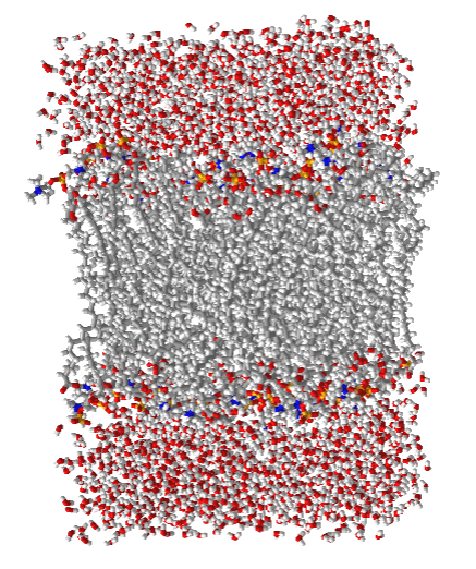
Figure \(\PageIndex{14}\): Hydrated di16:0 phosphatidycholine bilayer (Copyright; author via source).
Click the image for a popup.
We will consider two general types of fatty acid-containing membrane lipids, glycerolipids, with two fatty acids esterified to a glycerol base, and sphingolipids with one fatty acid in amide link to a different base, sphingosine. Sphingosine comes with its own built-in long alkyl chain that provides the "second" nonpolar chain. There are many different polar/charged head groups for these membrane lipids.
Now let's look in more detail at the double-chain amphiphiles comprising these membrane bilayers.
Glycerolipids
There are two main types of glycerolipids, glycerophospholipids, and glyceroglycolipids, which are the most common lipids in membranes.
Glycerophospholipids
Figure \(\PageIndex{15}\) shows the structural features and nomenclature for glycerophospholipids.

These lipids have enormous structural variability given the large number of different fatty acids (both saturated and unsaturated) and head groups that can be attached to a phosphate attached to the carbon 3 of glycerol. The structures of the most common glycerophospholipids are shown in Figure \(\PageIndex{16}\).

Phosphatidylcholine (PC) has the common name lecithin while phosphatidylserine (PS) is called cephalin.
Note that the head groups all have charges since they all have a negatively charged phosphate. PS has two additional charged atoms which would effectively cancel out. PE has a charged amine but could become uncharged at pH values approaching its pKa. PC has a quaternary amine which is charged independent of pH, which would give PC a net 0 charge but with two discrete charges.
Glyceroglycolipids
These do not have a phosphate group attached to the oxygen on C3 of glycerol. Rather they have a mono- or oligosaccharide or, more loosely, a betaine group, each attached by an ether linkage to the glycerol C3 carbon. Figure \(\PageIndex{17}\) Structural features and nomenclature for glyceroglycolipids.

Figure \(\PageIndex{18}\) shows some examples of glyceroglycolipids.

Again there are an enormous number of different glycoglycerolipids, owing to the diversity of head groups and fatty acids esterified to glycerol at C1 and C2.
The above figure (right) shows an example that has no phosphate but also doesn't have a mono- or polysaccharide for a head group. Rather it has a betaine group. Betaine is the common name for trimethylglycine but is used for any N-trimethylated amino acids. Betaine glycerolipids are found in lower eukaryotic organisms (algae, fungi, and some protozoa), in photosynthetic bacteria, and in some spore-producing plants like ferns. Some would call these lipoamino acids.
It is been extremely difficult to develop a vaccine against malaria. The resident time of the plasmodium parasite in the blood after a mosquito bite is quite short (just a few hours) so any immune response against it must be very fast. The sporozoite parasite soon migrates to the liver where it proliferates before leaving the liver and infecting red blood cells. In the liver, it is more sequestered from the antibody response engendered by typical vaccination which promotes the differentiation and proliferation of B-cells into antibody-producing cells. Another type of lymphocyte, T cells, can also be activated in an immune response. In particular, CD8 memory T cells (Trm cells) are produced in the liver against parasites but high levels are needed. Mouse liver cells make modest levels of Trm cells when vaccinated with mRNAs encoding the large ribosomal subunit protein L6 (RPL6) of the plasmodium. This is found in the liver for about a week after infection. However, when an "adjuvant" (something that increases the immune response usually in a nonspecific way) containing α-galactosylceramide (αGC)is used, there is an increase in the production of liver Trm cells. Chemically modifying the αGC adjuvant greatly increases the production of Trm cells. The structure of αGC and its derivatives are shown below.

The adjuvant interacts as an agonist with type I natural killer T cells which activates them and amplifies the immune response in the liver. Mice vaccinated with an mRNA vaccine containing the modified αGC adjuvant were afforded protection against the plasmodium parasite. The protection extended to mice previously exposed to the parasite as well.
Membrane Lipids - Sphingolipids
Sphingolipids contain a sphingosine backbone and a fatty acid linked through an amide bound. The simplest sphingolipids are the ceramides, which are double-chain amphiphiles but without further modification of the functional groups on the polar head of sphingosine. The structures of both sphingosine and ceramide are shown below in Figure \(\PageIndex{19}\):

Figure \(\PageIndex{19}\): Structures of sphingosine and ceramide
Ceramides are abundant in the skin where they help provide a protective barrier. Skin creams also contain ceramides. These lipids make up only about 1% of body lipids and there are over 200 different ceramides in humans arising from the different fatty acids linked to sphingosine. They have a key role in cell signaling and can also affect cardiovascular health in ways that are just being appreciated. Since they are double-chain amphiphiles, they are found in membranes where they alter the properties of the bilayer (including membrane fluidity). Ceramides with long acyl tails (especially those with 16, 18, or 24 carbons) seem especially deleterious.
Phosphosphingolipids and glycosphingolipids
These membrane lipids have a ceramide base but also contain modifications at the polar functional groups of the sphingosine head. Let's consider the phosphosphingolipids and the glycosphingolipids together. These groups do not use glycerol as a base for the attachment of fatty acids and head groups. Rather they use molecule sphingosine. Figure \(\PageIndex{20}\) shows the structural features and nomenclature of sphingolipids.

Examples of both classes are shown in Figure \(\PageIndex{21}\). Note the base sphingosine (in red) provides an amine to attach a fatty acid through an amide bond and an OH for attachment of the head group.

Sugar-containing glycosphingolipids are found largely in the outer face of plasma membranes. The primary lipid of myelin, which coats neuronal axons and insulates them from loss of electrical signaling down the axon, is galactocerebroside
Figure \(\PageIndex{22}\) shows a summary of all of the different types of fatty acid-containing lipids

Over a 1000 different lipids are found in eukaryotic cells. This complexity has led to the development of an even more comprehensive classification system for lipids. In this system, lipids are given a very detailed as well as all-encompassing definition: "hydrophobic or amphipathic small molecules that may originate entirely or in part by carbanion-based condensations of thioesters (fatty acyl, glycerolipids, glycerophospholipids, sphingolipids, saccharolipids, and polyketides) and/or by carbocation-based condensations of isoprene units (prenol lipids and sterol lipids)." Eight different categories of lipids are listed in the parentheses above. We will stick to the definition used throughout this chapter.
Shapes of membrane lipids
Let's look at the general shape of the double-chain amphiphiles that make bilayers. We saw the long-chain fatty or sulfate acids form conical structures which fit nicely together when they self-aggregate to form micelles. In contrast, membrane-forming double-chain amphiphiles have more cylindrical shapes that can't be fitted together in micelles but rather form a less curved bilayer structure, as shown in Figure \(\PageIndex{23}\). We will consider the variety of membrane structures in the next section.

Triacylglyceride/Phospholipid Stereochemistry
Glycerol is an achiral molecule since C2 has two identical substituents, -CH2OH. Glycerol in the body can be chemically converted to triglycerols and phospholipids (PL) which are chiral, and exist in one enantiomeric form. How can this be possible if the two CH2OH groups on C2 of glycerol are identical? It turns out that even though these groups are stereochemically equivalent, we can differentiate them as described in the figure below. Let's replace the -CH2OH in one of the end carbons with -CH2OD. With this simple change, glycerol is now chiral. Look at the top half of Figure \(\PageIndex{24}\).

Glycerol is oriented with the OH on C2 (the middle carbon) pointing to the left. The OH of the top carbon in this orientation, C1, is replaced with OD, where D is deuterium to make the molecule chiral (four different groups attached to C2). By rotating the molecule such that the H on C2 points to the back, and assigning priorities to the other substituents on C2 (OH =1, DOCH2 =2, and CH2OH = 3), it can be seen that the resulting molecule is in the S configuration. We simply name the C1 carbon which we modified with deuterium as the proS carbon. Likewise, if we replaced the OH on C3 with OD, we will form the R enantiomer. Hence C3 is the proR carbon. This shows that in reality, we can differentiate between the two identical CH2OH substituents. We say that glycerol is not chiral, but prochiral. (Think of this as glycerol has the potential to become chiral by modifying one of two identical substituents.)
In the bottom half of Figure \(\PageIndex{23}\), we can relate the configuration of glycerol above, (when OH on C2 is pointing to the left) to the absolute configuration of L-glyceraldehyde, a simple sugar (a polyhydroxyaldehyde or ketone), another 3C glycerol derivative. This molecule is chiral with the OH on C2 (the only chiral carbon) pointing to the left. It is easy to remember that any L sugar has the OH on the Last chiral carbon pointing to the Left. The enantiomer (mirror image isomer) of L-glyceraldehyde is D-glyceraldehyde, in which the OH on C2 points to the right. Biochemists use L and D for lipid, sugar, and amino acid stereochemistry, instead of the R, S nomenclature you used in organic chemistry. The stereochemical designation of all the sugars, amino acids, and glycerolipids can be determined from the absolute configuration of L- and D-glyceraldehyde.
Now let's see how an enzyme can take a prochiral molecule like glycerol and phosphorylate only one of the -CH2OHs to make one specific isomer, glycerol-3-phosphate, a key intermediate in the biosynthesis of phosphatidic acid (PA), a glycerophospholipid, as well as chiral triacylglycerols, shown in Figure \(\PageIndex{25}\). The far left part of the pathway shows how the proR CH2OH of glycerol is phosphorylated to produce one specific enantiomer, L-glycerol-3-phosphate. (The top part of the figure shows another way to make this molecule from glucose through the glycolytic pathway we will encounter in a future chapter.

The first step (above figure) involves the phosphorylation of the OH on C3 by ATP (a phosphoanhydride similar in structure to acetic anhydride, an excellent acetylating agent) to produce the chiral molecule glycerol phosphate. Based on the absolute configuration of L-glyceraldehyde, and using this to draw glycerol (with the OH on C2 pointing to the left), we can see that the phosphorylated molecule can be named L-glycerol-3-phosphate. However, by rotating this molecule 180 degrees, without changing the stereochemistry of the molecule, we don't change the molecule at all, but using the D/L nomenclature above, we would name the rotated molecule D-glycerol-1-phosphate. This is illustrated in Figure \(\PageIndex{26}\).

We can’t give the same molecule two different names. Hence biochemists have developed the stereospecific numbering system (sn), which assigns the 1-position of a prochiral molecule to the group occupying the proS position. The proS C1 is hence at the sn-1 position. With that designation, C2 is at the sn-2 position, and C3 is at the sn-3 position. Using this nomenclature, we can see that the chiral molecule described above, glycerol-phosphate, can be unambiguously named as sn-glycerol-3-phosphate. The hydroxyl substituent on the proR carbon was phosphorylated.
It is interesting to note that archaea use isoprenoid chains linked by ether bonds to sn-glycerol 1-phosphate in their synthetic pathways. As noted above, bacteria and eukaryotes use fatty acids attached by ester bonds to sn-glycerol 3-phosphate
The enzymatic phosphorylation of the proR CH2OH of glycerol to form sn-glycerol-3-phosphate is illustrated in Figure \(\PageIndex{27}\). As we were able to differentiate the 2 identical CH2OH substituents as containing either the proS or proR carbons, so can the enzyme. The enzyme can differentiate identical substituents on a prochiral molecule if the prochiral molecule interacts with the enzyme at three points. Another example of a prochiral reactants/enzyme system involves the oxidation of the prochiral molecule ethanol by the enzyme alcohol dehydrogenase, in which only the proR H of the 2 H’s on C2 is removed. (We will discuss this later.)

Isoprenoid-containing lipids
This is the last class of lipids we will consider. They do not contain fatty acids. Rather they contain isoprene, a small branched alkadiene, which can polymerize into larger molecules containing isoprene monomer to form isoprenoids, often called terpenes. Instead of using isoprene as the polymerization monomer, either dimethylallyl pyrophosphate (DMAPP) or isopentenylpyrophosphate (IPP) are used biologically.
Figure \(\PageIndex{28}\) shows how DMAPP and IPP (both containing 5Cs) are used in a polymerization reaction to form geranyl-pyrophosphate (C10), farnesyl pyrophosphate (C15) and geranyl-geranyl pyrophosphate (C20).

Many isoprenoid lipids are made from farnesyl pyrophosphate. For membrane purposes, the most important of these is cholesterol. Figure \(\PageIndex{29}\) shows an overview of the synthesis of cholesterol from two farnesyl pyrophosphates linking together in a "tail-to-tail" reaction to form squalene, a precursor of cholesterol. Each isoprene unit (5Cs) is shown in different colors to make it easier to see.

Other biologically important isoprenoid-containing vitamins are shown in Figure \(\PageIndex{30}\).

Everyone who has walked in the woods after a fresh rain knows the smell of a terpene called geosmin, "an Earthy-smelling substance" found in abundance in a group of substances collectively called petrichor, derived from the Greek petra (rock) and ichor (blood of gods). The structure of geosmin is shown below.

These substances are released when water falls or soils and rocks, and seeps into pores from which aerosols are released. Too hard of a rain will saturate pores in rocks and prevent the release. Some insects like the springtail are attracted to geosmin. The synthesis of geosmin by bacteria and blue-green algae is under the control of transcription factors which also affect bacterial spore formation. The attracted insects carry away the spores. Flies are repelled by geosmin, which is detected by a receptor at very low geosmin concentration. In contrast, the Aedes aegypti mosquito is attracted to geosmin which parallels the fact that the mosquito is not affected by bacterial toxins.
One last look at lipid structure and shapes
With the exclusion of waxes and triacylglycerols, the other lipids we have discussed, including the mostly planar molecule cholesterol, are amphiphilic. We have seen that single-chain fatty acids form micelles while lipids with two nonpolar chains and a polar/charged head group form bilayers. Given the relative size of the head group and the degree of unsaturation of the double bonds in fatty acids, the overall shapes of the membrane-forming lipids differ as illustrated below. They are arranged like dominos in the membrane based on their geometric volumes. Preferential clustering of identical types can cause local and extended changes in a prototypical bilayer structure.
Membranes and their components must be dynamic to enable all the functions and activities of a membrane. Ligands bind to membrane receptors (usually proteins), which can invaginate and pinch off to form an intracellular vesicle containing the receptor for processing. Likewise, vesicles can pinch off into the extracellular space. Cells must divide. Think of the membrane changes necessary for that! Also, consider that the length of cholesterol is half that of a typical double-chain amphiphile so it fits into just one of the bilayer lipid leaflets where it modulates lipid bilayer property. Figure \(\PageIndex{31}\) shows a series of lipids and their shape profiles.

We will consider membranes in greater detail in section 10.3. Next, however, we will explore in more detail the properties of micelle and lipid droplet systems before addressing the more structurally complicated lipid bilayers.
End-of-Chapter Questions
x) A cell membrane has the ability to remodel in response to stress to promote membrane integrity. In the situations below, how could the membrane be remodeled to prevent damage? (I.e. what types of lipids could be added/removed to ensure homeostasis)
a) Increase in temperature
b)
x) What are the three classes of lipids? Explain their similarities and differences.
x) For each lipid below, name the type of lipid (membrane lipid, triacylglycerol, storage lipid, sphingolipid, wax, sterol, membrane glycerolipid, none of these), if it could be found in membrane, and if it is fatty acid or isoprene derived.
(Insert pics of lipids)
Answer
-
Add texts here. Do not delete this text first.