
13.2A: Opsonization
- Page ID
- 3313

- Discuss how antibodies defend the body by way of opsonization. (Include what classes or isotypes of immunoglobulins are involved, the role of the Fab portion of the antibody, the role, if any, of the Fc portion of the antibody, and the role of any complement proteins, if any, involved.)
- Briefly describe two different ways bacteria may resist opsonization.
Opsonization, or enhanced attachment, refers to the antibody molecules IgG and IgE, the complement proteins C3b and C4b, and other opsonins attaching antigens to phagocytes. This results in a much more efficient phagocytosis.
Opsonization with IgG, IgA, C3b, and C4b
The process starts with antibodies of the isotype IgG, IgA, or IgM being made against a surface antigen of the organism or cell to be phagocytosed. The Fab portions of the antibody react with epitopes of the antigen. The Fc portion of IgG (but not IgM) can then bind to receptors on neutrophils and macrophages thus sticking the antigen to the phagocyte (Figure \(\PageIndex{1}\)). The Fc portion of secretory IgA can also bind to macrophages and neutrophils for opsonization.
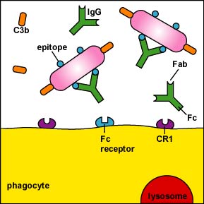
The Fc portion of secretory IgA can also bind to macrophages and neutrophils for opsonization.
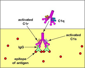
Alternately, IgG, IgA, and IgM can activate the complement pathways (Figure \(\PageIndex{2}\)) and C3b or C4b can stick the antigen to phagocytes (Figure \(\PageIndex{1}\)). Like IgG, C3b, and to a lesser extent C4b, can function as opsonins, that is, they can attach antigens to phagocytes.One portion of the C3b binds to proteins and polysaccharides on microbial surfaces; another portion attaches to CR1 receptors on phagocytes, B-lymphocytes, and dendritic cells for enhanced phagocytosis (Figure \(\PageIndex{3}\)). (Remember that C3b and C4b are also produced during the alternative complement pathway and the lectin pathway as was discussed in Unit 5.) Activation of the complement pathway also promotes inflammation to bring phagocytes and defense chemicals from the bloodstream to the infection site as discussed later under this topic.
Actually, C3b molecule can bind to pretty much any protein or polysaccharide. Human cells, however, produce Factor H that binds to C3b and allows Factor I to inactivate the C3b. On the other hand, substances such as LPS on bacterial cells facilitate the binding of Factor B to C3b and this protects the C3b from inactivation by Factor I. In this way, C3b does not interact with our own cells but is able to interact with microbial cells.
Attachment then promotes destruction of the antigen. Microorganisms are placed in phagosomes (Figure \(\PageIndex{4}\)) where they are ultimately digested by lysosomes (Figure \(\PageIndex{5}\)). If the antigen is a cell too large to be ingested - such as virus-infected host cells, transplant cells, and cancer cells - the phagocyte empties the contents of its lysosomes directly on the cell for extracellular killing (Figure \(\PageIndex{6}\) and Figure \(\PageIndex{7}\)).
Opsonization is especially important against microorganisms with antiphagocytic structures such as capsules since opsonizing antibodies made against the capsule are able to stick capsules to phagocytes (Figure \(\PageIndex{8}\)). In vaccines against pneumococccal pneumonia and Haemophilus influenzae type b, it is capsular polysaccharide that is given as the antigen in order to stimulate the body to make opsonizing antibodies against the encapsulated bacterium.
Opsonization with IgE and the promotion of inflammation
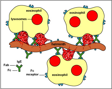
The antibody isotype IgE is made against parasitic worms (helminths) and arthropods. The Fab portions of IgE bind to epitopes on the helminth or arthropod while the Fc portion binds to receptors on eosinophils enabling opsonization. In other words, IgE sticks phagocytic eosinophils to helminths and arthropods for the extracellular killing of that organism (Figure \(\PageIndex{9}\)).
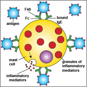
The Fc portion of IgE also binds to receptors on mast cells and basophils to trigger the release of inflammatory mediators (Figure \(\PageIndex{10}\)). The inflammatory response then enables phagocytes and defense chemicals to leave the bloodstream and go to the infected site as will be discussed later under this topic.
Compare and contrast how IgG, IgM, and IgE promote opsonization..
Because of a particular immunodeficiency disorder, a person is unable to produce C3 convertase. Which of the above antibody isotypes could still participate in opsonization? Briefly explain why.
How Bacteria Resist Attachment to Phagocytes
As we learned previously, some bacteria by means of the activities described below are able to resist phagocytic attachment :
- An outer membrane molecule of Neisseria gonorrhoeae called Protein II and the M-protein of Streptococcus pyogenes allow these bacteria to be more resistant to phagocytic engulfment. The M-protein of S. pyogenes, for example, binds factor H of the complement pathway and this leads to the degradation of the opsonin C3b by factor I and the formation of C3 convertase.
- Some capsules simply cover the C3b that does bind to the bacterial surface and prevent the C3b receptor on phagocytes from making contact with the C3b (Figure \(\PageIndex{11}\)). This is seen with the capsule of Streptococcus pneumoniae.
- Capsules can also resist unenhanced attachment by preventing the glycoprotein receptors on phagocytes from recognizing the bacterial cell wall components and mannose-containing carbohydrates.
- S. pneumonia activates the classical complement pathway, but resists C3b opsonization, and complement causes further inflammation in the lungs.
- Neisseria meningitidis has a capsule composed of sialic acid while Streptococcus pyogenes (group A beta streptococci) has a capsule made of hyaluronic acid. Both of these polysaccharides closely resemble carbohydrates found in human tissue polysaccharides and because they are not recognized as foreign by the lymphocytes that carry out the immune responses, antibodies are not made against these capsules.
- Some bacteria are able to coat themselves with host proteins such as fibronectin, lactoferrin, or transferrin. This prevents antibody molecules from binding to epitopes on the bacterial surface.
- Staphylococcus aureus produces protein A while Streptococcus pyogenes produces protein G. Both of these proteins bind to the Fc portion of antibodies, the portion that normally binds to receptors on phagocytes (Figure \(\PageIndex{12}\)). In this way the bacteria become coated with antibodies in a way that does not result in opsonization (Figure \(\PageIndex{13}\)).
- Streptococcus pyogenes produces Mac proteins that are able to bind to the receptors on phagocytes to which IgG and C3b normally attach (Figure \(\PageIndex{14}\).and Figure \(\PageIndex{15}\)). This blocks opsonization.
- Pathogenic Yersinia, such as the one that causes plague, contact phagocytes and, by means of a type III secretion system, deliver proteins which depolymerize the actin microfilaments needed for phagocytic engulfment into the phagocytes. Another Yersinia protein degrades C3b and C5a.
Summary
Opsonization, or enhanced attachment, refers to the antibody molecules IgG and IgE, the complement proteins C3b and C4b, and other opsonins attaching antigens to phagocytes. The Fab portions of the antibody IgG react with epitopes of the antigen. The Fc portion of IgG can then bind to neutrophils and macrophages thus sticking the antigen to the phagocyte. The Fc portion of secretory IgA can also bind to macrophages and neutrophils for opsonization. IgG and IgM can activate the classical complement pathway and C3b or C4b can stick the antigen to phagocytes. IgE is made against parasitic worms (helminths) and arthropods. The Fab portions of IgE bind to epitopes on the helminth or arthropod while the Fc portion binds to receptors on eosinophils enabling opsonization.
Questions
Study the material in this section and then write out the answers to these questions. Do not just click on the answers and write them out. This will not test your understanding of this tutorial.
- Discuss how antibodies defend the body by way of opsonization. (Include what classes or isotypes of immunoglobulins are involved, the role of the Fab portion of the antibody, the role, if any, of the Fc portion of the antibody, and the role of any complement proteins, if any, involved.) (ans)
- We know Streptococcus pneumoniae is encapsulated and capsules resist phagocytosis. Yet the body is eventually able to phagocytose this organism. Describe how. (ans)
- Staphylococcus aureus produces an exotoxin called Protein A. Protein A is able to react with the Fc portion of IgG. In terms of humoral immunity, discuss how Protein A may help the Staphylococcus resist phagocytosis. (ans)
- The M-protein of Streptococcus pyogenes binds factor H of the complement pathway and allows these bacteria to be more resistant to phagocytic engulfment. Explain how. (ans)
- Multiple Choice (ans)