
D1. Glycoprotein Biosynthesis
- Page ID
- 4900

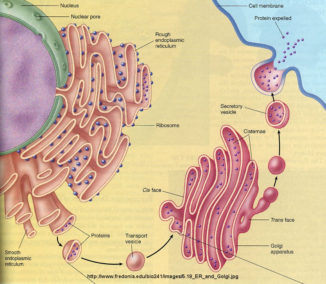
Carbohydrates are added to proteins in a very complicated process which involves two organelles, the endoplasmic reticulum and the Golgi apparatus. CHO addition to proteins occurs both co- and post-translationally. The RNA coding the protein sequence enters the cytoplasm where it binds to ribosomes (large RNA-protein complexes) which are the site for protein synthesis. Cytoplasmic proteins are synthesized on free ribosomes, but for future glycoproteins, the ribosomes bind to an elongated, extensive organelle in the cell called the endoplasmic reticulum (ER).
 Figure: ribosomes bind to an elongated, extensive organelle in the cell called the endoplasmic reticulum (ER)
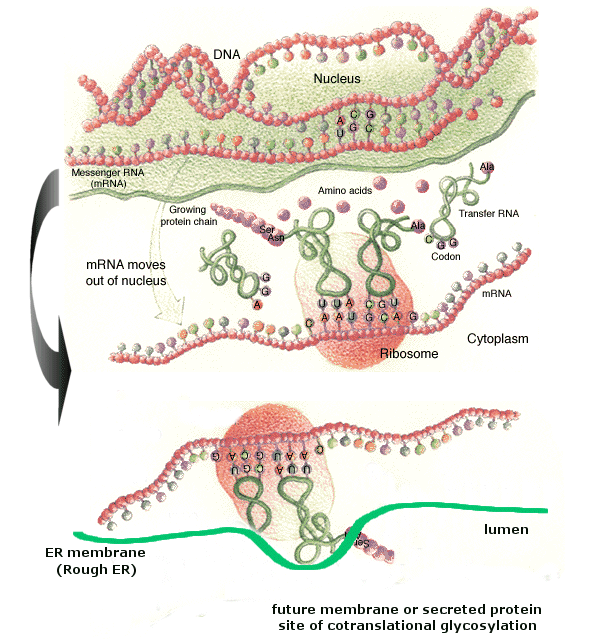
(from web, searching for reference; note: anticodon mislabeled as codon)
The nascent protein chain enters the lumen of the ER and a core oligosaccharide is added to the protein. Further additions and removal (trimming and processing) of monosaccharides are preformed in the lumen until a final core mannose structure has been added. This is demonstrated in the animation below. (The pink two-lobbed structure represents the ribosome which is bound to the ER membrane. The newly synthesized protein is shown in the lumen side of the ER membrane. That is where glycan transfer occurs.) The ER lumen contains high concentrations of molecular chaperones to assist protein folding.
- Animations of Glycoprotein Biosynthesis: Endoplasmic reticulum
Now additional carbohydrate modifications (post-translational) are made as the protein moves from the lumen of the ER (probably by a budding process) to another series of stacked, pancake-like organelles called the Golgi apparatus. Here terminal carbohydrate modification is completed. The Golgi does not contain molecular chaperons since protein folding is complete when the proteins arrive. Rather they have high concentrations of membrane bound enzymes, including glycosidases, and glycosyltransferases.
- Animations of Glycoprotein Biosynthesis: Golgi