
43.3A: Male Reproductive Anatomy
- Page ID
- 14122

- Diagram the structures of human male reproductive anatomy
Human Reproductive Anatomy
The reproductive tissues of male and female humans develop similarly in utero until a low level of the hormone testosterone is released from male gonads. Testosterone causes the undeveloped tissues to differentiate into male sexual organs. Primitive gonads become testes; other tissues produce a penis and scrotum in males.
Male Reproductive Anatomy
In the male reproductive system, the scrotum houses the testicles or testes, providing passage for blood vessels, nerves, and muscles related to testicular function. The testes, a pair of male reproductive organs, produce sperm and male sex hormones, including the steroid testosterone. Coiled in each testis are seminiferous tubules that produce sperm.
Sperm
Sperm are immobile at body temperature; therefore, the scrotum and penis are external to the body so that a proper temperature is maintained for motility. In land mammals, the pair of testes must be suspended outside the body at about 2° C lower than body temperature to produce viable sperm.
Sperm develop in the seminiferous tubules that are coiled inside the testes. The walls of the seminiferous tubules are composed of the developing sperm cells, with the least-developed sperm at the periphery of the tubule and the fully-developed sperm in the lumen. The sperm cells are mixed with “nursemaid” cells called Sertoli cells which protect the germ cells and promote their development. Other cells mixed in the wall of the tubules are the interstitial cells of Leydig; these cells produce high levels of testosterone once the male reaches adolescence.
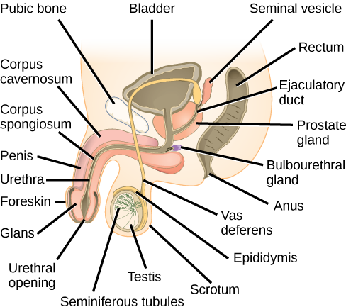
Sperm consist of a flagellum (as a tail), a neck that contains the cell’s energy-producing mitochondria, and a head that contains the genetic material. When the sperm have developed flagella, or lash-like appendages that protrude from the cell body, and are nearly mature, they leave the testicles and enter the epididymis. This structure lies along the top and posterior portion of the testes; it is the site of sperm maturation. The sperm leave the epididymis and enter the vas deferens, which is the duct in the testicle that carries sperm from the epididymis to the ejaculatory duct.
Semen is a mixture of sperm and spermatic duct secretions (about 10 percent of the total), along with fluids from accessory glands, that contribute most of the semen’s volume. An ejaculate will contain from two to five milliliters of fluid with from 50–120 million sperm per milliliter. The bulk of the semen comes from the accessory glands associated with the male reproductive system, including the seminal vesicles, the prostate gland, and the bulbourethral gland.
Seminal vesicles, penis, prostate, and bulbourethral gland
The seminal vesicles are a pair of glands that lie along the posterior border of the urinary bladder. The glands make a solution that is thick, yellowish, and alkaline. As sperm are only motile in an alkaline environment, a basic pH is important to reverse the acidity of the vaginal environment. The solution also contains mucus, fructose (a sperm mitochondrial nutrient), a coagulating enzyme, ascorbic acid, and local-acting hormones called prostaglandins.
The penis is an organ that drains urine from the renal bladder and functions as a copulatory organ during intercourse. The penis contains three tubes of erectile tissue running through the length of the organ. These consist of a pair of tubes on the dorsal side, called the corpus cavernosum, and a single tube of tissue on the ventral side, called the corpus spongiosum. This tissue, when engorged with blood, becomes erect and hard, in preparation for intercourse. The organ is inserted into the vagina, culminating with an ejaculation, which is the forcible ejection of semen from the urethra. An orgasm is a two-stage process: first, glands and accessory organs connected to the testes contract; second, semen (containing sperm) is expelled through the urethra during ejaculation. After intercourse, the blood drains from the erectile tissue and the penis becomes flaccid.
The walnut-shaped prostate gland surrounds the urethra, the connection to the urinary bladder. It has a series of short ducts that directly connect to the urethra. The gland is a mixture of smooth muscle and glandular tissue. The muscle provides much of the force needed for ejaculation to occur.
The bulbourethral gland, or Cowper’s gland, is an exocrine gland which secretes a clear fluid known as pre-ejaculate that is generated upon sexual arousal. This gland releases its secretion prior to the release of the bulk of the semen. It neutralizes any acid residue in the urethra left over from urine. This usually accounts for a couple of drops of fluid in the total ejaculate and may contain a few sperm. Withdrawal of the penis from the vagina before ejaculation to prevent pregnancy may not work if sperm are present in the bulbourethral gland secretions.
Key Points
- The male gonads, or testes, produce sperm within the seminiferous tubules; the sperm are housed here until they are nearly mature, at which point they enter the epidydimis for full maturation.
- The testes are housed in the scrotum, an external sac that keeps the sperm at a temperature lower than that of the body.
- At ejaculation, sperm leave the epidydimis and enter the vas deferens, a duct which carries the sperm out of the body through the urethra, along with the fluids of various glands of the male reproductive system.
- The seminal vesicles produce a thick fluid that is alkaline in order to protect sperm from the acidic nature of the female vagina; it also contains sugars to nourish the sperm.
- The prostate gland produces the force necessary to push the sperm out of the epididymis at ejaculation, while the bulbourethral gland emits a fluid just prior to ejaculation that neutralizes acid from any urine left over in the urethra.
- During sexual arousal, the spongy tissue inside the penis (the corpus spongiosum) fills with blood, causing the penis to become erect and hard; after ejaculation, the blood flows back out of the penis, leaving it flaccid.
Key Terms
- epididymis: a narrow, tightly-coiled tube connecting the efferent ducts from the rear of each testicle to its vas deferens, where sperm are stored during maturation
- prostate gland: a gland in male mammals surrounding the urethra just below the urinary bladder that controls the release of urine from the bladder and produces a secretion that is the fluid part of semen
- seminiferous tubule: any of many threadlike structures, located in the testes, that are the specialized areas of sperm production