
11.2: DNA Fingerprinting
- Page ID
- 24186

DNA fingerprinting is routinely used today to establish paternity, in the diagnosis of inherited disorders, and for use in criminal cases. DNA fingerprinting enables forensic investigators to determine whether two DNA samples originate from the same individual. Not all of the DNA present is used in this analysis. Restriction enzymes act as molecular scissors and are used to cleave DNA molecules at specific points. Over 2,500 different restriction enzymes have been identified. These enzymes are produced by bacteria and are used to destroy foreign DNA such as bacteriophages - viruses that infect and replicate within a bacterium.
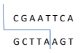
For example the restriction enzyme EcoR1, isolated from E. coli, cuts DNA at the sequence GAATTC.
The length and the number of the fragments produced depends upon the frequency and the distance between the recognition sites. This distinct pattern is known as restriction fragment length polymorphisms (RFLP’s) which are unique to each individual therefore forming a DNA fingerprint. After DNA samples are cut by restriction enzymes, the fragments are separated using gel electrophoresis. PCR, polymerase chain reaction, can be used to analysis very small or degraded samples. This enzyme amplifies even trace amounts of DNA
present. The length of the segments analyzed are much smaller and the repeat sites are called microsatellites.
The phosphate group of the DNA molecule is negatively charged which gives the fragments an overall negative charge. In an electrical current the negatively charged fragments will be attracted to the positive pole. Smaller fragments will migrate faster and further in a given time period. Therefore the fragments are separated by size. After separation radioactive markers are added which are complementary to the separated fragments. Photographic film is placed over the gel and the areas that are exposed to the radioactive markers darken. This generates a series of lines that resemble a bar code. The film then becomes the DNA fingerprint. In this experiment we will be using a mixture of dyes that will simulate the migration of DNA fragments.