
4.2: Breathing Lab Teaching Preparation Notes
- Page ID
- 25221
Overview
This minds-on, hands-on activity begins with analysis and discussion questions that develop student understanding of homeostasis and negative feedback, the difference between negative and positive feedback, and the cooperation between the respiratory and circulatory systems to provide O2 and remove CO2 for cells all over the body. Then, students carry out and analyze an experiment which investigates how the rate and depth of breathing are affected by negative feedback regulation of blood levels of CO2 and O2. Finally, students formulate a question concerning the effects of exercise on breathing, design and carry out a relevant experiment, analyze and interpret their data, and relate their results to homeostasis during exercise.
Learning Goals
In accord with the Next Generation Science Standards:
- This activity helps students to prepare for the Performance Expectation:
- HS-LS1-3. "Plan and conduct an investigation to provide evidence that feedback mechanisms maintain homeostasis."
- Students learn the following Disciplinary Core Idea:
- LS1.A "Feedback mechanisms maintain a living system's internal conditions within certain limits and mediate behaviors, allowing it to remain alive and functional even as external conditions change within some range. Feedback mechanisms can encourage (through positive feedback) or discourage (negative feedback) what is going on inside the living system."
- Students engage in recommended Scientific Practices, including:
- "asking questions"
- "planning and carrying out investigations"
- "analyzing and interpreting data"
- "constructing explanations".
- This activity provides the opportunity to discuss the Crosscutting Concept, "Stability and Change".
Additional Specific Learning Goals
- Homeostasis refers to the maintenance of relatively constant internal conditions.
- Negative feedback occurs when a change in a regulated variable triggers a response which reverses the initial change and brings the regulated variable back to the setpoint. Negative feedback plays an important role in maintaining homeostasis. For example, negative feedback helps to maintain a relatively constant internal body temperature.
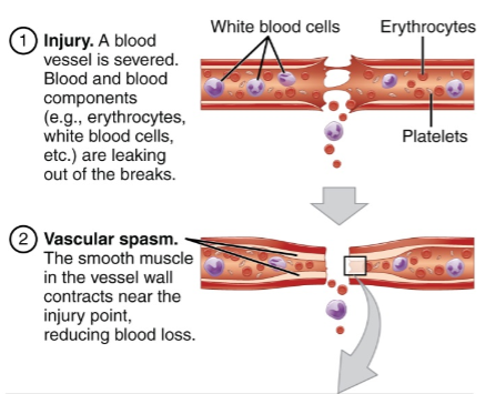
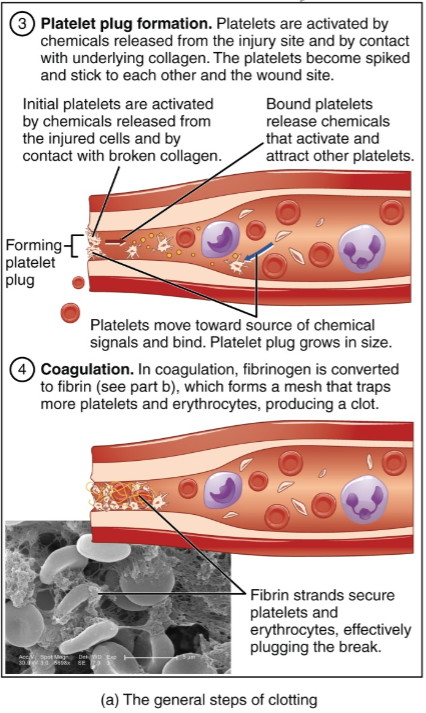
- Positive feedback occurs when a change in a variable triggers a response which causes more change in the same direction. Positive feedback is useful when there is an advantage in making a rapid change. For example, positive feedback facilitates the rapid formation of a platelet plug which helps to prevent excessive blood loss when a blood vessel is injured.
- Cells carry out cellular respiration to make ATP, a molecule that provides energy in a form that cells can use. Cellular respiration requires O2 and produces CO2.
- The respiratory and circulatory systems work together to bring O2 to cells all over the body and get rid of CO2. When a person inhales, air with O2 is brought into the lungs. O2 diffuses from the air in the tiny air sacs of the lungs into the blood. The O2-carrying blood is pumped by the heart to blood vessels near all the cells in the body. O2 diffuses from the blood into the cells where O2 is used in cellular respiration. CO2 produced by cellular respiration moves through the blood to the lungs where it is exhaled.
- Negative feedback regulation of blood levels of CO2 and O2 helps to ensure that enough O2 is delivered to meet the cells’ needs for cellular respiration and enough CO2 is removed to prevent harmful effects. Increased blood levels of CO2 stimulate increased breathing (especially increased depth of breathing).
- When a person exercises, his or her muscle cells use much more ATP per second than when he or she is resting. This requires a substantially increased rate of cellular respiration. To maintain homeostasis during exercise, breathing rate and depth increase to supply more O2 and remove more CO2.
- For a scientific investigation to yield accurate results, scientists need to begin by developing reliable, valid methods of measuring the variables in the investigation.
Supplies
For section III. Negative Feedback and the Regulation of Breathing:
- One 8 gallon plastic garbage bag per student
- Some way of timing 8 consecutive 30-second intervals for each group of four students
For section IV. Homeostasis and Breathing during Exercise:
- Some way of timing 30-second intervals plus supplies for whatever method of measuring breathing rate and depth you choose (see pages 10-11)
- One or two pages of notebook paper and one page of graph paper per student
- You may also want to have available resistance bands or instructions for and pictures of yoga poses in case students want to include those types of exercise in their experiment.
Instructional Suggestions and Background Information
The following timeline may be appropriate for this multi-part activity (assuming you have 50-minute class periods). If time is limited, you may want to use just sections I-III and omit section IV, Homeostasis and Changes in Breathing Due to Exercise; if you have some teaching time after final tests have been administered at the end of the year, you may want to use section IV during that time.
|
Day |
Activities |
Student Handout Pages |
|
1 |
Analysis and discussion questions on "Homeostasis and Negative Feedback" and "Respiration and Circulation" + Prepare for negative feedback experiment |
1-4 |
|
2 |
"Negative Feedback and the Regulation of Breathing" experiment and begin analysis |
5-6 |
|
3 |
Analyze and interpret negative feedback experiment and plan experiments for "Homeostasis and Changes in Breathing Due to Exercise" |
7-top part of 9 |
|
4 |
Finish planning and carry out exercise experiments and analyze and interpret results; you may want to assign question 29 as homework. |
9 |
In the Student Handout, numbers in bold indicate questions for the students to answer.
A key is available upon request to Ingrid Waldron (iwaldron@sas.upenn.edu). The following paragraphs provide additional instructional suggestions and background information – some for inclusion in your class discussions and some to provide you with the relevant background that may be useful for your understanding and/or for responding to student questions.
For the analysis and discussion questions, you can maximize student participation and learning, by having your students work individually, in pairs, or in small groups to complete groups of related questions and then having a class discussion after each group of related questions. In each discussion, you can probe student thinking and help them develop a sound understanding of the concepts and information covered before moving on to the next group of related questions.
I. Homeostasis and Negative Feedback
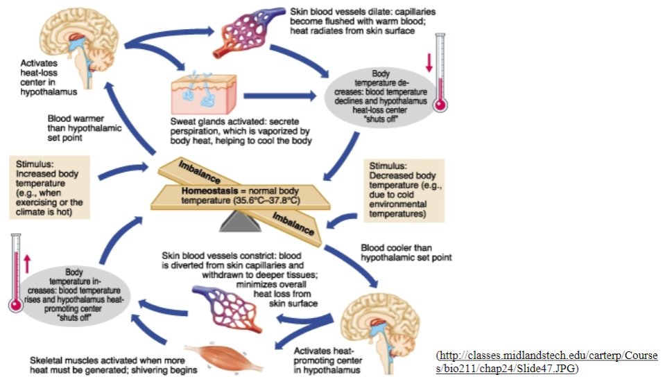
The figure below shows another way of diagramming negative feedback regulation of internal body temperature. This figure provides additional information about this negative feedback and illustrates several important points which you may want to include in your discussion of the bottom half of page 1 of the Student Handout:
- Negative feedback maintains body temperature within a narrow range by changing other aspects of body physiology (sweating, shivering, blood flow to the skin). These changes persist until body temperature is restored to the setpoint range and then the sweating or shivering and change in blood flow are turned off.
- The key stimulus for these changes is the discrepancy between the setpoint temperature and the actual body temperature.
- Negative feedback often operates via more than one type of physiological response.
Negative feedback regulation does not imply having a constant temperature at all times. You can change the set point on the thermostat in a home and, similarly, physiological responses can change your body's set point for temperature regulation. For example, when you have an infection, the phagocytic cells that defend against bacteria and viruses send a chemical signal to the region of the brain which functions as a thermostat. This chemical signal increases the set point for temperature regulation, so you develop a fever. The increase in body temperature can help your immune system fight the infection since the increase in temperature generally increases the immune response and decreases the growth of many infectious microorganisms. When you have a fever, normal body temperature may result in shivering and feeling chills because the body temperature is below the fever setpoint temperature.
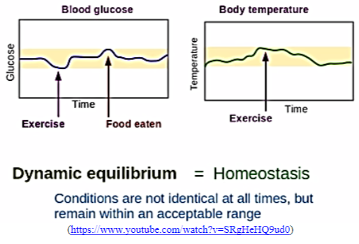
During exercise, body temperature tends to increase because the increased energy expenditure (up to 15-fold above resting levels) results in increased heat production which may exceed the ability of the body to get rid of heat. Usually, this results in fluctuation of body temperature within an acceptable range (see figure on right on the next page). In this case, the rise in temperature is not due to a change in setpoint but instead is due to the inability of the negative feedback mechanisms to cope with the amount of temperature stress.
In mammals, negative feedback regulation maintains a relatively high body temperature which allows mammals to move rapidly even when environmental temperatures are low. This type of thermoregulation depends on a relatively high metabolic rate which requires a high caloric intake.
A brief video introducing homeostasis and temperature regulation is available at https://www.khanacademy.org/partner-content/mit-k12/mit-k12-biology/v/homeostasis. An introduction to homeostasis, negative feedback, and positive feedback is available at http://www.lionden.com/homeostasis.htm.
Positive feedback is useful when there is an advantage to a rapid transition between two states, e.g. from blood flowing freely in a blood vessel to the formation of a platelet plug and blood clot in an injured blood vessel. Another example where positive feedback helps to speed up a transition is childbirth (the transition from a fetus in the uterus receiving oxygen via the placenta to a baby that has been born and is breathing on its own) (see http://www.johnwiley.net.au/highered/interactions/media/Foundations/conte nt/Foundations/homeo4a/bot.htm). Of course, positive feedback is not the only way that the body achieves rapid change; for example, neural control of muscles or secretory organs can also produce rapid responses.
This figure provides additional information about positive feedback in the formation of a platelet plug. Note that positive feedback in platelet plug formation contributes to homeostasis by preventing excessive loss of blood and thus conserving fluid and helping to maintain blood pressure.
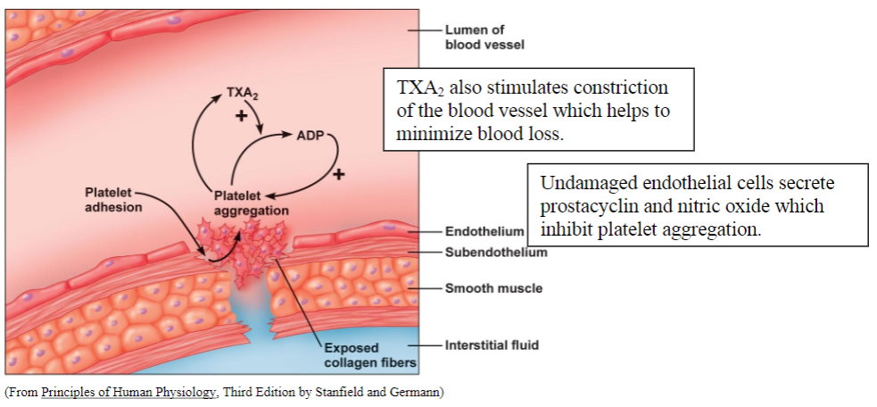
The platelet plug provides the basis for the formation of a blood clot (see figure on the last page). Undamaged endothelial cells in the lining of the blood vessels secrete chemical signals that inhibit platelet aggregation and blood clot formation, so the platelet plug and blood clot are limited to the location where the endothelium has been damaged. A description of the process of clot formation and the processes that prevent excessive clotting is available at http://www.biosbcc.net/doohan/sample/htm/Hemosta sis.htm.
II. Respiration and Circulation
This section provides an important background that your students will need in order to understand the negative feedback regulation of breathing and interpret the experiment in Section III. This background will also be helpful for your students as they think about breathing and exercise in Section IV.
If your students are not familiar with cellular respiration and ATP, you may want to introduce these topics with the analysis and discussion activity, "How do biological organisms use energy?" (available at http://serendip.brynmawr.edu/exchange/bioactivities/energy).
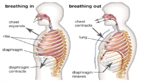
If your students are not familiar with how people breathe, you may want to provide some additional explanation. During inhalation, the lung is expanded by contraction of the diaphragm and certain rib muscles; as shown in the figure on the next page, the diaphragm pulls downward as the muscle shortens. The expansion of the lungs reduces the pressure inside the lungs below the air pressure in the surrounding environment and air moves into the lungs. During exhalation, the diaphragm and rib muscles relax and the elasticity of the lungs causes the lungs to get smaller. This increases the air pressure inside the lungs above the external air pressure so air moves out of the lungs. Thus, quiet breathing is due to the alternation between the contraction of breathing muscles which results in inhalation and relaxation of breathing muscles which results in exhalation. This rhythmic pattern of contraction and relaxation of the breathing muscles is due to a rhythmic pattern of stimulation that originates in the medulla in the brainstem. In deep breathing, contraction of certain rib muscles contributes to exhalation. A simple animation showing inhalation and exhalation is available at http://www.smm.org/heart/lungs/b reathing.htm.
The figure below provides additional information about the structure of the respiratory system. If your students are familiar with the terms alveolus/alveoli, you may want to use these terms to replace the terms air sac/air sacs on page 3 of the Student Handout.

A description and a helpful video that explain how the respiratory and circulatory systems work together to provide O2 and remove CO2 from the body's cells are available at http://www.nhlbi.nih.gov/health/health-topics/topics/hlw/whathappens. You may want to show this video to your students before they answer Question 11 or during your discussion of student answers to question 11. You may want to mention to your students that the circulatory system has multiple additional important functions such as transport of hormones, food molecules (e.g. glucose), heat, and antibodies and white blood cells to fight infection.
III. Negative Feedback and the Regulation of Breathing
Changes in the amount of air breathed into the lungs per minute (pulmonary ventilation in milliliters per minute) can result from changes in breathing rate (breaths per minute) and/or changes in depth of breathing (tidal volume in milliliters per breath). By simple algebra:
Pulmonary Ventilation = Breathing Rate x Tidal Volume
As altitude increases and the concentration of O2 in air decreases, mean arterial blood O2 levels decrease. Above ~10,000 feet, mean arterial blood O2 levels decrease to low enough levels to stimulate peripheral chemoreceptors that stimulate the breathing center in the medulla in the brain; this input stimulates increased pulmonary ventilation. If the transition to high altitude is rapid, a person is likely to experience acute mountain sickness; one major reason is that the increased pulmonary ventilation removes CO2 faster than it is produced by cellular respiration and, as CO2 levels fall, alkalosis develops. CO2 dissolved in the water of the blood is a major source of acidity:
H2O + CO2 ⇔ H2CO3 ⇔ H+ + HCO3-
Over the long term, acclimatization to living at high altitudes results in several adaptive changes:
- Increased red blood cell production so the blood carries more O2 per mL
- Increased number of capillaries within the tissues so O2 has a shorter distance to diffuse to reach the cells
- More mitochondria in cells so available O2 is used more efficiently
- Kidneys retain more H+.
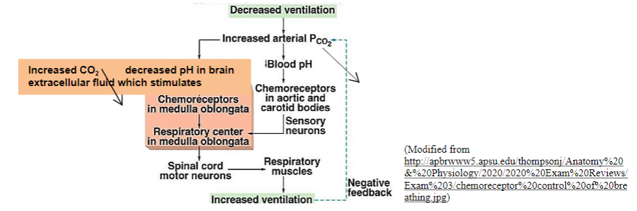
It should be noted that under most circumstances breathing is regulated primarily by the concentration of CO2 in arterial blood and the associated changes in pH. This is useful because the percent O2 saturation of hemoglobin is relatively insensitive to O2 concentrations, whereas changes in CO2 concentration in the blood have an immediate effect on pH. The following diagram shows the negative feedback regulation of CO2 levels.
You may want to introduce the section on "Developing Your Experimental Procedures" (page 5 of the Student Handout) by emphasizing that scientists need to develop reliable and valid methods of measurement in order to get accurate results. Reliable methods produce the same, consistent results on different repetitions of the same experiment. Valid methods produce results that accurately reflect the variable the scientist is trying to measure.
As your students practice breathing into the bag, emphasize the importance of:
- Making sure to fill the bag with as much air as possible
- Making sure to have a tight seal between the bag and the person’s face so no air is leaking in and out of the bag
- Maintaining a tight seal throughout the entire test interval (3 minutes for developing the procedure for evaluating the depth of breathing and 4 minutes for the actual experiment).
The observers need to be able to see the whole bag to evaluate the breathing rate and depth. Sometimes this is best achieved by having the subject stand up while breathing into the bag. A demo video is available at https://www.youtube.com/watch?v=UjzBRiX1Jpc&feature=youtu.be.
A student who has a serious respiratory or heart problem probably should not be a subject in the experiments. It may be advisable for a participating student with asthma to keep his/her inhaler close at hand for use if needed.
Results of the experiment vary for different subjects (and even for the same subject in repeated trials). One reason for this variation is that breathing is highly subject to voluntarily control. Trends may differ because of distractions in the environment, emotional influences, or other types of brain activity that may influence breathing. The role of the brain is also reflected in the subjective response your students may experience toward the end of the four-minute interval when the air in the bag has increased levels of CO2 and decreased levels of O2 (see question 15b). Due to the variability in results, you may want to collect individual data from all the students in your class and calculate class averages for the number of breaths and depth of breathing (taking into account the methodological issue discussed in the next paragraph).
Question 16 is included in case one or more of the subjects in a group fails to complete the entire four minutes of breathing into the bag. In that case, students should not compare the earlier averages of breathing rates and depth for four students with later averages of breathing rates and depth for fewer students. Possible alternative approaches include:
- Using only the data for the 30-second intervals when all students were breathing into the bag
- Calculating averages for only the students who completed all four minutes of breathing into the bag
- Plotting individual trends for each student.
To choose the best approach, students should consider the pattern of when the subjects in their group stopped breathing into the bag.
In our experience, changes in the rate of breathing are inconsistent both within and between subjects. In contrast, most subjects show a relatively consistent trend to the increased depth of breathing. These observations are in accord with scientific research results which show that increased CO2 is associated with more consistent increases in depth of breathing and smaller, inconsistent increases in the rate of breathing. You may want to relate these findings to the observation that deeper breathing is more efficient than more rapid breathing as a way to increase the intake of O2 and release of CO2. To understand the reason for this, consider what happens when you begin to inhale. The first air to enter the air sacs in the lungs is the air that was just exhaled into the bronchioles, bronchi, trachea, pharynx, mouth, and nose (see bottom figure on page 6). This recently exhaled air has low O2 and high CO2, so it is less useful than fresh air for gas exchange in the air sacs of the lungs. A very shallow breath will bring only this recently exhaled air into the air sacs. A deeper breath will bring proportionately more fresh air with high O2 and low CO2 into the air sacs; this will increase the diffusion of O2 into the blood and diffusion of CO2 out of the blood.
If your results are similar to our findings, discussion of question 21 will provide the opportunity to talk about the importance of testing even predictions that seem eminently reasonable. Failure of experimental results to confirm a prediction can lead to new insights and an improved hypothesis – in this case, the recognition that negative feedback regulation of blood levels of CO2 affects primarily depth of breathing, with less effect or no effect on breathing rate. This result makes sense biologically since the increased depth of breathing is a more efficient way of improving gas exchange.
The experiment described in the Student Handout demonstrates the importance of high levels of CO2 and/or low levels of O2 in stimulating deeper breathing, but this experiment does not allow students to distinguish the relative importance of changes in levels of CO2 vs. O2. In order to estimate the effect of changes in O2 levels with relatively little change in CO2 levels, you may want to add the following activity.
Repeat the experiment described on page 5 of the Student Handout while breathing into a plastic bag that contains a small bowl with KOH (which absorbs CO2). You need to be very cautious in handling KOH since it is caustic. The specific procedures are as follows:
- Put a piece of filter paper in the bottom of a finger bowl, and use a spatula to put approximately 6-7 pieces of KOH in the finger bowl.
- Moisten the filter paper with a few scattered drops of water (KOH has to be moist in order to absorb CO2).
- Cut a piece of cheesecloth a few layers thick and big enough to surround the finger bowl; use a rubber band to close the cheesecloth over the finger bowl.
- Place the finger bowl in an 8-gallon plastic bag which has been filled with air.
- Carry out the breathing experiment.
- After the experiment, dispose of the KOH in the jar provided.
IV. Homeostasis and Changes in Breathing Due to Exercise
Question 24 provides the opportunity for students to recognize that they already know quite a bit about changes in breathing during and after exercise. For the first column, students should be encouraged to report observations concerning breathing, not recommendations or interpretations. Interpretations can be provided in the third column, in which students link their observations to the understanding of homeostasis they have developed in earlier sections of this activity.
This figure shows a typical set of experimental results for exercise-related changes in pulmonary ventilation (= breathing rate x depth of breathing). Research evidence indicates that both breathing rate and depth of breathing increase during and after aerobic exercise. See pages 12-13 for an explanation of the rapid early rise in pulmonary ventilation, which can even begin slightly before the exercise begins
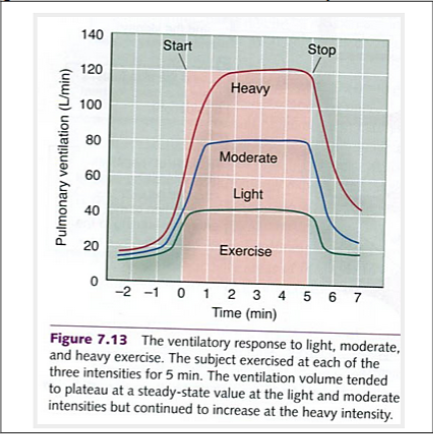
(http://completesoccertraining.blogspot.com/2012/06/respiratory-responses-to-acute-exercise.html )
Question 25 is designed to stimulate students to develop a question that can expand their understanding beyond what they already know and provide new information about changes in breathing due to exercise. Scientists do need to replicate previous findings to ensure their reliability but the observation that rate and depth of breathing increase during vigorous physical activity is sufficiently well-established that you should encourage students to think about how to expand their understanding beyond this simple observation. Appropriate questions might include:
- Is the change in breathing greater for aerobic exercise (e.g. jogging in place) vs. strength training (e.g. using resistance bands) or yoga?
- Does breathing rate double if a person exercises twice as fast (e.g. doubling the number of jumping jacks in a minute) or twice as hard (e.g. two resistance bands instead of one for strength training exercise)?
- How long does it take for breathing to return to resting levels after different types and durations of exercise?
- Does the rate of breathing at rest and after exercise differ between athletes and non-athletes? (If a student group wants to investigate this type of question, they should find at least one other student group to cooperate with in order to get a meaningful sample size.)
There are several possible ways that your students can measure the rate and depth of breathing before and after exercise. You may want to pilot test one or both of the methods described below and choose one to recommend for your students or you may want to have students record their own breathing before and after exercise. If you develop any improvement for either of the methods described below or a good alternative method for measuring the rate and depth of breathing, please let me know (iwaldron@sas.upenn.edu). Thank you!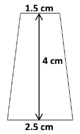
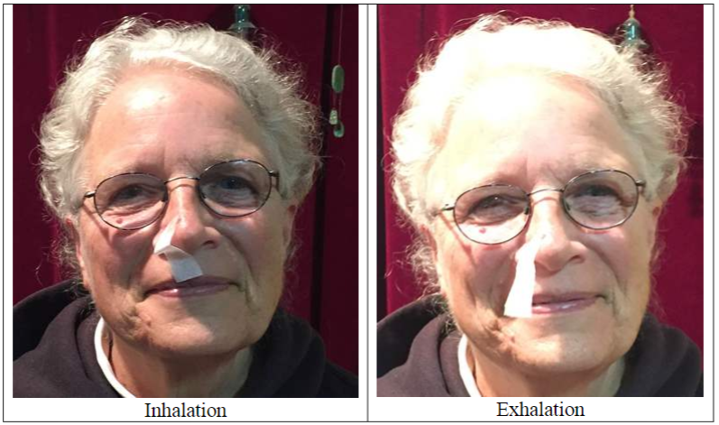
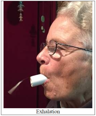
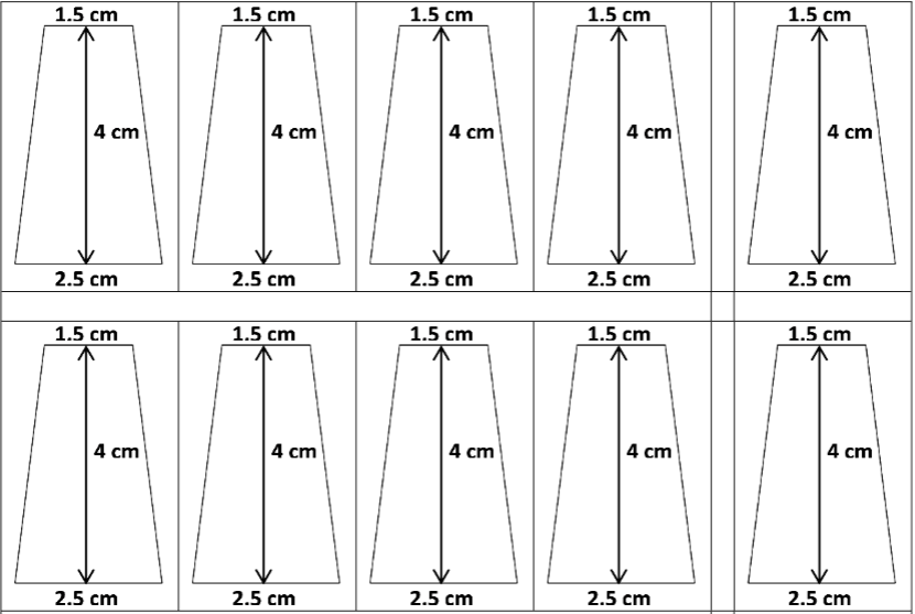
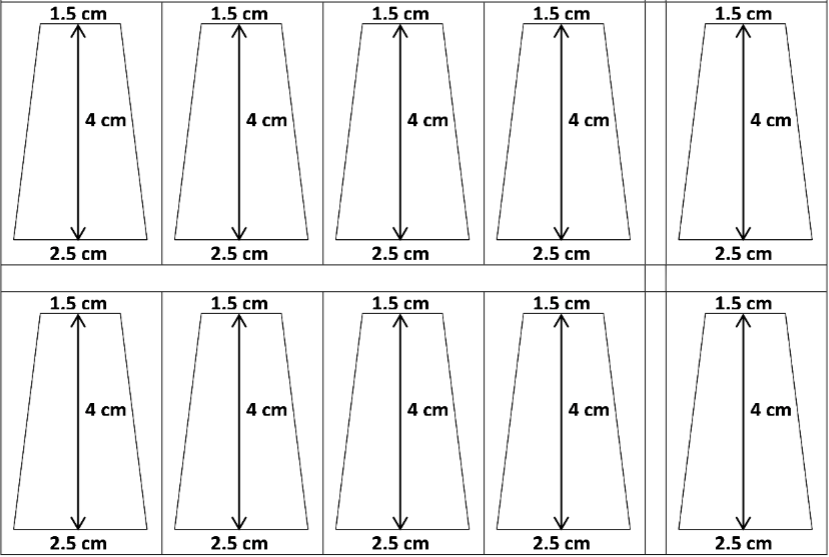
One low-tech, effective method for detecting each breath uses a small piece of facial tissue taped to the subject's nose so the tissue hangs over the edge of one nostril (see figure below). A piece of tissue the shape and size shown works well for measuring the rate and depth of breathing. This method only works if you keep your mouth closed and breathe through your nose. If you want to use this method, you may find it helpful to use the page of templates provided on the next to the last page of these Teacher Preparation Notes. If you use this method, you will probably want to have one scissors, one facial tissue, and one length of sensitive skin medical tape for each group of four students
An alternative approach to measuring breathing rate and depth is to use a 2-inch length of ¾ inch PVC pipe with a metallic streamer taped so that it flops over one edge. (PVC pipe diameter refers to the internal diameter, not the external diameter.) We have used a metallic streamer ~¼ inch wide, with a total length of ~3 ¾ inches; we obtained the streamers from a "foil fringe garland" obtained at a party store.
To prepare the 2-inch lengths of PVC pipe, we first used a hack saw to cut the needed number of pieces of pipe and then smoothed the edges of the ends using an X-Acto knife or a single-edge razor blade in a holder. For sanitary reasons, you will need one piece of pipe for each student in your largest class.
To disinfect the pieces of pipe for use in another class, we recommend the following procedure:
- Wash your hands with soap and water for at least 30 seconds. Rinse and then dry with a paper towel.
- Remove the streamers and tape and scrub the inside and outside of each PVC tube using a brush or pipe cleaner and soap and water until the tube is clean.
- Shake extra water off the tubes. Soak the tubes in 70% isopropyl alcohol for 5 minutes or in bleach (5 mL of 6% bleach in 8 ounces of water) for 3 minutes or microwave the tubes for 5 minutes.
- Rinse the tubes. Place the tubes on a clean surface to dry.
- Be sure to wash your hands with soap and water for at least 30 seconds before handling the dry tubes to store them in a plastic bag.
Any student who has been excused from physical education may need to be excused from participating as a subject in the experiments in this section. You may want a student with asthma to keep his/her inhaler close at hand for use if needed. Students should be advised to wear appropriate clothing and footwear for physical activity.
Before students begin to design their experiments, you may want to have them discuss basic methodological points such as:
- The importance of standardizing their physical activity and their methods for recording changes in breathing
- The importance of getting a valid baseline measure of breathing rate and depth before exercise for comparison to breathing rate and depth after exercise
- The importance of changing only the variable they are testing and controlling other variables
- The importance of replication (e.g., having each member of the group participates as a subject).
We have found it useful to check that each datasheet corresponds to the experimental design and clearly specifies the observations to be recorded. Each student group’s analysis plan should include some way of summarizing their results to answer their question.
Your students may notice sweating during the experiment, and you will probably want to point out that negative feedback regulation of body temperature occurs not only in response to environmental changes in temperature (section I), but also in response to internal changes in metabolism which result in changes in the amount of heat produced. For example, physical activity results in increased metabolism and production of heat. During cellular respiration, only about 50% of the energy in nutrient molecules is transferred to ATP and the other 50% is converted to heat. During ATP expenditure by cells, another 25% of the energy derived from food becomes heat. Internal friction during physical activity contributes to additional heat production. Overall, during muscle activity, only about 20-25% of the chemical energy expended is captured in the kinetic energy of muscle contraction and the rest of the energy is converted to heat. These observations provide the opportunity to reinforce the principle that all types of energy conversion result in the production of heat.
Your students will probably also notice that the heart beats faster and stronger during exercise. During exercise, the total amount of blood pumped per minute can increase as much as fourfold in an untrained person and eightfold in a trained athlete. Most of the increase in the amount of blood pumped per minute goes to the active muscles; at rest, ~20% of blood flow goes to skeletal muscles, whereas during vigorous exercise ~90% of blood flow goes to skeletal muscles. You may want to link student observations of faster and stronger heartbeats to the discussion in Section II of how the respiratory and circulatory systems cooperate to provide the O2 needed for cellular respiration and remove the CO2 produced by cellular respiration.
You may want to have each student group prepare a poster with their question or hypothesis, a summary of their procedures and results, and their conclusions. Then student groups can share their results for:
- A discussion of best practices in the design and interpretation of experiments
- A more complete picture of changes in breathing due to exercise.
You may want to follow-up with a discussion of the following question:
What do you think would be the most useful experiment to do next? Explain your reasoning.
The paragraphs below introduce complexities that may not be appropriate for your students. However, if this level of complexity will not overwhelm your students, this information provides the opportunity to reinforce the important principle that even when experimental results are compatible with a hypothesis, it is important to consider possible alternative interpretations before concluding that the results support the hypothesis.
During exercise, both breathing rate and depth of breathing increase as the intensity of exercise increases. Although this increase in breathing during exercise appears compatible with the negative feedback regulation discussed in section III, multiple lines of evidence indicate that this negative feedback is not the primary cause of increased breathing during exercise. For example, during exercise, blood levels of O2 and CO2 generally show only small and inconsistent changes from the levels observed at rest; these small and inconsistent changes in blood levels of O2 and CO2 are in sharp contrast to the substantial increases in breathing rate and depth during many types of exercise.
A broad range of additional evidence supports the conclusion that multiple mechanisms contribute to the increase in breathing during exercise.
- Available evidence indicates that the motor areas of the cerebral cortex simultaneously stimulate the motor neurons of the exercising muscles and the respiratory neurons in the medulla. The direct input from motor areas to the respiratory center is a major reason for the very rapid increase in breathing at or even slightly before the beginning of the exercise.
- Sensory receptors that respond to joint and muscle movement provide input that stimulates increased breathing during exercise. (This response can also be observed during passive movement of a person's limbs).
- During exercise, increases in body temperature and epinephrine levels in the blood help to stimulate increased breathing.
- During intense exercise, the production of lactic acid during anaerobic fermentation can result in a reduced pH which can help to stimulate increased breathing during and after exercise.
The multiple reinforcing mechanisms that contribute to the regulation of breathing are typical of the redundancy observed in many biological regulatory systems.
(http://philschatz.com/anatomy-book/resources/1909_Blood_Clotting.jpg)