
18.12: Laboratory Activities and Assignment
- Page ID
- 53799

Laboratory Activities and Assignment
Part 1: Review of Blood Vessels
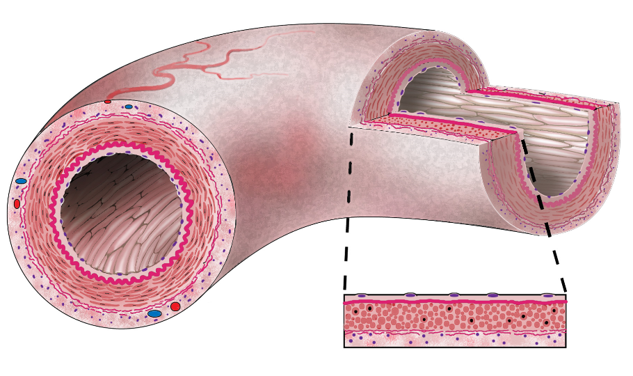
1. On the diagram of a blood vessel below, label the following:
|
|
|
2. For each description below, write one of the choices below in the blank spaces provided. Choices can be used more than once.
- arteries
- arterioles
- capillaries
- veins
- venules
a. __________________ These large blood vessels carry blood to the heart.
b. __________________ & __________________ These blood vessels contain valves.
c. __________________ These large blood vessels carry blood away from the heart.
d. __________________ These smaller blood vessels carry blood from the vessels of where materials are transferred between the blood and the tissues to the large blood vessels carrying blood to the heart.
e. __________________ These are the smallest blood vessels in the body.
f. __________________ These smaller blood vessels carry blood to the vessels where materials are transferred between the blood and the tissues.
g. __________________ The inferior vena cava is one of these.
h. __________________ The aorta is one of these.
i. __________________ The vessels where materials are transferred between the blood and the tissues.
j. __________________, __________________, & __________________ Blood vessels that do not have valves.
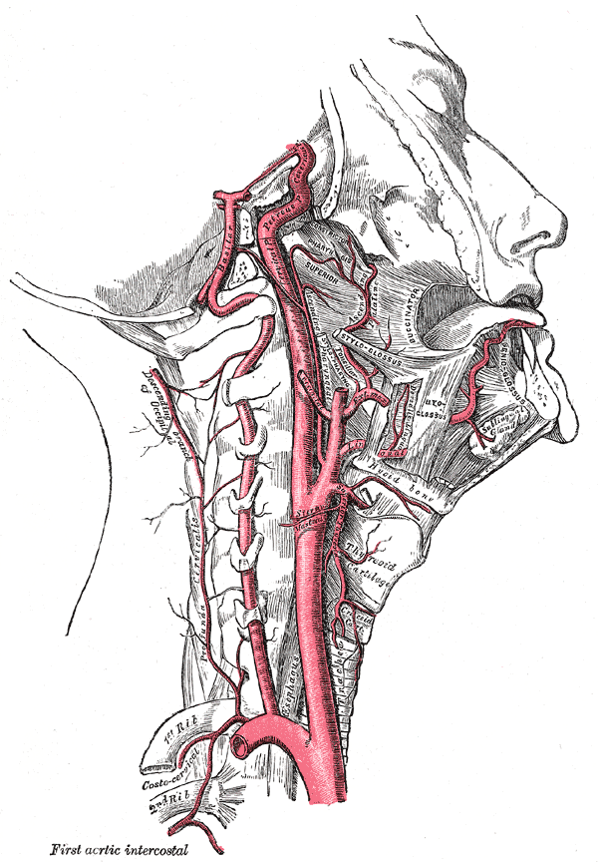
3. Name the arteries on the diagram below. Use the choices given to fill in each blank.
|
|
|
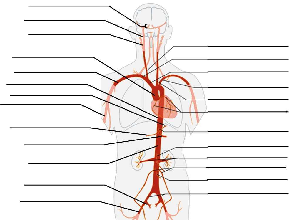
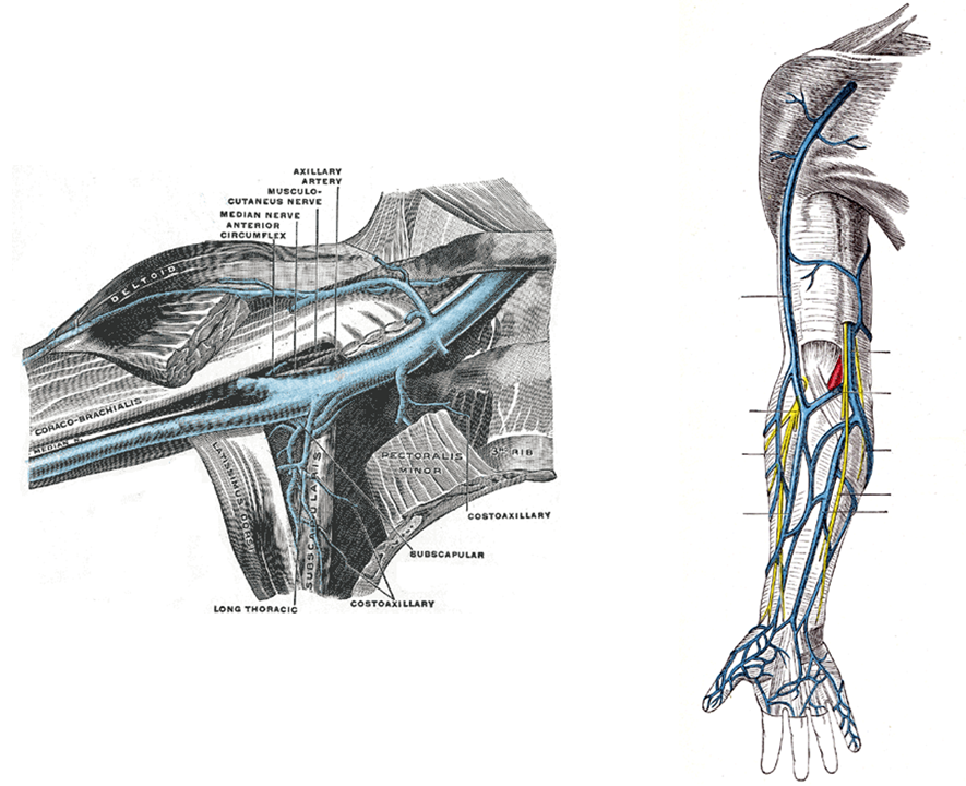
3. Label the following arteries on the diagram below:
|
|

4. Label the following arteries on the diagram below:
|
|
5. Label the following arteries on the diagram below:
|
|
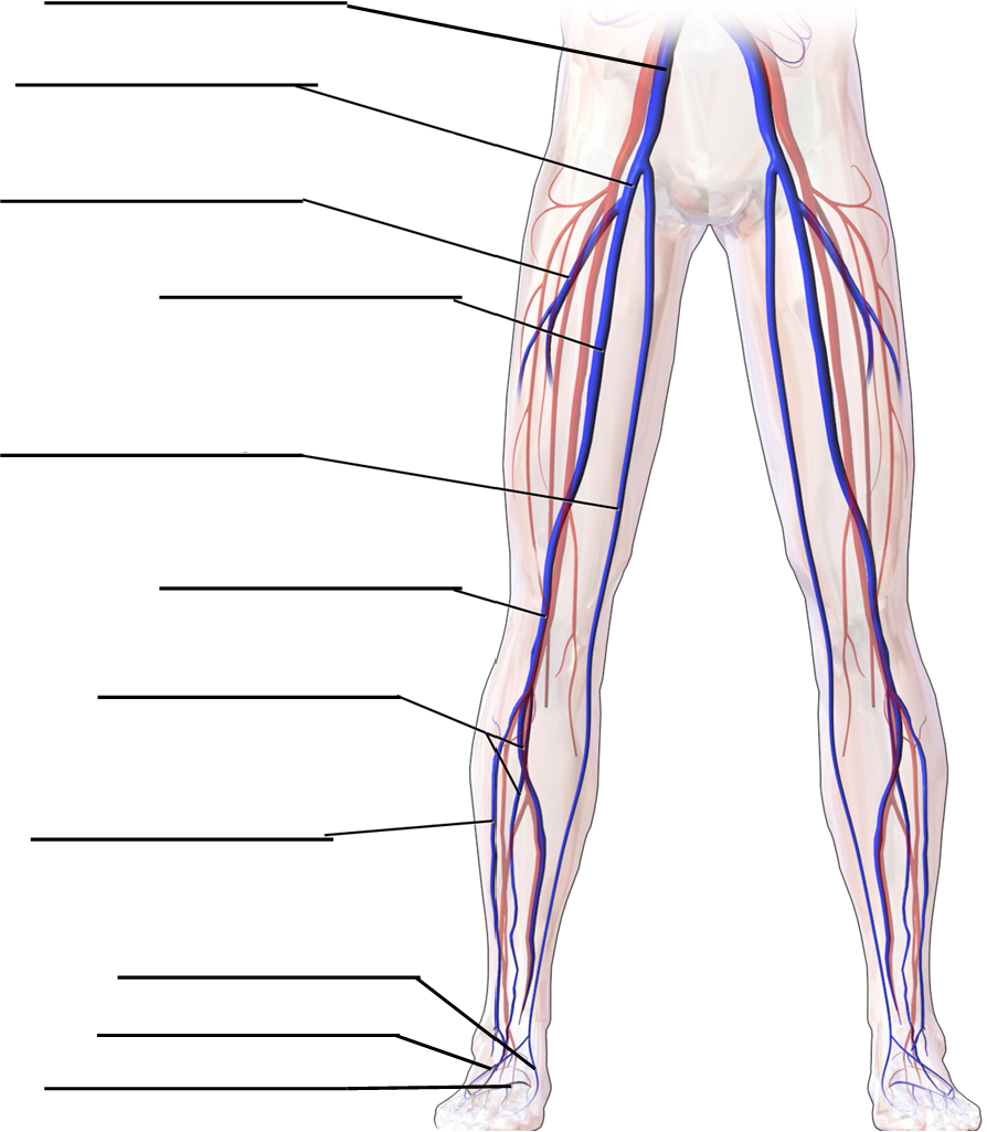
6. Write the number in each blank that corresponds with the name of each artery in the diagram.
|
_____ anterior tibial artery _____ deep femoral artery _____ external iliac artery |
_____ femoral artery _____ genicular artery _____ lateral femoral circumflex artery |

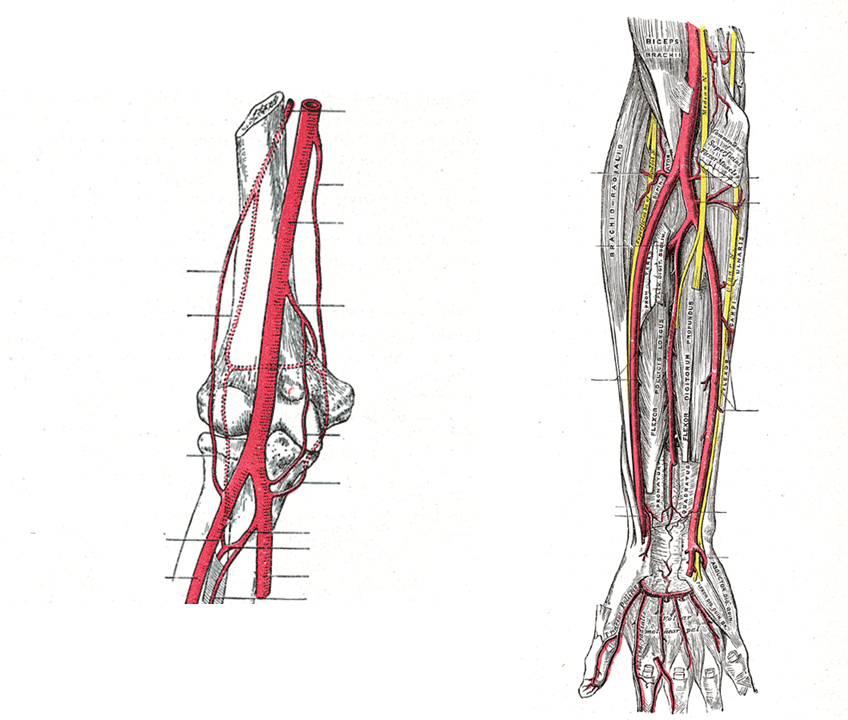
7. Label the following veins on the diagrams below:
|
|
|
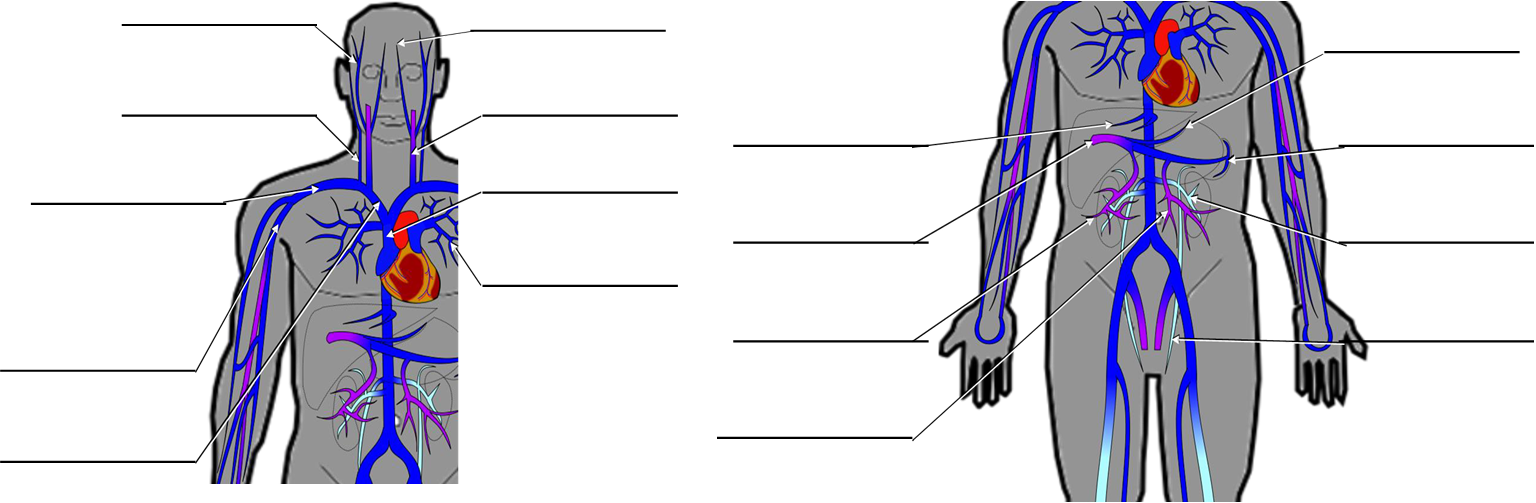
6. Label the following veins on the diagrams below:
|
|
7. Label the following veins on the diagrams below:
|
|
Part 2: Examining the Histology of the Blood Vessels
- Obtain the slides listed below that are available for today’s lab.
- Focus on each sample and identify the structures listed for each type of tissue.
- Indicate the total magnification you make each illustration at in the space provided.
- Illustrate each tissue you observe with the microscope at the magnification you listed.
- Label each illustration with the structures listed for each.
Large Elastic Artery Cross Section
Label the tissue with: tunica interma, tunica media, tunica adventitia, smooth muscle, elastic fibers, vasa vasorum

Capillary-Containing Tissue
Label the tissue with: capillary, nucleus of endothelial cell
Vein Cross Section
Label the tissue with: tunica interma, tunica media, tunica adventitia, nucleus of endothelial cell, valve
Part 3: The Arteries and Veins of the Systemic Circuit
- For each blood vessel in the table below, identify it on anatomical models, anatomical diagrams provided by your instructor, or on the virtual model at www.zygotebody.com.
- Fill out the table to tell the location of the blood vessel.
- Indicate whether this blood vessel supplies blood or drains it by checking the appropriate box.
- Tell what organs or body regions are supplied or drained by the blood vessel.
- Indicate whether this blood vessel is paired (right and left) or if there is only one by checking the appropriate box.
Blood Vessels of the Trunk
|
Blood Vessel |
Location |
Supplies |
Drains |
Organ(s) Supplied/Drained |
Paired |
Unpaired |
|---|---|---|---|---|---|---|
|
abdominal aorta |
||||||
|
azygous vein |
||||||
|
brachiocephalic trunk |
||||||
|
brachiocephalic vein |
||||||
|
common carotid artery |
||||||
|
common hepatic artery |
||||||
|
common iliac artery |
||||||
|
common iliac vein |
||||||
|
coronary arteries |
||||||
|
hemiazygous vein |
||||||
|
hepatic portal vein |
||||||
|
hepatic veins |
||||||
|
external jugular vein |
||||||
|
gonadal artery |
||||||
|
gonadal vein |
||||||
|
inferior mesenteric artery |
||||||
|
inferior mesenteric vein |
||||||
|
intercostal arteries |
||||||
|
intercostal veins |
||||||
|
internal jugular vein |
||||||
|
left gastric artery |
||||||
|
renal artery |
||||||
|
renal vein |
||||||
|
splenic artery |
||||||
|
splenic vein |
||||||
|
subclavian artery |
||||||
|
subclavian vein |
||||||
|
superior mesenteric artery |
||||||
|
superior mesenteric vein |
||||||
|
superior vena cava |
||||||
|
thoracic aorta |
Blood Vessels of the Head and Neck
|
Blood Vessel |
Location |
Supplies |
Drains |
Organ(s) Supplied/Drained |
Paired |
Unpaired |
|---|---|---|---|---|---|---|
|
basilar artery |
||||||
|
brachiocephalic trunk |
||||||
|
brachiocephalic vein |
||||||
|
cerebral arterial circle (aka circle of Willis) |
||||||
|
common carotid artery |
||||||
|
external carotid artery |
||||||
|
external jugular vein |
||||||
|
facial artery |
||||||
|
internal carotid artery |
||||||
|
internal jugular vein |
||||||
|
superficial temporal artery |
||||||
|
superior vena cava |
||||||
|
vertebral artery |
||||||
|
vertebral vein |
Blood Vessels of the Upper Limbs
|
Blood Vessel |
Location |
Supplies |
Drains |
Organ(s) Supplied/Drained |
Paired |
Unpaired |
|---|---|---|---|---|---|---|
|
axillary artery |
||||||
|
axillary vein |
||||||
|
basilic vein |
||||||
|
brachial artery |
||||||
|
brachial vein |
||||||
|
brachiocephalic trunk |
||||||
|
brachiocephalic vein |
||||||
|
cephalic vein |
||||||
|
deep palmar (arterial) arch |
||||||
|
digital arteries (of the hand) |
||||||
|
digital veins (of the hand) |
||||||
|
median cubital vein |
||||||
|
radial artery |
||||||
|
radial vein |
||||||
|
subclavian artery |
||||||
|
subclavian vein |
||||||
|
superficial (arterial) palmar arch |
||||||
|
ulnar artery |
||||||
|
ulnar vein |
Blood Vessels of the Lower Limbs
|
Blood Vessel |
Location |
Supplies |
Drains |
Organ(s) Supplied/Drained |
Paired |
Unpaired |
|---|---|---|---|---|---|---|
|
common iliac artery |
||||||
|
common iliac vein |
||||||
|
deep femoral artery |
||||||
|
digital arteries (of the foot) |
||||||
|
digital veins (of the foot) |
||||||
|
external iliac artery |
||||||
|
external iliac vein |
||||||
|
femoral artery |
||||||
|
femoral vein |
||||||
|
fibular artery |
||||||
|
great saphenous vein |
||||||
|
plantar artery |
||||||
|
plantar venous arch |
||||||
|
dorsal venous arch |
||||||
|
popliteal artery |
||||||
|
popliteal vein |
Attributions
Part 1: Review of Blood Vessels
- "Anatomy and Physiology Lab Homework" by Laird C Sheldahl is licensed under CC BY-SA 4.0
- "Aorta branches.svg" by Mikael Häggström is licensed under CC BY-SA 3.0 / A derivative from the original work
- "Digital Histology" by Department of Anatomy and Neurobiology and the Office of Faculty Affairs, Virginia Commonwealth University School of Medicine and the ALT Lab at Virginia Commonwealth University is licensed under CC BY 4.0
- "Gray's Anatomy plates" by Henry Vandyke Carte is in the Public Domain
- "Medical gallery of Blausen Medical 2014" by Blausen.com staff is licensed under CC BY 3.0
- "Thigh arteries schema numbered.svg" by Jecowa is licensed under CC BY 3.0 / A derivative from the original work
Part 2: Examining the Histology of the Blood Vessels
- "BIOL 250 Human Anatomy Lab Manual SU 19" by Yancy Aquino, Skyline College is licensed under CC BY-NC-SA 4.0
Part 3: The Arteries and Veins of the Systemic Circuit
- "Systemic Blood Vessels" by Dongho Kim is licensed under CC BY-NC-SA 4.0