
18.3: Branches of the Aorta
- Page ID
- 53790

Branches of the Aorta

Above: Generalized illustration of the structure of the aorta.
The aorta, the largest artery in the body, is separated from the heart by the aortic valve. The aorta ascends (ascending aorta), arches (aortic arch), and then descends (descending aorta) posterior to the heart through the thoracic cavity (thoracic aorta), through the diagram and then through the abdominal cavity (abdominal aorta). In the pelvic cavity, the aorta splits right and left and now is called the right common iliac artery and the left common iliac artery. All of the oxygenated blood leaving the heart leaves through the aorta and then enters the multiple arteries branching off of the aorta to reach all the tissues and organs of the body.
Along the aortic arch, arteries branch including the brachiocephalic trunk, the left common carotid artery, and the left subclavian artery. The brachiocephalic trunk (brachial = arm, cephalic = head) branches into the right common carotid artery bringing blood to the head and the right subclavian artery bringing blood to the right arm. When "common" is a part of a vessel's name, it means it splits into internal and external parts. For example, the right and left common carotid arteries both branch into internal carotid arteries and external carotid arteries. The descending thoracic aorta continues branching into eleven pairs (right and left) of posterior intercostal arteries. A pair of superior phrenic arteries (right and left) branch from the aorta to serve the superior diaphragm. The thoracic aorta continues to descend through a foramen in the diaphragm and into the abdominal cavity (inferior of the diaphragm, the aorta is now called the abdominal aorta).

Above: (Left) Diagram of the branches of the aorta located in the thoracic cavity. (Right) Diagram of the arteries branching from the abdominal aorta.
A pair of inferior phrenic arteries serve the inferior aspect of the diaphragm. The descending abdominal aorta has a large anterior branch called the celiac trunk which branches into three arteries: left gastric artery (superior branch, to the stomach), common hepatic artery (right branch, to the liver), and splenic artery (left branch, to the spleen and pancreas). A pair of adrenal arteries branch off the aorta (right and left, to the adrenal glands), followed by an anterior branch called the superior mesenteric artery (to the small intestine and colon) and then a pair of renal arteries traveling to the kidneys (right and left). Additional paired right and left branches occur including gonadal arteries (to reproductive organs) and the lumbar arteries (to the body wall of the posterior trunk) as well as an anterior branch called the inferior mesenteric artery (to the small intestine and colon). Finally, in the pelvic region, the aorta splits into two large arteries: the right common iliac artery and the left common iliac artery to provide blood to the pelvic organs (internal iliac arteries) and to the lower limbs (external iliac arteries).

Above: Arteries of the trunk, anterior view.

Above: Arteries of the celiac trunk shown with the stomach flipped up above of its normal position.
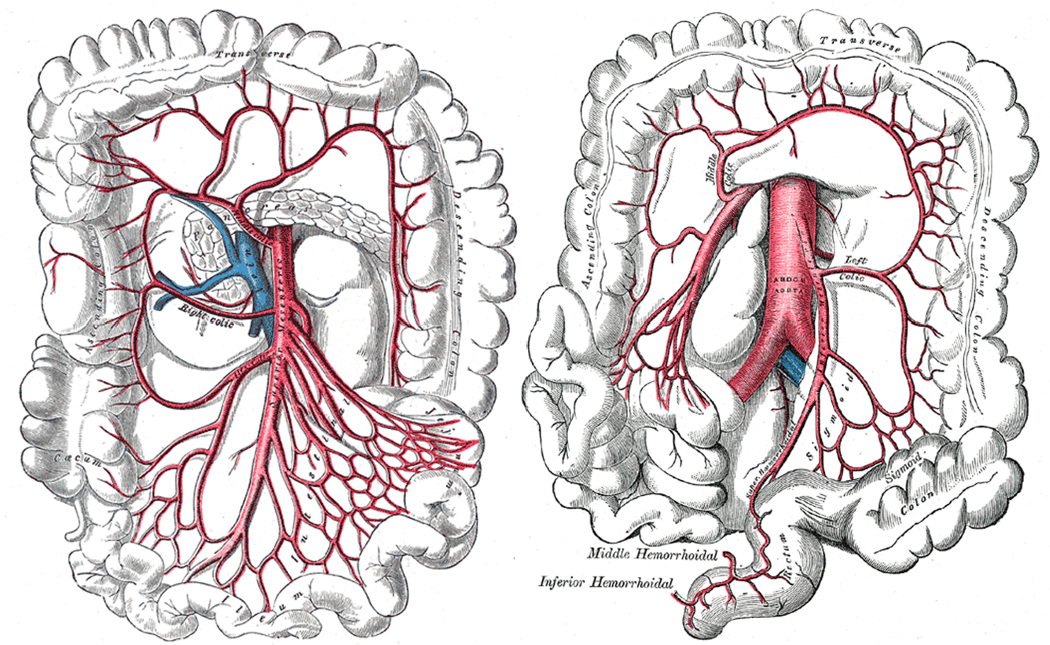
Above: (Left) Superior mesenteric artery and (right) inferior mesenteric artery.
Attributions
- "Anatomy 204L: Laboratory Manual (Second Edition)" by Ethan Snow, University of North Dakota is licensed under CC BY-NC 4.0
- "Anatomy and Physiology" by J. Gordon Betts et al., OpenStax is licensed under CC BY 4.0
- "Gray's Anatomy plates" by Henry Vandyke Carte is in the Public Domain