
16.2: Vision Anatomy
- Page ID
- 53757

Vision Anatomy
Vision (sight) is perception of light emitted or reflected from objects in the environment. Visible light is electromagnetic radiation with wavelengths from 400 to 750nm. Light must cause a photochemical reaction in order to produce a nerve signal that is sent to the occipital lobes of the brain for processing. Radiation below 400 nm has so much energy it kills cells (ultraviolet or UV light). Radiation above 750 nm has too little energy to cause photochemical reaction (it only warms the tissue) (infrared).
Macroscopic Anatomy of the Eye
The human eye is an organ that reacts to light and allows vision. The eye is the specialized organ of sight which has three principal layers, the fibrous tunic (tunica fibrosa), the vascular tunic (tunica vasculosa) and the neural tunic (tunica interna). Furthermore, there are two main chambers, the anterior chamber, containing aqueous humor and the posterior chamber, that contains vitreous humor. In the neural tunic of the retina, light propagates from the ganglionic cells through the bipolar cells to the rods cells and cone cells, which, somewhat paradoxically hyperpolarize opposite the direction of light.
|
Tunic of the Eyeball |
Layer Type |
Structures |
Function |
|---|---|---|---|
|
tunica fibrosa (fibrous tunic) |
fibrous layer |
sclera, cornea |
provides protection |
|
tunica vasculosa (vascular tunic) |
vascular layer (contains blood vessels) |
choroid, ciliary body, iris |
provides nourishment |
|
tunica interna (neural tunic) |
internal layer |
retina, optic nerve |
converts light to nerve signal and sends to the occipital lobes of the brain |

Above: Transverse section of the eye with a superior view (left) image of eye section and (right) illustration of eye structures.
|
Cavity of the Eye |
Location |
Fluid (Humor) |
Function |
|---|---|---|---|
|
anterior cavity |
anterior to the lens of the eye (cornea to lens) |
aqueous humor (watery fluid) |
aqueous humor helps to maintain the intraocular pressure and supply of nutrients to the lens and the cornea |
|
posterior cavity |
posterior to the lens of the eye (between the lens to retina) |
vitreous humor (jelly-like substance) |
vitreous humor holds the retina against the choroid layer and prevents the eyeball from collapsing |
Similar to the eyes of other mammals, the human eye's non-image-forming photosensitive ganglion cells in the retina receive light signals which affect adjustment of the size of the pupil, regulation and suppression of the hormone melatonin and entrainment of the body clock.
The eye is not shaped like a perfect sphere, rather it is a fused two-piece unit, composed of the anterior segment and the posterior segment. The anterior segment is made up of the cornea, iris and lens. The cornea is transparent, is more curved, and is linked to the larger posterior segment, composed of the vitreous humor, retina, choroid and the outer white shell called the sclera. The cornea is typically about 11.5 mm (0.3 in) in diameter, and 0.5 mm (500 μm) in thickness near its center. The posterior chamber constitutes the remaining five-sixths; its diameter is typically about 24 mm. The cornea and sclera are connected by an area termed the limbus. The iris is the pigmented circular structure concentrically surrounding the center of the eye, the pupil, which appears to be black. The size of the pupil, which controls the amount of light entering the eye, is adjusted by the iris' dilator and sphincter muscles.
Light enters the eye through the cornea, through the pupil and then through the lens. The lens shape is changed for near focus (accommodation) and is controlled by the ciliary muscle. Photons of light falling on the light-sensitive cells of the retina (photoreceptor cone cells and rod cells) are converted into electrical signals that are transmitted to the brain by the C.N. II optic nerve and interpreted as sight and vision.
|
Structure |
Location |
Function |
|---|---|---|
|
choroid |
posterior portion of tunica vasculosa |
contains melanin (pigment) that absorbs light to prevent reflection back into the eyeball, which could cause blurred vision; provides nourishment to the retina |
|
ciliary body |
composed of ciliary process and ciliary muscle, connected by suspensory ligaments |
helps in adjusting the shape of the lens for near and far vision; produces aqueous humor |
|
conjunctiva (bulbar/ocular) |
covers the sclera (white part of the eye) |
lubrication and protection by producing fluid and mucus; monitors microorganisms present to prepare immune response |
|
conjunctiva (palpebral) |
thin mucus membrane that lines the inside of the eyelids |
secretes mucus to reduce friction and moisten the eyeball surface |
|
cornea |
the transparent, curved coat of the eyeball that covers the iris and pupil |
admits light to the interior; responsible for bending light to focus toward the back of the eye; dense fibrous nature provides protection |
|
fovea centralis |
a small depression in the macula lutea |
area of the sharpest vision because of the abundance of cone cells |
|
iris |
pigments portion around the central aperture (the pupil) |
constrictor and dialator muscle of the iris change the diameter of the pupil to regulate the amount of light striking the retina |
|
lens |
posterior to the iris and pupil |
fine-tunes bending of light to focus image on the retina; divides the eyeball into two cavities/chambers: anterior cavity/chamber and posterior cavity/chamber |
|
macula lutea |
oval-shaped central region of the retina containing higher concentration of photoreceptors; fovea centralis is located in its center |
region of highest-quality vision due to the presence of lots of photoreceptors |
|
optic disc |
location where the optic nerve exits the eyeball; lacks photoreceptors |
the blind spot of the eye where no photoreceptors are located due to the presence of the optic nerve |
|
optic nerve (C.N. II) |
exits the eyeball at the optic disc, through the optic canal of the skull, into the cranial cavity (optic chiasma, to optic tract, to occipital lobe which contains the visual cortex) |
transmits light information from the eye to the visual cortex in the occipital lobe of the brain |
|
ora serrata |
where the retina meets the ciliary body; has a serrated (jagged) appearance |
location where photosensitive tissue transitions into non-photosensitive tissue |
|
plica semilunaris |
a semilunar (crescent-shaped) fold in the bulbar/ocular conjunctiva in the medial corner of the eye |
aids in movement of lacrimal fluid across the eye (for lubrication of the eye) |
|
pupil |
contractile hole located in the center of the iris of the eye |
allows light to enter the retina |
|
retina |
innermost layer of the posterior eye; contains two layers (outer pigmented layer and inner neuronal layer) |
outer pigmented layer prevents light scattering and absorbs light; inner neuronal layer contains two types of photoreceptors, rod cells and cone cells, that collect light information and transmit to neurons |
|
sclera |
covers the eyeball except at the cornea; covered by the bulbar/ocular conjunctiva; white part of the eye |
resists punctures and protects the eye; helps maintain pressure in the eye to keep its shape and keep the retina adhered to the back of the eye; attachment point of extrinsic eye muscles |

Above: Structures of the eye.
Clinical Application: "Blind Spot"

Above: Photograph of left human retina, anterior view.
There are no rods or cones (visual receptors) at the optic disc (the structure formed from where CN II (Optic n.) connects to the retina) because that space is being occupied up by the optic nerve fibers and retinal vasculature. Therefore, as light passes through the eye and onto the retina for visual processing, the light that shines on the optic disc is not processed into eyesight, causing a “blind spot” in each eye’s normal field of vision. You can “see” your blind spot for yourself using the image below. Close your right eye and look directly at the center of the plus sign with your left eye (don’t look away or at the circle). Hold the image about one foot away from your face (how close you need to view the image will depend on the size of the image (whether in print or digital format which is dependent on the zoom in your view)). While looking directly at the plus sign with your left eye, the white circle should disappear from your peripheral vision because that area in your field of vision is shining on your optic disc. You typically don’t notice your blind spots because your brain takes vision from your other eye and “fills in” your blind spots for a full field of view. Unlike the optic disc, the fovea centralis, a structure on the retina near the optic disc, has the highest concentration of cones and processes the most acute vision necessary for reading, driving, etc.

Above: Diagram for "seeing" your blind spot.
Clinical Application: Conjunctivitis
Conjunctivitis (or pink eye) is a common inflammation of the conjunctiva of the eye. Symptoms typically include redness, itching, increased tear production, and crusting around the eyes. It is highly contagious, and can be spread by direct contact (i.e. handshake or hug) or indirect contact (i.e. touching a contaminated pencil or doorknob). Although the inflammation is usually not severe and will resolve on its own, the process can be sped up with antibiotic eye drops.
Microscopic Anatomy of the Eye
The human eye can differentiate between about 10 million colors and is possibly capable of detecting a single photon. Rod and cone cells in the retina allow conscious light perception and vision including color differentiation and the perception of depth.
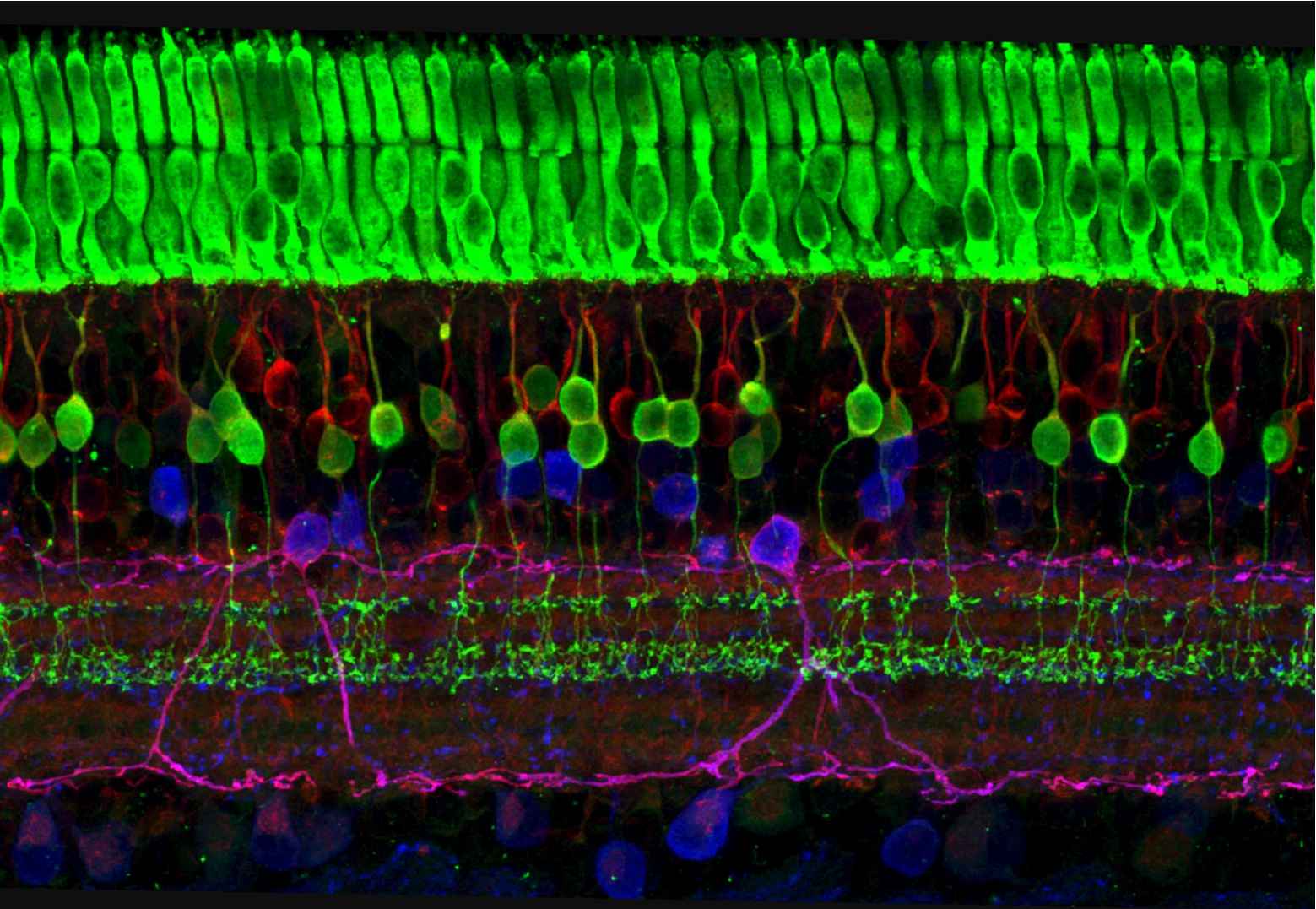
Above: This image captures the many layers of nerve cells in the retina. The top layer (green) is made up of cells called photoreceptors that convert light into electrical signals to relay to the brain. The two best-known types of photoreceptor cells are rod- and cone-shaped. Rods help us see under low-light conditions but can't help us distinguish colors. Cones don't function well in the dark but allow us to see vibrant colors in daylight. The top of the image is the posterior of the retina and the bottom of the image is positioned toward the posterior cavity filled with vitreous humor.

Above: Diagram of the cells of the retina. Light would enter the eye from the right of the figure and strike the retina and be detected by the rods and cones which send light information to bipolar cells and ganglion cells.

Above: Tissue of the retina magnified by 400x. The top of the image shows the posterior cavity of the eye where light is coming in toward the retina. Light is sensed by the rods and cones that pass the information to bipolar cells and then to ganglion cells. The axons of ganglion cells are bundled in C.N. II optic nerve.
|
Structure |
Location |
Function |
|---|---|---|
|
amacrine cells |
retina, inner neuronal layer |
inhibit some light signals from being transmitted (inhibitory neurons); interneurons |
|
bipolar cells |
retina, inner neuronal layer |
transmit light information from rod and cone cells to ganglion cells; interneurons |
|
cone cells |
retina, inner neuronal layer |
a type of photoreceptor; detects color vision and visual acuity; used in bright light |
|
ganglion cells |
retina, inner neuronal layer, optic nerve |
transmit light information from bipolar cells and amacrine cells; axons are bundled in the optic nerve (C.N. II); sensory neurons |
|
rod cells |
retina, inner neuronal layer |
a type of photoreceptor; determines motion and general shape of objects in dim light; black and white vision |

Above: Microscopic image of the tissue of the optic disc (blind spot), magnified by 100x.
Vision Neuron Pathway
Axons of ganglion cells in the retina are bundled into the optic nerve (C.N. II). The path of these axons is dependent on which part of the eye they come from. Axons of ganglion cells arising from the lateral retinas remain on the same side of the brain as the eye the they came from. The axons of medial ganglion cells however cross to the opposite side of the brain as that eye in the optic chiasma. Axons of ganglion cells terminate in the thalamus which filters out unneeded or extraneous visual information, aiding the cerebral cortex in focusing on important visual signals. Light information that pass through the "gate" of the thalamus is transmitted to the primary visual cortex in the occipital lobe, a region of the brain that processes visual information. Additionally, some of the ganglion cell axons terminate in the superior colliculi of the midbrain which collects visual information and modifies movements of the eyes.

Above: Diagram showing the path of axons of ganglion cells of the retina.
Lacrimal Apparatus
The lacrimal apparatus frames the eye and coats the sclera and cornea in lacrimal fluid, a bactericide, which lubricates and protects them. Lacrimal fluid is produced consistently to maintain lubrication and protection for the eye, but increases production when the eye is irritated or when an individual is emotional (tears). The lacrimal apparatus is made of the lacrimal gland, lacrimal canaliculi, lacrimal sac and nasolacrimal duct. This network of structures allows tears produced by the lacrimal gland to cover the eye, drain through the lacrimal puncta into the lacrimal canaliculi, collect in the lacrimal sac, travel down the nasolacrimal duct and finally empty into the nose. This is why crying leads to a runny nose.

Above: Structures of the lacrimal apparatus of the right eye showing the passage of lacrimal fluid (1-7).
|
Structure |
Location |
Function |
|---|---|---|
|
lacrimal canaliculi (superior duct and inferior duct) |
paired canals (superior duct and inferior duct) at the medial aspect of the eye between the lacrimal puncta and nasolacrimal duct |
transfers lacrimal fluid from lacrimal puncta to the nasolacrimal duct |
|
lacrimal caruncle |
red fleshy globe-like nodule in the medial commissure of the eye that contains sebaceous glands and sweat glands |
eye lubrication; dried secretions forms sandy residue |
|
lacrimal ducts |
small passageways connecting the lacrimal gland with the surface of the eye |
carries newly-formed lacrimal fluid to the eye's surface |
|
lacrimal fluid |
produced in the lacrimal gland and travels to the eye surface through lacrimal ducts and then across the eye from lateral to medial before draining into the lacrimal puncta, then lacrimal canaliculi, to the nasolacrimal duct, and into the nasal cavity |
lubricates the eye and protects it from debris and bacteria; aids in O2 and CO2 diffusion; creates tears during emotional response |
|
lacrimal gland |
superior lateral edge of the orbit |
production of lacrimal fluid (tears) to lubricate and protect the eye (fluid contains lysozyme, an antibiotic, antibodies, and mucus) |
|
lacrimal puncta |
paired openings at the medial aspect of the eye leading to the lacrimal canaliculi (superior duct and inferior duct) |
collection of lacrimal fluid from the eye's surface into lacrimal canaliculi (draining fluid) |
|
lacrimal sac |
a broad region of the nasolacrimal duct, located at the superior aspect of the nasolacrimal duct |
receives lacrimal fluid from the lacrimal canaliculi |
|
nasolacrimal duct |
a passage connecting the lacrimal canaliculi with the nasal cavity |
carries lacrimal fluid from lacrimal canaliculi into the nasal cavity |
|
palpebrae (eyelids) |
one superior and one inferior per eye |
blinking the palpebrae spreads lacrimal fluid and protects the eye |
Other Accessory Eye Structures

Above: Structures of the eye. (Top) Left eye. (Bottom) Closed right eye without skin to show the tarsal plates.
|
Structure |
Details |
|---|---|
|
ciliary gland / Moll's gland |
glands located at the base of the eyelashes producing sebum to lubricate the eyelashes; when infection of a ciliary gland occurs it results in a sty |
|
eyebrows |
hair superior to the eye, protection from light and sweat |
|
lateral commissure |
lateral corner of the eye |
|
levator palpebrae superioris muscle |
lifts the upper palpebra (eyelid) |
|
medial commissure |
medial corner of the eye, houses the lacrimal caruncle |
|
tarsal glands / Meibomian glands |
glands found in the tarsal plate, produce oily substance to prevent eyes from drying out |
|
tarsal plates |
located within the superior and inferior palpebrae (eyelids); comparatively thick, elongated plates of dense connective tissue that contribute to eyelids form and support |
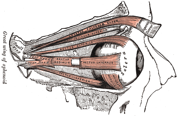
Above: Diagram of the ocular muscles of the right eye. The lateral rectus is shown as broken only to allow view of medial rectus.
|
Extrinsic Eye Muscles |
Location |
Action |
Innervation |
|---|---|---|---|
|
inferior oblique |
originating at the inferior medial aspect of the orbit and extending laterally and posteriorly to the eyeball |
looking up and laterally (eye roll) |
C.N. III oculomotor nerve |
|
inferior rectus |
inferior aspect of the eyeball approximately in the sagittal plane |
looking down (depression) |
C.N. III oculomotor nerve |
|
lateral rectus |
lateral aspect of the eyeball approximately in the sagittal plane |
looking laterally (abduction) |
C.N. VI abducens nerve |
|
medial rectus |
medial aspect of the eyeball approximately in the sagittal plane |
looking medially (adduction) |
C.N. III oculomotor nerve |
|
superior oblique |
originates medial to the eyeball approximately along the sagittal plane and then loops obliquely through a pully (trochlea of superior oblique) to from a v-like shape |
looking down and laterally (eye roll) |
C.N. IV trochlear nerve |
|
superior rectus |
superior aspect of the eyeball approximately in the sagittal plane |
looking up (elevation) |
C.N. III oculomotor nerve |

Above: Human eye and its extrinsic muscles (left) anterior view of the left eye with surrounding tissues including the lacrimal gland, (middle) anterior view of the left eyeball and its extrinsic eye muscles, and (right) posterior view of the left eyeball and its extrinsic eye muscles.
Attributions
- "1204 Optic Nerve vs Optic Tract.jpg" by OpenStax is licensed under CC BY 4.0
- "Anatomy 204L: Laboratory Manual (Second Edition)" by Ethan Snow, University of North Dakota is licensed under CC BY-NC 4.0
- "Anatomy and Physiology I Lab" by Victoria Vidal is licensed under CC BY 4.0
- "Anatomy and Physiology Lab Reference" by Laird C Sheldahl, OpenOregonEducational Resources, Mt. Hood Community College is licensed under CC BY-SA 4.0
- "Digital Histology" by Department of Anatomy and Neurobiology and the Office of Faculty Affairs, Virginia Commonwealth University School of Medicine and the ALT Lab at Virginia Commonwealth University is licensed under CC BY 4.0
- "Gray's Anatomy plates" by Henry Vandyke Carte is in the Public Domain
- "Human Anatomy Lab Manual" by Malgosia Wilk-Blaszczak, Mavs Open Press, University of Texas at Arlington is licensed under CC BY 4.0
- "Special Senses – Anatomy of the Eye" by Dongho Kim is licensed under CC BY-NC-SA 4.0
- "Tear system-pt.svg" by Jmarchn is licensed under CC BY-SA 3.0
- "The eye uses many layers of nerve cells to convert light into sight" by Wei Li is licensed under CC BY-NC-SA 3.0