14.3: Spinal Cord
- Page ID
- 53723
Spinal Cord
The spinal cord is a long, thin, tubular structure made up of nervous tissue, which extends from the medulla oblongata in the brainstem to the lumbar region of the vertebral column. It encloses the central canal of the spinal cord, which contains cerebrospinal fluid. In humans, the spinal cord begins at the occipital bone, passing through the foramen magnum and entering the spinal canal at the beginning of the cervical vertebrae. The spinal cord extends down to between the first and second lumbar vertebrae, where it tapers to from the conus medullaris near the second lumbar vertebra as it spreads out into a horse tail-like structure called cauda equina and terminating in a fibrous extension known as the filum terminale. The enclosing bony vertebral column protects the relatively shorter spinal cord. It is around 45 cm (18 in) in men and around 43 cm (17 in) long in women. The diameter of the spinal cord ranges from 13 mm (1⁄2 in) in the cervical and lumbar regions to 6.4 mm (1⁄4 in) in the thoracic area.

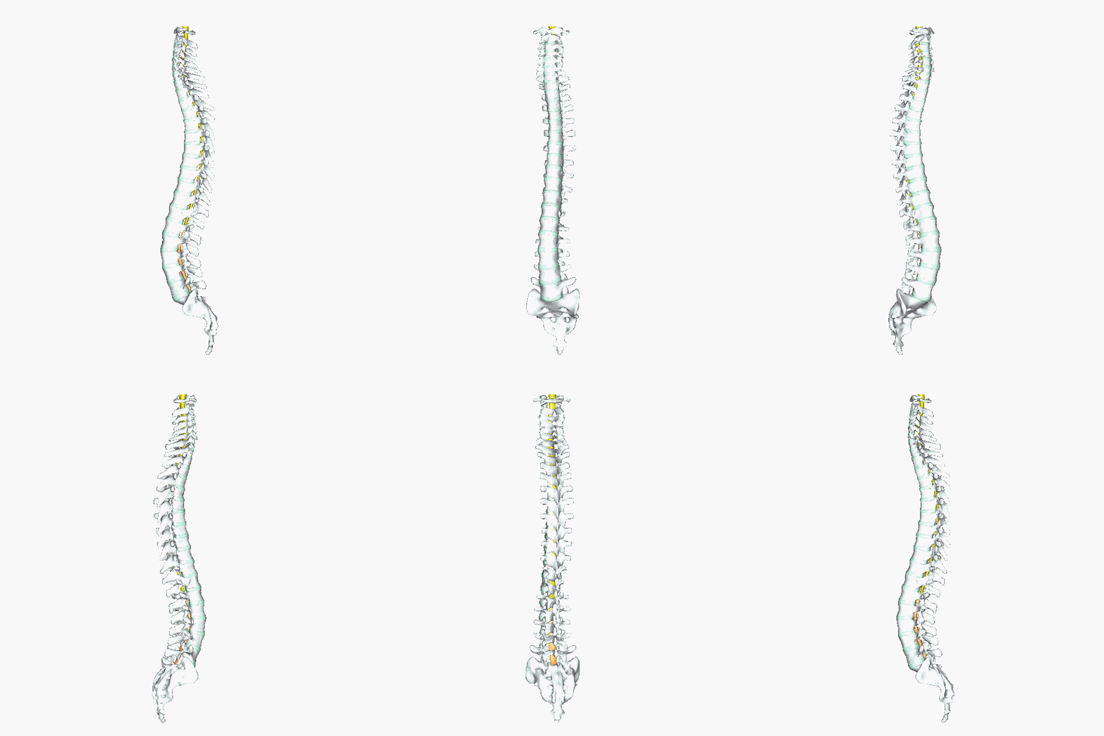
Above: The spinal cord. (Top left) Illustration and (top middle) image from a cadaver of the spinal cord without or with only part of the bones of the vertebral column present. (Top right) Transverse sections of the spinal cord show a butterfly-pattern in the neural tissue of the spinal cord and the shape and width of the cord differs as the spinal cord descends through the vertebral foramina of the vertebral column. (Bottom) Within the vertebral foramina of the vertebrae, the condensed spinal cord can be seen shown in yellow with cauda equina shown in orange.
Above: Superior view of a cervical vertebra with the spinal cord located in the vertebral foramen surrounded by the three layers of meninges: the dura mater, arachnoid mater, and the pia mater.
The spinal cord functions primarily in the transmission of nerve signals from the motor cortex to the body, and from the afferent fibers of the sensory neurons to the sensory cortex. It is also a center for coordinating many reflexes and contains reflex arcs that can independently control reflexes. It is also the location of groups of spinal interneurons that make up the neural circuits known as central pattern generators. These circuits are responsible for controlling motor instructions for rhythmic movements such as walking.
Meninges
Above: Illustration of the meninges surrounding the spinal cord, transverse section with a superior view.
The spinal cord (and brain) are protected by three layers of connective tissue or membranes called meninges. The dura mater is the outermost layer, and it forms a tough protective coating. Between the dura mater and the surrounding bone of the vertebrae is a space called the epidural space. The epidural space is filled with adipose tissue, and it contains a network of blood vessels. The arachnoid mater, the middle protective layer, is named for its open, spiderweb-like appearance. The space between the arachnoid and the underlying pia mater is called the subarachnoid space. The subarachnoid space contains cerebrospinal fluid (CSF), which can be sampled with a lumbar puncture, or "spinal tap" procedure. The delicate pia mater, the innermost protective layer, is tightly associated with the surface of the spinal cord. The cord is stabilized within the dura mater by the connecting denticulate ligaments, which extend from the enveloping pia mater laterally between the dorsal and ventral roots. The dural sac ends at the vertebral level of the second sacral vertebra.
Above: Cadaver image of the spinal cord shown cut in the transverse plane, posterior view. The dura mater and arachnoid mater on the anterior of the spinal cord are shown and the transparent pia mater can be seen on the posterior of the spinal cord.
Organization of the Spinal Cord
Above: Transverse section of the spinal cord and spinal nerve. The blue lines represent axons of sensory neuron and blue dots are cell bodies of sensory neurons. The red lines represent axons of motor neurons. You can see that motor neurons exit the spinal cord from the ventral root (anteriorly) and sensory neurons enter the spinal cord at the dorsal root (posteriorly).
In cross-section, the peripheral region of the cord contains neuronal white matter tracts containing sensory and motor axons. Internal to this peripheral region is the gray matter, which contains the nerve cell bodies arranged in the three gray columns that give the region its butterfly-shape. This central region surrounds the central canal, which is an extension of the fourth ventricle and contains cerebrospinal fluid.
The spinal cord is elliptical in cross section, being compressed dorsolaterally. Two prominent grooves, or sulci, run along its length. The posterior median sulcus is the groove in the dorsal side, and the anterior median fissure is the groove in the ventral side.
Above: the structures of the spinal cord, transverse section with a superior view.
The gray matter is divided into posterior (dorsal) gray horns which contain sensory neurons, and lateral gray horns and anterior (ventral) gray horns that contain the cell bodies of motor neurons. The surrounding white matter is divided into anterior (ventral) white columns, lateral white columns, and posterior (dorsal) white columns. The gray commissure is the gray matter posterior to the central canal where the neurons from either side of the spinal cord crossover. The same principle applies to the white commissure which lies anteriorly to the gray matter.
Gray Matter of the Spinal Cord
In cross-section, the gray matter of the spinal cord has the appearance of an ink-blot test, with the spread of the gray matter on one side replicated on the other—a shape reminiscent of a bulbous capital “H” or a butterfly. The gray matter is subdivided into regions that are referred to as horns. The posterior gray horn is responsible for sensory processing. The anterior gray horn sends out motor signals to the skeletal muscles. The lateral gray horn, which is only found in the thoracic, upper lumbar, and sacral regions, is the central component of the sympathetic division of the autonomic nervous system.
Above: The gray matter of the spinal cord is organized such that the posterior gray horns contain sensory nuclei and the anterior gray horns and lateral gray horns contain motor nuclei. These are further subdivided into visceral and sensory. Somatic sensory located in the posterior aspects of the posterior gray horns (green), visceral sensory located in the anterior aspects of the posterior gray horns (red), somatic motor located in the anterior aspects of the anterior gray horns (orange), and visceral motor located in the lateral gray horns (light green).
Some of the largest neurons of the spinal cord are the multipolar motor neurons in the anterior horn. The fibers that cause contraction of skeletal muscles are the axons of these neurons. The motor neuron that causes contraction of the big toe, for example, is located in the sacral spinal cord. The axon that has to reach all the way to the belly of that muscle may be a meter in length. The neuronal cell body that maintains that long fiber must be quite large, possibly several hundred micrometers in diameter, making it one of the largest cells in the body.
White Matter of the Spinal Cord
Just as the gray matter is separated into horns, the white matter of the spinal cord is separated into columns. Ascending tracts of nervous system fibers in these columns carry sensory information up to the brain, whereas descending tracts carry motor commands from the brain. Looking at the spinal cord longitudinally, the columns extend along its length as continuous bands of white matter. Between the two posterior horns of gray matter are the posterior columns. Between the two anterior horns, and bounded by the axons of motor neurons emerging from that gray matter area, are the anterior columns. The white matter on either side of the spinal cord, between the posterior horn and the axons of the anterior horn neurons, are the lateral columns. The posterior columns are composed of axons of ascending tracts. The anterior and lateral columns are composed of many different groups of axons of both ascending and descending tracts—the latter carrying motor commands down from the brain to the spinal cord to control output to the periphery.
Above: Functional organization of the tracts of white matter in the spinal cord.
Attributions
- "Anatomy 204L: Laboratory Manual (Second Edition)" by Ethan Snow, University of North Dakota is licensed under CC BY-NC 4.0
- "Anatomy and Physiology" by J. Gordon Betts et al., OpenStax is licensed under CC BY 4.0
- "Anatomy and Physiology I Lab" by Victoria Vidal is licensed under CC BY 4.0
- "Gray's Anatomy plates" by Henry Vandyke Carte is in the Public Domain
- "Human Anatomy Lab Manual" by Malgosia Wilk-Blaszczak, Mavs Open Press, University of Texas at Arlington is licensed under CC BY 4.0
- "Spinal Cord - 4742.jpg" by Amada44 is licensed under CC BY-SA 3.0
- "Spinal cord and roots and dural tube which covers them. Wellcome L0002010.jpg" by Wellcome Images is licensed under CC BY 4.0
- "Spinal cord tracts - English.svg" by Polarlys and Mikael Häggström is licensed under CC BY-SA 3.0
- "Spinal dura mater 1.jpg" by Anatomist90 is licensed under CC BY-SA 3.0


