
13.3: Nerves are Bundles of Neuron Axons in the PNS
- Page ID
- 53709

Nerves are Bundles of Neuron Axons in the PNS
Nerves are bundles of axons in the PNS. Nerves can be classified as sensory (afferent) for signals coming into the CNS from the PNS, or motor (efferent) for signals from the CNS to the PNS. Most nerves are mixed, and therefore carry both sensory and motor information.
Ganglia are clusters of neuron cell bodies in the nerves of the PNS. Ganglia have various functions controlling and monitoring body functions. For example, dorsal root ganglia are sensory neurons clustered lateral to the spinal cord.

Above: Nerves (bundles of axons in the PNS) connect with the spinal cord in the CNS at spinal cord (spinal nerves) or at the brain (cranial nerves). The diagram above shows the structure of spinal nerves. Notice that nerves are composed of axons of both sensory and motor neurons (mixed nerves) and then are organized sensory neurons at the dorsal root of the spinal cord and motor neurons at the ventral root. Cell bodies of sensory neurons are bundled together in an enlarged region of the dorsal root called dorsal root ganglion.

Above: Microscopic image of a nerve with a longitudinal view. Axons appear as long strands in this view. Tissue is magnified by 40x.

Above: Structure of a nerve.
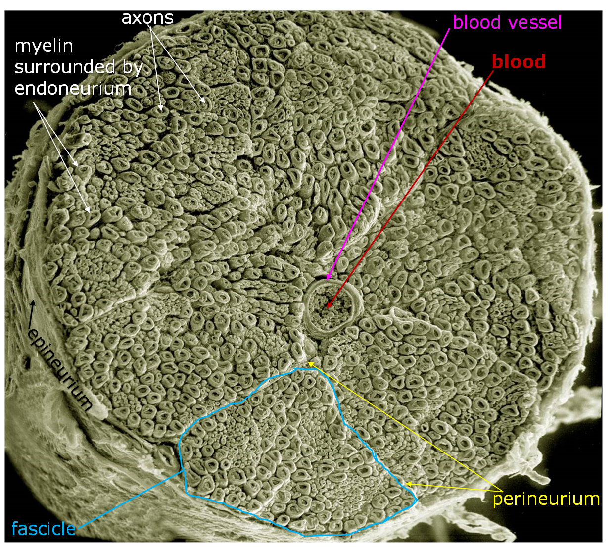
Above: SEM image of a spinal nerve cross section.
The arrangements of axons in a nerve is similar to the organization of muscle fibers in a skeletal muscle (see Chapter 11: Introduction to Skeletal Muscles). Each axon is surrounded by a sheath of tissue called endoneurium. The axons surrounded by endoneurium are bundled together into sections called fascicles. Each fascicle is surrounded by a sheath of tissue called perineurium. Multiple fascicles, each surrounded by perineurium, are bundled together and wrapped with epineurium to for a nerve.

Above: Light microscope cross section of a nerve. Tissue is magnified 100x.
Attributions
- "Anatomy and Physiology Lab Reference" by Laird C Sheldahl, OpenOregonEducational Resources, Mt. Hood Community College is licensed under CC BY-SA 4.0
- "Animal Tissues and Organs" by Berkshire Community College Bioscience Image Library is in the Public Domain, CC0
- "Digital Histology" by Department of Anatomy and Neurobiology and the Office of Faculty Affairs, Virginia Commonwealth University School of Medicine and the ALT Lab at Virginia Commonwealth University is licensed under CC BY 4.0
- "NCMIR human spinal nerve" by Mark Ellisman and Tom Deerinck is licensed under CC BY-NC-SA 3.0
- "Principles of Biology I and II lab manuals" by Dalton State University is licensed under CC BY-SA 4.0
- "Spinal nerve no text.svg" by jmarchn is licensed under CC BY-SA 3.0