
13.2: Neurons
- Page ID
- 53708

Neurons
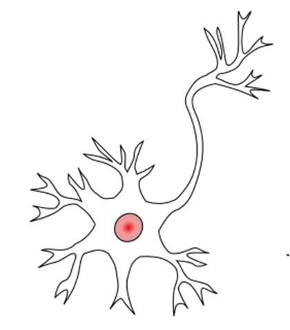
The nervous system is composed of more than 100 billion cells known as neurons. A neuron is a cell in the nervous system whose function it is to receive and transmit information. Neurons are made up of three major parts: a cell body, or soma, which contains the nucleus of the cell and keeps the cell alive; a branching tree-like fiber known as the dendrite, which collects information from other cells and sends the information to the cell body (soma); and a long, segmented fiber known as the axon, which transmits information away from the cell body toward other neurons or to the muscles and glands. An excited neuron transmits signals down its axon toward the axon terminals through a wave of electricity known as an action potential. Think of an axon as being an electric wire transmitting electricity from one place to another.

Above: Generalized structure of a neuron.
Once a signal is received by the dendrite, it then travels to the cell body that may be excited enough to stimulate an action potential down the axon. An axon is a tube-like structure that propagates (increases) the integrated signal to specialized endings called axon terminals. These terminals in turn synapse on other neurons, or other cells such as muscle cells. Chemicals released at axon terminals allow signals to be communicated to these other cells.
Above: Animation comparing the speed of action potentials along myelinated and unmyelinated neurons.
Some axons are covered with myelin, which acts as an insulator to minimize dissipation of the electrical signal as it travels down the axon, greatly increasing the speed on conduction. This insulation is important as the axon from a human motor (efferent) neuron can be as long as a meter (100 cm)—from the base of the spine to the toes - and therefore some signals have long distances to travel, making the speed of the action potential particularly important. The myelin sheath is not actually part of the neuron. Myelin is produced by specific types of glial cells. Along the axon there are periodic gaps in the myelin sheath. These gaps are called nodes of Ranvier and are sites where the signal is “recharged” as it travels along the axon.

Above: Nervous tissue. Tissue is magnified by 200x.
It is important to note that a single neuron does not act alone—neuronal communication depends on the connections that neurons make with one another (as well as with other cells, like muscle cells). Dendrites from a single neuron may receive synaptic contact from many other neurons.
Synapses are physical junctions between two neurons, as well as between neurons and other cells (e.g. neuron-muscle cell, neuron-sensory receptor cell), that allow for communication. Chemical synapses (most common type) use an action potential to signal the exocytosis of neurotransmitter chemicals (over 100 different neurotransmitters known in humans). Each neurotransmitter requires a receptor protein (multiple subtypes known for each different neurotransmitter). Electrical synapses are direct membrane junctions between cells that allow continuations of action potentials. A neuron passes signals to another neuron or cell through synapses at its axon terminals. Neurons receive signals from other neurons or cells through synapses at its cell body or dendrites.

Above: Illustration of a synapse between two neurons. A chemical synapse is depicted here.
Structural Classifications of Neurons
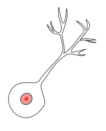
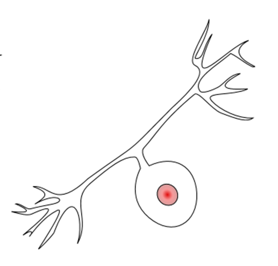
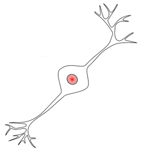
A neuron is composed of one cell body, one axon, and usually one or more dendrites, but there are different ways these components may be organized resulting is some structural differences among neurons. There are four main structural classifications of neurons: multipolar, bipolar, unipolar, and pseudounipolar.
|
Neuron Structural Type |
Number of Branches off Cell Body |
Description of Branches off Cell Body |
Function/Info |
|---|---|---|---|
|
unipolar
|
1 |
1 neurite (functions as dendrite and axon) |
not present in humans found in invertebrate animals |
|
pseudounipolar
|
1, splits into two later |
branch connects to axon branching two directions (has no dendrites) |
sensory neurons (signals from PNS to CNS) one axon end branches to CNS; the other axon end branches to PNS |
|
bipolar
|
2 |
1 axon + 1 dendrite |
|
|
multipolar
|
3+ |
1 axon + 2 or more dendrites |
motor neurons (signals from CNS to PNS) or interneurons (pass signals between two neurons) most common type of neuron in humans |
Sensory Neurons, Motor Neurons, & Interneurons

Above: Sensory neurons collect sensory information about the environment in the PNS and transmit information to the CNS. Motor neurons transmit signals from the CNS to the PNS to stimulate a change. In this example, a person tests the water temperature before getting in the shower (sensed by sensory neurons) and adjusts the water temperature accordingly using motor neurons to cause skeletal muscle contractions.
Within the nervous system, there are 3 fundamental functional types of neurons:
- sensory (afferent) neurons conduct signals from receptors to the CNS
- motor (efferent) neurons conduct signals from the CNS to effectors such as muscles and glands (PNS)
- interneurons (association neurons) are confined to the CNS
Sensory information such as touch, temperature, smell, sight, sounds, vibrations, and pain is received by sensory neurons, also called afferent neurons, in the PNS and transmitted to the CNS. The example in the diagram below shows how a sensory neuron in the hand senses the temperature of shower water and transmits that information to the CNS.
Motor neurons, also called efferent neurons, give commands from the CNS to cause changes including muscular contractions (skeletal, cardiac, and smooth) and gland secretions. Continuing the example diagramed below, motor neurons cause the muscles of the hand and forearm to contract to adjust the shower temperature knobs based on the sensory information received from the sensory neurons.
Mediating communication between sensory neurons and motor neurons are interneurons. Interneurons are found in the CNS and are neither sensory nor motor, they act as intermediaries between sensory neurons and motor neurons.
Clinical Application: Neurogenesis
At one time, scientists believed that people were born with all the neurons they would ever have. Research performed during the last few decades indicates that neurogenesis, the birth of new neurons, continues into adulthood. Neurogenesis was first discovered in songbirds that produce new neurons while learning songs. For mammals, new neurons also play an important role in learning: about 1,000 new neurons develop in the hippocampus (a brain structure involved in learning and memory) each day. While most of the new neurons will die, researchers found that an increase in the number of surviving new neurons in the hippocampus correlates with how well rats learn a new task. Interestingly, both exercise and some antidepressant medications also promote neurogenesis in the hippocampus. Stress has the opposite effect. While neurogenesis is quite limited compared to regeneration in other tissues, research in this area may lead to new treatments for disorders such as Alzheimer’s, stroke, and epilepsy.
Attributions
- "Anatomy 204L: Laboratory Manual (Second Edition)" by Ethan Snow, University of North Dakota is licensed under CC BY-NC 4.0
- "Anatomy and Physiology" by J. Gordon Betts et al., OpenStax is licensed under CC BY 4.0
- "Chemical synapse schema cropped.jpg" by Officer or employee of the United States is in the Public Domain
- "Introduction to Psychology" by University of Minnesota is licensed under CC BY-NC-SA 4.0
- "Neuron or Nerve cell - Labelled Image.png" by DrJanaOfficial is in the Public Domain, CC0
- "Neurons & Nervous System" by Mohamed Awad is licensed under CC BY-NC-SA 4.0
- "Neurons uni bi multi pseudouni.svg" by Juoj8 is licensed under CC BY-SA 3.0 / A derivative from the original work
- "Principles of Biology I and II lab manuals" by Dalton State University is licensed under CC BY-SA 4.0
- "Saltatory Conduction.gif" by DrJanaOfficial is in the Public Domain, CC0
- "UGA Anatomy and Physiology 1 Lab Manual, 3rd Edition" by DeLoris Hesse, Deanna Cozart, Brett Szymik, Rob Nichols, University of Georgia is licensed under CC BY 4.0