
12.2: Muscles of the Head and Neck
- Page ID
- 53685

Muscles of the Head and Neck
Head
The epicranius muscle is also very broad and covers most of the top of the head. The epicranius muscle includes a middle section which is all aponeurosis (white, fibrous, flat, tendon-like tissue). The actual muscle tissue is only found over the forehead (the portion of the muscle called the epicranius frontalis, or frontal belly of epicranius) and the back of the head (the portion of the muscle called the epicranius occipitalis, or occipital belly of the epicranius).
The buccinator muscles, one on each side of the face, compress the cheeks when contracted. The name is from the Latin for trumpet, which requires blowing air out of the cheeks to play, and also reflects the anatomical adjective for the cheek, buccal.
The two masseter muscles are also on each side of the face. They close the jaw when contracted. Its name is derived from the same Greek root as mastication, which means to chew.
The zygomaticus major muscles and the zygomaticus minor muscles are found on each side of the face both have their origins on the zygomatic bone. They both can change the shape of the mouth by elevating it.

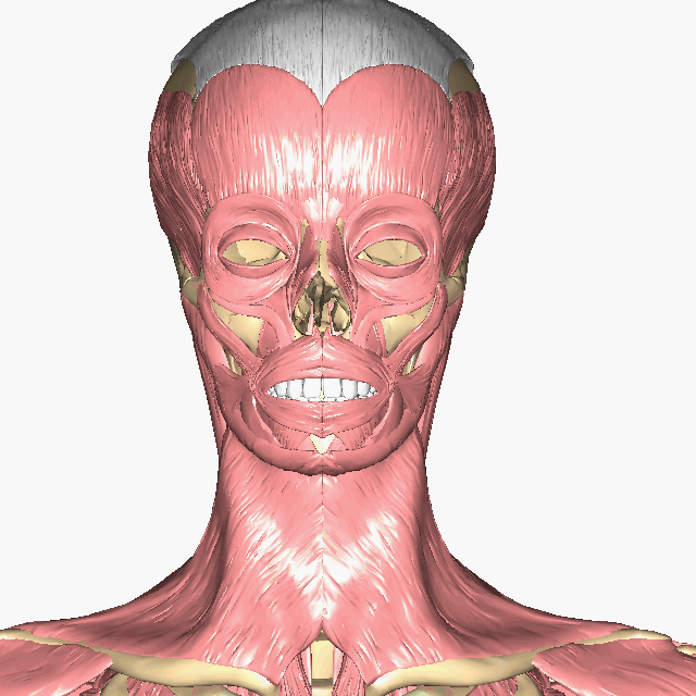
Above: Muscles of the head.

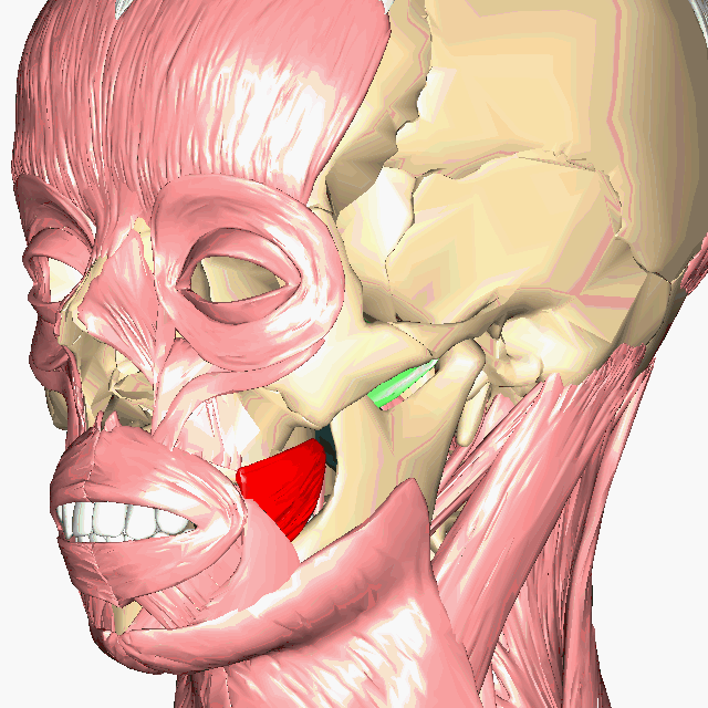
Above: Deeper muscles of the head. This diagram shows the masseter removed (superficial and deep), temporalis removed, risorius removed, and zygmaticus major removed. Buccinator is shown in red, lateral pterygoid in green, and medial pterygoid in black.

Above: Cadaver images of the muscles of the head.
Clinical Application: The Buccinator Muscle
The buccinator muscle is a deep muscle of the cheek. Its primary role is to assist in mastication (chewing) by keeping food between upper and lower teeth. If you’ve ever bitten your cheek bad enough, you may have bitten down on your buccinator (although under most instances, you only bite down on the epithelium and superficial fascia that lines the innermost surfaces of your cheeks). The buccinator muscle also assists infants with suckling and adults with whistling.
Eye
The movements of the eyeball are controlled by six muscles. The muscles "rectus" are straight muscles and the "oblique" muscles are positioned at an angle in relationship to the straight muscles. Each of the ocular muscles are named for their positions (superior, inferior, lateral and medial): superior rectus, superior oblique, inferior rectus, inferior oblique, lateral rectus, and medial rectus. Additionally, levator palpebrae superioris, located superiorly to the eye, raises the eyelid.
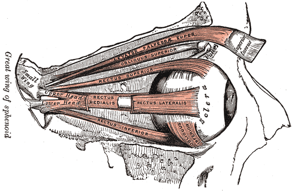
Above: Diagram of the ocular muscles of the right eye. The lateral rectus is shown as broken only to allow view of medial rectus.
Anterior Neck

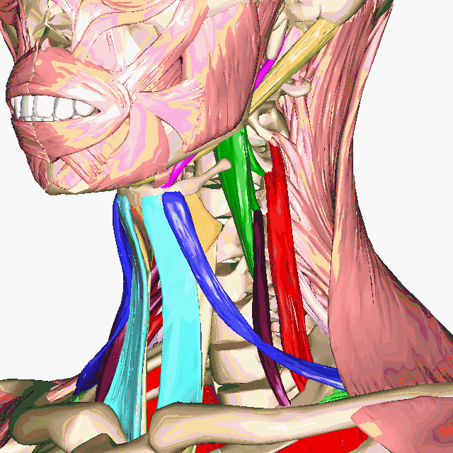
Above: (Left) Anterior and (right) lateral views of superficial neck muscles with deeper anterior neck muscles shown in different colors.
A single platysma muscle is only shown in the lateral view of the head muscles in. There are two platysma muscles, one on each side of the neck. Each is a broad sheet of a muscle that covers most of the anterior neck on that side of the body. The other anterior neck muscles are below them, and most anatomical models and figures have the platysma muscles cut away to show the deeper muscles. The platysma muscles help pull down the lower jaw (mandible.)
Under the platysma are two sternocleidomastoid muscles. One on each side of the neck. These muscles have two origins, one on the sternum and the other on the clavicle. They insert on the mastoid process of the temporal bone. They can flex or extend the head, or can rotate the towards the shoulders.

Above: Deep anterior neck muscles shown with platysma and sternocleidomastoid removed (top) anterior, (middle) anterolateral, and (bottom) lateral views.

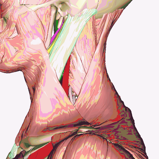
Above: Muscles of the anterior neck, platysma, sternohyoid, and sternothyroid removed. (Top) Anterior and slightly inferior view of deep neck muscles. (Bottom) Inferior view of the deep muscles of the neck.

Above: Cadaver image of the muscles of the anterior neck.
Attributions
- "Anatomy 204L: Laboratory Manual (Second Edition)" by Ethan Snow, University of North Dakota is licensed under CC BY-NC 4.0
- "BIOL 250 Human Anatomy Lab Manual SU 19" by Yancy Aquino, Skyline College is licensed under CC BY-NC-SA 4.0
- "BodyParts3D/Anatomography" by The Database Center for Life Science is licensed under CC BY-SA 2.1
- "Gray's Anatomy plates" by Henry Vandyke Carte is in the Public Domain