Biology LibreTexts

6.5: Laboratory Activities and Assignment

  • Page ID
    53592
  • \( \newcommand{\vecs}[1]{\overset { \scriptstyle \rightharpoonup} {\mathbf{#1}} } \) \( \newcommand{\vecd}[1]{\overset{-\!-\!\rightharpoonup}{\vphantom{a}\smash {#1}}} \)\(\newcommand{\id}{\mathrm{id}}\) \( \newcommand{\Span}{\mathrm{span}}\) \( \newcommand{\kernel}{\mathrm{null}\,}\) \( \newcommand{\range}{\mathrm{range}\,}\) \( \newcommand{\RealPart}{\mathrm{Re}}\) \( \newcommand{\ImaginaryPart}{\mathrm{Im}}\) \( \newcommand{\Argument}{\mathrm{Arg}}\) \( \newcommand{\norm}[1]{\| #1 \|}\) \( \newcommand{\inner}[2]{\langle #1, #2 \rangle}\) \( \newcommand{\Span}{\mathrm{span}}\) \(\newcommand{\id}{\mathrm{id}}\) \( \newcommand{\Span}{\mathrm{span}}\) \( \newcommand{\kernel}{\mathrm{null}\,}\) \( \newcommand{\range}{\mathrm{range}\,}\) \( \newcommand{\RealPart}{\mathrm{Re}}\) \( \newcommand{\ImaginaryPart}{\mathrm{Im}}\) \( \newcommand{\Argument}{\mathrm{Arg}}\) \( \newcommand{\norm}[1]{\| #1 \|}\) \( \newcommand{\inner}[2]{\langle #1, #2 \rangle}\) \( \newcommand{\Span}{\mathrm{span}}\)\(\newcommand{\AA}{\unicode[.8,0]{x212B}}\)

    Laboratory Activities and Assignment

    Part 1: Review of the Anatomy of the Integumentary System

    1. Label the anatomical model of the skin below with the terms listed:

    • epidermis
    • dermis
    • hypodermis
    • adipose tissue
    • basement membrane
    • outer root sheath (of hair)
    • inner root sheath (of hair)
    • dermal papilla
    • skin surface
    • sebaceous gland
    • duct of sebaceous gland
    • arrector pili muscle
    • hair matrix
    • blood vessels
    • sensory nerve
    • cuticle (of hair)
    • cortex (of hair)
    • medulla (of hair)
    • eccrine sweat gland
    • duct of eccrine sweat gland
    • hair bulb
    • Pacinian corpuscle
    • hair papilla

    Image of skin anatomical model for labeling.

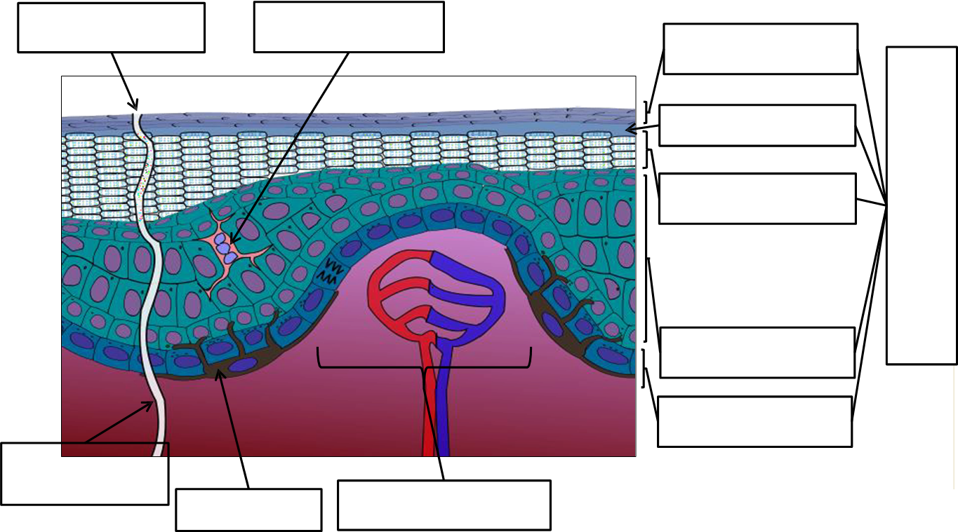
    2. Label the diagram of the skin cross section below with the terms listed:

    • hair follicle
    • sebaceous gland
    • epidermis
    • dermis (papillary layer)
    • dermis (reticular layer)
    • hypoderms
    • arrector pili muscle
    • sweat gland

    Illustration of skin cross section for labeling.

    3. Label the diagram of the skin cross section below with the terms listed:

    • stratum corneum
    • stratum lucidum
    • stratum granulosum
    • stratum spinosum
    • stratum basale
    • dermal papilla
    • sweat duct
    • sweat pore
    • stratified squamous epithelium
    • dendritic cell
    • melanocyte

    Illustration of superficial layers of the skin for labeling.

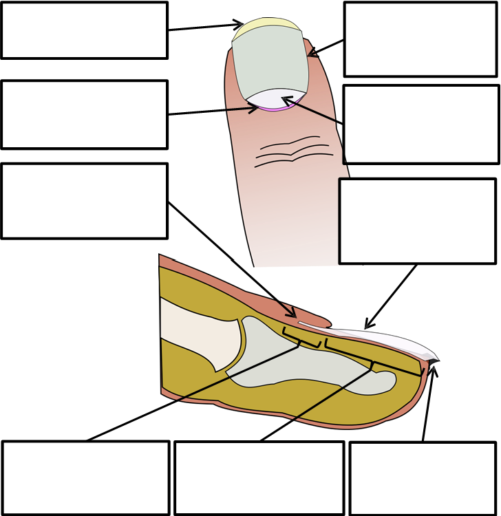
    4. Label the diagram of the hair and skin cross section below with the terms listed:

    • sebaceous gland
    • hair shaft
    • hair bulb
    • hair root
    • hair papilla

    Illustration of a hair cross section for labeling.

    5. Label the diagram of the skin cross section below with the terms listed:

    • § free edge
    • § hyponychium
    • § lunula
    • § nail matrix
    • § nail root
    • § lateral nail fold
    • § nail bed
    • § nail plate
    • § eponychium

    Illustration of a nail cross section for labeling.

    Part 2: Histology of the Integumentary System

    1. Obtain the slides listed below that are available for today’s lab.
    2. Focus on each sample and identify the structures listed for each type of tissue.
    3. Indicate the total magnification you make each illustration at in the space provided.
    4. Illustrate each tissue you observe with the microscope at the magnification you listed.
    5. Label each illustration with the structures listed for each.

    Skin (cross section)

    Label the tissue with: epidermis, dermis, papillary layer of the dermis, reticular layer of the dermis, basement membrane, stratum basale, stratum corneum, dermal papilla, epithelial tissue, areolar tissue, dense irregular connective tissue, skin surface

    Location for sample illustration.

    Human Scalp with Hair Follicle

    Label the tissue with: epidermis, dermis, arrector pili muscle, sebaceous gland, dermal papilla, hair follicle, hair bulb

    Location for sample illustration.

    Attributions

    Part 1: Review of the Anatomy of the Integumentary System

    Part 2: Histology of the Integumentary System


    This page titled 6.5: Laboratory Activities and Assignment is shared under a CC BY-NC-SA 4.0 license and was authored, remixed, and/or curated by Rosanna Hartline.

    • Was this article helpful?