
5.4: Muscle Tissues
- Page ID
- 53578

Muscle Tissues
Muscle tissues are responsible for movements. Muscle tissue is subdivided into three broad categories: skeletal muscle, cardiac muscle, and smooth muscle. The three types of muscle can be distinguished by both their locations and their microscopic features.
Skeletal Muscle Tissue
Skeletal muscle is largely found attached to bones. It consists of long multinucleate (cells that contain multiple nuclei) fibers. The fibers run the entire length of the muscle they come from and so are usually too long to have their ends visible when viewed under the microscope. The fibers are relatively wide and very long, but unbranched. The nuclei are usually up against the edge of the fiber. There are striations (different colored stripes) in skeletal muscle. These are alternating dark and light bands perpendicular to the edge of the fiber that are present all along the fiber.

Above: Skeletal muscle, teased, longitudinal view. Tissue magnified by 200x.
Cardiac Muscle Tissue
Cardiac muscle is only found in the heart. Its fibers are longer than they are wide, and they are striated, like skeletal muscle fibers. But, unlike skeletal muscle fibers, cardiac muscle fibers have distinct ends to them, called intercalated discs. These are dark lines that run from one side of the fiber to the other. The intercalated discs are not much thicker than the striations, but they are usually darker and so distinct for that reason. One cardiac muscle fiber is the material between two intercalated discs. Cardiac muscle fibers are mononucleate, with only one nucleus per fiber, and they can sometimes be branched.

Above: Cardiac muscle, longitudinal view. Tissue magnified by 400x
Smooth Muscle Tissue
Smooth muscle is found in the walls of internal organs, such as the organs of the digestive tract, blood vessels, and others. It consists of mononucleate fibers with tapered edges. No striations are visible in smooth muscle under the microscope. Because smooth muscle often is wrapping around the organ it is associated with, it can be hard to find an entire smooth muscle fiber in profile in a tissue slice on a microscope slide. Most of the fibers will be sectioned at angles or will be difficult to get into a single plane of focus, but a little bit of searching can usually turn up some with all of the defining characteristics visible.
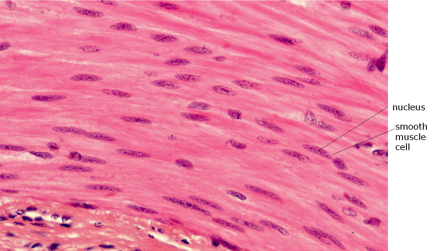
Above: Smooth muscle, longitudinal view. Tissue magnified by 1000x
Attributions
- "Animal Tissues and Organs" by Berkshire Community College Bioscience Image Library is in the Public Domain, CC0
- "BIOL 250 Human Anatomy Lab Manual SU 19" by Yancy Aquino, Skyline College is licensed under CC BY-NC-SA 4.0
- "Digital Histology" by Department of Anatomy and Neurobiology and the Office of Faculty Affairs, Virginia Commonwealth University School of Medicine and the ALT Lab at Virginia Commonwealth University is licensed under CC BY 4.0