Biology LibreTexts

2020_Winter_Bis2A_Facciotti_Lecture_17

  • Page ID
    27624
  •  

    \( \newcommand{\vecs}[1]{\overset { \scriptstyle \rightharpoonup} {\mathbf{#1}} } \) \( \newcommand{\vecd}[1]{\overset{-\!-\!\rightharpoonup}{\vphantom{a}\smash {#1}}} \)\(\newcommand{\id}{\mathrm{id}}\) \( \newcommand{\Span}{\mathrm{span}}\) \( \newcommand{\kernel}{\mathrm{null}\,}\) \( \newcommand{\range}{\mathrm{range}\,}\) \( \newcommand{\RealPart}{\mathrm{Re}}\) \( \newcommand{\ImaginaryPart}{\mathrm{Im}}\) \( \newcommand{\Argument}{\mathrm{Arg}}\) \( \newcommand{\norm}[1]{\| #1 \|}\) \( \newcommand{\inner}[2]{\langle #1, #2 \rangle}\) \( \newcommand{\Span}{\mathrm{span}}\) \(\newcommand{\id}{\mathrm{id}}\) \( \newcommand{\Span}{\mathrm{span}}\) \( \newcommand{\kernel}{\mathrm{null}\,}\) \( \newcommand{\range}{\mathrm{range}\,}\) \( \newcommand{\RealPart}{\mathrm{Re}}\) \( \newcommand{\ImaginaryPart}{\mathrm{Im}}\) \( \newcommand{\Argument}{\mathrm{Arg}}\) \( \newcommand{\norm}[1]{\| #1 \|}\) \( \newcommand{\inner}[2]{\langle #1, #2 \rangle}\) \( \newcommand{\Span}{\mathrm{span}}\)\(\newcommand{\AA}{\unicode[.8,0]{x212B}}\)

    Learning objectives associated with 2020_Winter_Bis2A_Facciotti_Lecture_17

    • Explain the difference between cyclic and non-cyclic photophosphorylation.  Be able to illustrate the flow of electrons in both processes, highlighting the source and sink in both cases.
    • List the advantages of having two photosystems and the ability to use water (H2O) as an electron donor.
    • Express the functional links between carbon fixation and light-driven ATP and NADPH production.
    • Create an energy story for the Calvin cycle.
    • Create a sketch model and an energy story for photosynthesis that includes the flow of electrons from a “low energy” source to an electron transporter (e.g. NADP), and finally to a reduced carbon compound.
    • Compare and contrast oxidative phosphorylation (respiration) and photophosphorylation.

    Photophosphorylation

    Photophosphorylation an overview

    Photophosphorylation is the process of transferring the energy from light into chemicals, particularly ATP. The evolutionary roots of photophosphorylation are likely in the anaerobic world, between 3 billion and 1.5 billion years ago, when life was abundant in the absence of molecular oxygen. Photophosphorylation probably evolved relatively shortly after electron transport chains (ETC) and anaerobic respiration provided metabolic diversity. The first step of the process involves the absorption of a photon by a pigment molecule. Light energy transfers to the pigment and promotes electrons (e-) into a higher quantum energy state—something biologists term an "excited state". Note the use of anthropomorphism here; the electrons are not "excited" in the classic sense and aren't suddenly hopping all over or celebrating their promotion. They are simply in a higher energy quantum state. In this state, the electrons are colloquially said to be "energized". While in the "excited" state, the pigment now has a much lower reduction potential and can donate the "excited" electrons to other carriers with greater reduction potentials. These electron acceptors may become donors to other molecules with greater reduction potentials and, in doing so, form an electron transport chain.

    As electrons pass from one electron carrier to another via red/ox reactions, enzymes can couple these exergonic electron transfers to the endergonic transport (or pumping) of protons across a membrane to create an electrochemical gradient. This electrochemical gradient generates a proton motive force (PMF).  Enzymes can couple the exergonic drive of these protons to reach equilibrium to the endergonic production of ATP, via ATP synthase. As we will see in more detail, the electrons involved in this electron transport chain can have one of two fates: (1) they may return to their initial source in a process called cyclic photophosphorylation; or (2) they can transfer onto a close relative of NAD+ called NADP+. If electrons return to the original pigment in a cyclic process, the whole process can start over. If, however, the electron transfers onto NADP+ to form NADPH (**shortcut note—we didn't explicitly mention any protons but assume that they are also involved**), the original pigment must regain an electron from somewhere else. This electron must come from a source with a smaller reduction potential than the oxidized pigment and depending on the system there are different sources, including H2O, reduced sulfur compounds such as SH2 and even elemental S0.

    What happens when a compound absorbs a photon of light?

    When a compound absorbs a photon of light, the compound is said to leave its ground state and become "excited".

    excited_electron.png

    Figure 1. A diagram depicting what happens to a molecule that absorbs a photon of light. Attribution: Marc T. Facciotti (original work)

    What are the fates of the "excited" electron? There are four possible outcomes, which are schematically diagrammed in the figure below. These options are:

    1. The e- can relax to a lower quantum state, transferring energy as heat.
    2. The e- can relax to a lower quantum state and transfer energy into a photon of light—a process known as fluorescence.
    3. The energy can be transferred by resonance to a neighboring molecule as the e- returns to a lower quantum state.
    4. The energy can change the reduction potential such that the molecule can become an e- donor. Linking this excited e- donor to a proper e- acceptor can lead to an exergonic electron transfer. The excited state can be involved in red/ox reactions.

    figurePPenergy.jpg

    Figure 2. What can happen to the energy absorbed by a molecule.

    As the excited electron decays back to its lower energy state, it can transfer its energy in a variety of ways. While many so-called antenna or auxiliary pigments absorb light energy and transfer it to something known as a reaction center (by mechanisms depicted in option III in Figure 2), it is what happens at the reaction center that we are most concerned with (option IV in the figure above). Here a chlorophyll or bacteriochlorophyll molecule absorbs a photon's energy, and an electron is excited. This energy transfer suffices to allow the reaction center to donate the electron in a red/ox reaction to a second molecule. This starts the electron transport reactions. The result is an oxidized reaction center that must now be reduced to start the process again. How this happens is the basis of electron flow in photophosphorylation and we describe this below.


    Simple photophosphorylation systems: anoxygenic photophosphorylation

    Early in the evolution of photophosphorylation, these reactions evolved in anaerobic environments where there was very little molecular oxygen available. Two sets of reactions evolved under these conditions, both directly from anaerobic respiratory chains as described previously. We know these as the light reactions because they require the activation of an electron (an "excited" electron) from the absorption of a photon of light by a reaction center pigment, such as bacteriochlorophyll. We classify the light reactions either as cyclic or as noncyclic photophosphorylation, depending upon the final state of the electron(s) removed from the reaction center pigments. If the electron(s) returns to the original pigment reaction center, such as bacteriochlorophyll, this is cyclic photophosphorylation; the electrons make a complete circuit. We diagram this in Figure 4. If the electron(s) are used to reduce NADP+ to NADPH, the electron(s) are removed from the pathway and end up on NADPH; we refer to this process as noncyclic since the electrons are no longer part of the circuit. Here the reaction center must be re-reduced before the process can happen again. Therefore, an external electron source is required for noncylic photophosphorylation. In these systems reduced forms of Sulfur, such as H2S, which can be used as an electron donor and is diagrammed in Figure 5. To help you better understand the similarities of photophosphorylation to respiration, we provided a red/ox tower that contains many commonly used compounds involved with photosphosphorylation.


    oxidized form

    reduced form

    n (electrons)

    Eo´ (volts)

    PS1* (ox)

    PS1* (red)

    -

    -1.20

    ferredoxin (ox) version 1

    ferredoxin (red) version 1

    1

    -0.7

    PSII* (ox)

    PSII* (red)

    -

    -0.67

    P840* (ox)

    PS840* (red)

    -

    -0.67

    acetate

    acetaldehyde

    2

    -0.6

    CO2

    Glucose

    24

    -0.43

    ferredoxin (ox) version 2

    ferredoxin (red) version 2

    1

    -0.43

    CO2

    formate

    2

    -0.42

    2H+

    H2

    2

    -0.42 (at [H+] = 10-7; pH=7)

    NAD+ + 2H+

    NADH + H+

    2

    -0.32

    NADP+ + 2H+

    NADPH + H+

    2

    -0.32

    Complex I

    FMN (enzyme bound)

    FMNH2

    2

    -0.3

    Lipoic acid, (ox)

    Lipoic acid, (red)

    2

    -0.29

    FAD+ (free) + 2H+

    FADH2

    2

    -0.22

    Pyruvate + 2H+

    lactate

    2

    -0.19

    FAD+ + 2H+ (bound)

    FADH2 (bound)

    2

    0.003-0.09

    CoQ (Ubiquinone - UQ + H+)

    UQH.

    1

    0.031

    UQ + 2H+

    UQH2

    2

    0.06

    Plastoquinone; (ox)

    Plastoquinone; (red)

    -

    0.08

    Ubiquinone; (ox)

    Ubiquinone; (red)

    2

    0.1

    Complex III Cytochrome b2; Fe3+

    Cytochrome b2; Fe2+

    1

    0.12

    Complex III Cytochrome c1; Fe3+

    Cytochrome c1; Fe2+

    1

    0.22

    Cytochrome c; Fe3+

    Cytochrome c; Fe2+

    1

    0.25

    Complex IV Cytochrome a; Fe3+

    Cytochrome a; Fe2+

    1

    0.29

    1/2 O2 + H2O

    H2O2

    2

    0.3

    P840GS (ox)

    PS840GS (red)

    -

    0.33

    Complex IV Cytochrome a3; Fe3+

    Cytochrome a3; Fe2+

    1

    0.35

    Ferricyanide

    ferrocyanide

    2

    0.36

    Cytochrome f; Fe3+

    Cytochrome f; Fe2+

    1

    0.37

    PSIGS (ox)

    PSIGS (red)

    .

    0.37

    Nitrate

    nitrite

    1

    0.42

    Fe3+

    Fe2+

    1

    0.77

    1/2 O2 + 2H+

    H2O

    2

    0.816

    PSIIGS (ox)

    PSIIGS (red)

    -

    1.10

    * Excited State, after absorbing a photon of light

    GS Ground State, state prior to absorbing a photon of light

    PS1: Oxygenic photosystem I

    P840: Bacterial reaction center containing bacteriochlorophyll (anoxygenic)

    PSII: Oxygenic photosystem II

    Figure 3. Electron tower that has a variety of common photophosphorylation components. PSI and PSII refer to Photosystems I and II of the oxygenic photophosphorylation pathways.


    Cyclic photophosphorylation

    In cyclic photophosphorylation the bacteriochlorophyllred molecule absorbs enough light energy to energize and eject an electron to form bacteriochlorophyllox. The electron reduces a carrier molecule in the reaction center which in turn reduces a series of carriers via red/ox reactions. These carriers are the same carriers found in respiration. If the change in reduction potential from the various red/ox reactions are sufficiently large, H+ protons can be translocated across a membrane. Eventually, the electron is used to reduce bacteriochlorophyllox (making a complete loop) and the whole process can start again. This flow of electrons is cyclic and is therefore said to drive a processed called cyclic photophosphorylation. The electrons make a complete cycle: bacteriochlorophyll is the initial source of electrons and is the final electron acceptor. ATP is produced via the F1F0 ATPase. The schematic in Figure 4 demonstrates how cyclic electrons flow and thus how cyclic photophosphorylation works.

    figurePPcyclic.jpg

    Figure 4. Cyclic electron flow. The reaction center P840 absorbs light energy and becomes excited, denoted with an *. The excited electron is ejected and used to reduce an FeS protein leaving an oxidized reaction center. The electron its transferred to a quinone, then to a series of cytochromes, which in turn reduces the P840 reaction center. The process is cyclical. Note the gray array coming from the FeS protein going to a ferridoxin (Fd), also in gray. This represents an alternative pathway the electron can take and will be discussed below in noncyclic photophosphorylation. Note that the electron that initially leaves the P840 reaction center is not necessarily the same electron that eventually finds its way back to reduce the oxidized P840.


    Possible NB Discussion nb-sticker.pngPoint

    The figure of cyclic photophosphorylation above depicts the flow of electrons in a respiratory chain. How does this process help generate ATP? Why might running the process in a cyclical fashion be advantageous for a cell?


    Noncyclic photophosphorylation

    In cyclic photophosphorylation, electrons cycle from bacteriochlorophyll (or chlorophyll) to a series of electron carriers and eventually back to bacteriochlorophyll (or chlorophyll); there is theoretically no net loss of electrons and they stay in the system. In noncyclic photophosphorylation, electrons leave from the photosystem and red/ox chain and eventually end up on NADPH. That means there needs to be a source of electrons, a source that has a smaller reduction potential than bacteriochlorophyll (or chlorophyll) that can donate electrons to bacteriochlorophyllox to reduce it. From looking at the electron tower in Figure 3, you can see what compounds can reduce the oxidized form of bacteriochlorophyll. The second requirement is that, when bacteriochlorophyll becomes oxidized by ejecting its excited electron, it must reduce a carrier that has a greater reduction potential than NADP/NADPH (see the electron tower). Here, electrons can flow from energized bacteriochlorophyll to NADP forming NADPH and oxidized bacteriochlorophyll. The system loses electrons and they end up on NADPH; to complete the circuit, bacteriochlorophyllox is reduced by an external electron donor such as H2S or elemental S0.

    Noncyclic electron flow

    figurePPnoncyclic.jpg

    Figure 5. Noncyclic electron flow. In this example, the P840 reaction center absorbs light energy and becomes energized; the emitted electron reduces a FeS protein and reduces ferridoxin. Reduced ferridoxin (Fdred) can now reduce NADP to form NADPH. The electrons are now removed from the system, finding their way to NADPH. The electrons need to be replaced on P840, which requires an external electron donor. Here, H2S serves as the electron donor.

    We note that for bacterial photophosphorylation pathways, for each electron donated from a reaction center [remember only one electron is actually donated to the reaction center (or chlorophyl molecule)], the resulting output from that electron transport chain is either  the formation of NADPH (requires two electrons) or ATP can be made but NOT not both. The path the electrons take in the ETC can have one or two outcomes. This puts limits on the versatility of the bacterial anoxygenic photosynthetic systems. But what would happen if a process evolved that used both systems?  More precicely, a cyclic and noncyclic photosynthetic pathway which could form both ATP and NADPH from a single input of electrons? A second limitation is that these bacterial systems require compounds such as reduced sulfur to act as electron donors to reduce the oxidized reaction centers, but they are not necessarily widely found compounds. What would happen if a chlorophyll ox molecule would have a reduction potential higher (more positive) than that of the molecular O2/H2O reaction? Answer: a planetary game changer.


    Oxygenic photophosphorylation

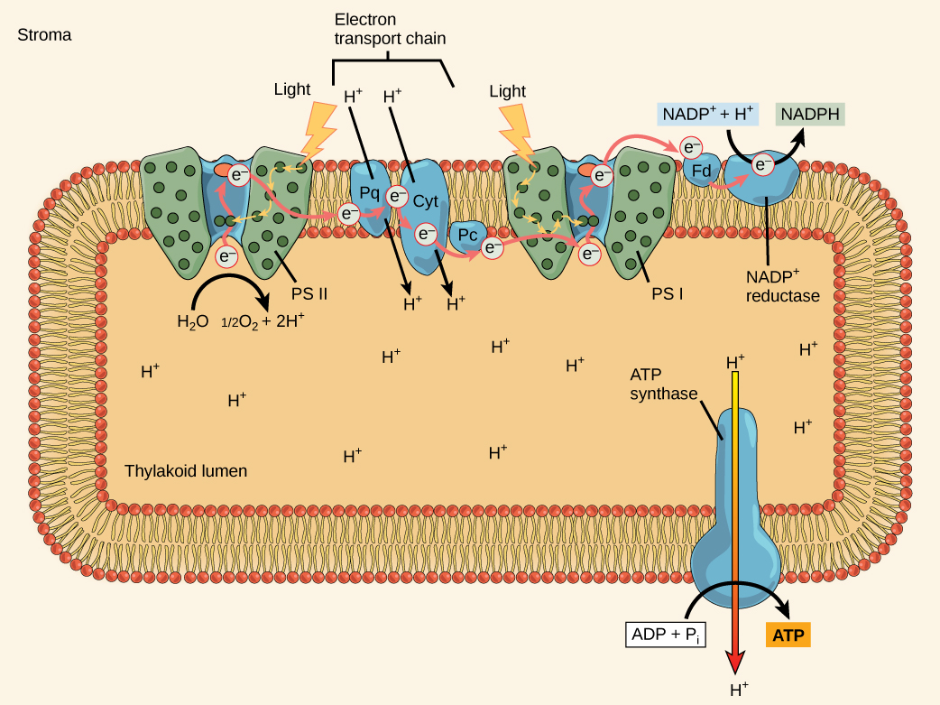
    Generation of NADPH and ATPgre_connection_icon.png

    The overall function of light-dependent reactions is to transfer solar energy into chemical compounds, largely the molecules NADPH and ATP. This energy supports the light-independent reactions and fuels the assembly of sugar molecules. We depict the light-dependent reactions in Figures 6 and 7. Protein complexes and pigment molecules work together to produce NADPH and ATP.
     

    Figure_08_02_07ab.png

    Figure 1. A photosystem consists of a light-harvesting complex and a reaction center. Pigments in the light-harvesting complex pass light energy to two special chlorophyll a molecules in the reaction center. The light excites an electron from the chlorophyll a pair, which passes to the primary electron acceptor. The excited electron must then be replaced. In (a) photosystem II, the electron comes from the splitting of water, which releases oxygen as a waste product. In (b) photosystem I, the electron comes from the chloroplast electron transport chain discussed below.

    The actual step that transfers light energy into a biomolecule takes place in a multiprotein complex called a photosystem, two types of which are found embedded in the thylakoid membrane, photosystem II (PSII) and photosystem I (PSI). The two complexes differ based on what they oxidize (that is, the source of the low energy electron supply) and what they reduce (the place to which they deliver their energized electrons).

    Both photosystems have the same basic structure; several antenna proteins to which the chlorophyll molecules bind surround the reaction center in which the photochemistry takes place. Each photosystem associates with the light-harvesting complex, which passes energy captured from sunlight to the reaction center; it comprises multiple antenna proteins that contain a mixture of 300–400 chlorophyll a and b molecules and other pigments like carotenoids. The absorption of a single photon—a distinct quantity or “packet” of light—by any of the chlorophylls pushes that molecule into an excited state. In short, the biological molecule has now captured light energy. However, the energy is not yet stored in any useful form. The captured energy transfers from chlorophyll to chlorophyll until, eventually (after about a millionth of a second), it arrives to the reaction center. Up to this point, only energy has been transferred between molecules, not electrons.

    photosynthesiscell.jpg

    Figure 2. In the photosystem II (PSII) reaction center, energy from sunlight is used to extract electrons from water. The electrons travel through the chloroplast electron transport chain to photosystem I (PSI), which reduces NADP+ to NADPH. The electron transport chain moves protons across the thylakoid membrane into the lumen. At the same time, splitting of water adds protons to the lumen, and reduction of NADPH removes protons from the stroma. The net result is a low pH in the thylakoid lumen, and a high pH in the stroma. ATP synthase uses this electrochemical gradient to make ATP.

    The reaction center contains a pair of chlorophyll a molecules with a special property. Those two chlorophylls can undergo oxidation upon excitation; they can give up an electron in a process called a photoactivation. It is at this step in the reaction center, this step in photophosphorylation, that light energy transfers into an excited electron. All the subsequent steps involve a getting of that electron onto the energy carrier NADPH for delivery to the Calvin cycle where the electron can be deposited onto carbon for long-term storage in the form of a carbohydrate.


    The Z-scheme

    PSII and PSI are two major components of the photosynthetic electron transport chain, which also include the cytochrome complex. The reaction center of PSII (called P680) delivers its high-energy electrons, one at a time, to a primary electron acceptor called pheophytin (Ph), and then sequentially to two bound plastoquinones QA and QB. Electrons then transfer off of PSII onto a pool of mobile plastoquinones (Q pool) which then transfer the electrons to a protein complex called Cytochromeb6f. The cytochrome complex uses the red/ox transfers to pump proton across the thylakoyd membrane establishing a proton-motive force that can be used for the synthesis of ATP. Electrons leaving the Cytochrome transfer to a copper-containing protein called plastocyanin (PC) which then transfers electrons to PSI (P700). P680’s missing electron is replaced by extracting an electron from water; thus, water splits and PSII is re-reduced after every photoactivation step. Just for the sake of sharing some numbers: Splitting one H2O molecule releases two electrons, two hydrogen atoms, and one atom of oxygen. The formation of one molecule of diatomic O2 gas requires the splitting two water molecules. In plant tissue, mitochondria use about ten percent of that oxygen to support oxidative phosphorylation. The rest escapes to the atmosphere where it is used by aerobic organisms to support respiration.

    As electrons move through the proteins that live between PSII and PSI, they take part in exergonic red/ox transfers. The free energy associated with the exergonic red/ox reaction is coupled to the endergonic transport of protons from the stromal side of the membrane to the thylakoid lumen by the cytochrome complex. Those hydrogen ions, plus the ones produced by splitting water, accumulate in the thylakoid lumen and create a proton motive force that will drive the synthesis of ATP in a later step. Since the electrons on PSI now have a greater reduction potential than when they started their trek (it is important to note that unexcited PSI has a greater red/ox potential than NADP+/NADPH), they must be re-energized in PSI before getting deposited onto NADP+. Therefore, to complete this process, another photon must be absorbed by the PSI antenna. That energy transfers to the PSI reaction center (called P700). P700 then oxidizes and sends an electron through several intermediate red/ox steps to NADP+ to form NADPH. Thus, PSII captures the energy in light and couples its transfer via red/ox reactions to the creation of a proton gradient. As already noted, the exergonic and controlled relaxation of this gradient can be coupled to the synthesis of ATP. PSI captures energy in light and couples that, through a series of red/ox reactions, to reduce NADP+ into NADPH. The two photosystems work in concert, in part, to guarantee that the production of NADPH will be in the right proportion to the production of ATP. Other mechanisms exist to fine tune that ratio to match the chloroplast’s constantly changing energy needs.

    oxygenic_photorespiration.png

    Figure 3. A diagram depicting the flow of electrons and the red/ox potentials of their carriers in oxygenic photosynthetic systems expressing both photosystem I (boxed in blue) and photosystem II (boxed in green). Ph = pheophytin; QA = bound plastoquinone, QB = more loosely associated plastoquinone; Q pool = mobile plastoquinone pool; Cyt bf = Cytochrome b6f complex; PC = plastocyanin; Chla0 = special chrolophyl; A1 = vitamin K; Fx and FAB = iron-sulfur centers; Fd = ferredoxin; FNR = ferredoxin-NADP reductase. Attribution: Marc T. Facciotti (own work)


    Light Independent Reactions and Carbon Fixationgre_connection_icon.png

    A short introduction

    The general principle of carbon fixation is that some cells under certain conditions can take inorganic carbon, CO2 (also referred to as mineralized carbon), and reduce it to a usable cellular form. Most of us know that green plants can take up CO2 and produce O2 in a process known as photosynthesis. We have already discussed photophosphorylation, the ability of a cell to transfer light energy onto chemicals and ultimately to produce the energy carriers ATP and NADPH in a process known as the light reactions. In photosynthesis, the plant cells use the ATP and NADPH formed during photophosphorylation to reduce CO2 to sugar, (as we will see, specifically G3P) in what we call the dark reactions. While we appreciate that this process happens in green plants, photosynthesis had its evolutionary origins in the bacterial world. In this module we will go over the general reactions of the Calvin Cycle, a reductive pathway that incorporates CO2 into cellular material.

    In photosynthetic bacteria, such as Cyanobacteria and purple non-sulfur bacteria, as well plants, the energy (ATP) and reducing power (NADPH) - a term used to describe electron carriers in their reduced state - obtained from photophosphorylation, is coupled to "Carbon Fixation", incorporating inorganic carbon (CO2) into organic molecules; initially as glyceraldehyde-3-phosphate (G3P) and eventually into glucose. We refer to organisms that can get all of their required carbon from an inorganic source (CO2) as autotrophs, while we refer to those organisms that require organic forms of carbon, such as glucose or amino acids, as heterotrophs. The biological pathway that leads to carbon fixation is called the Calvin Cycle and is a reductive pathway (consumes energy/uses electrons) which leads to the reduction of CO2 to G3P.


    The Calvin Cycle: the reduction of CO2 to Glyceraldehyde 3-Phosphate

    calvin.jpg

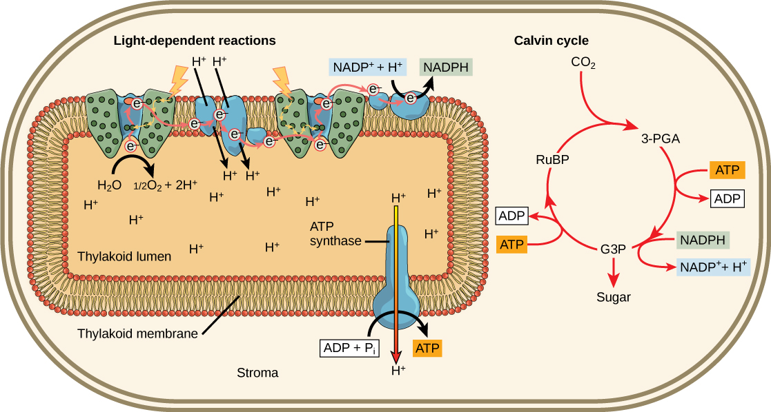
    Figure 1. Light reactions harness energy from the sun to produce chemical bonds, ATP, and NADPH. These energy-carrying molecules are made in the stroma where carbon fixation takes place.

    In plant cells, the Calvin cycle is located in the chloroplasts. While the process is similar in bacteria, there are no specific organelles that house the Calvin Cycle and the reactions occur in the cytoplasm around a complex membrane system derived from the plasma membrane. This intracellular membrane system can be quite complex and highly regulated. There is strong evidence that supports the hypothesis that the origin of chloroplasts from a symbiosis between cyanobacteria and early plant cells.

    Stage 1: Carbon Fixation

    In the stroma of plant chloroplasts, besides CO2, two other components are present to start the light-independent reactions: an enzyme called ribulose-1,5-bisphosphate carboxylase/oxygenase (RuBisCO), and three molecules of ribulose bisphosphate (RuBP), as shown in the figure below. Ribulose-1,5-bisphosphate (RuBP) is composed of five carbon atoms and includes two phosphates.
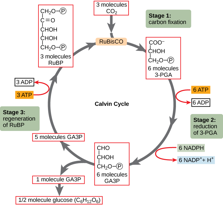
    Figure_08_03_02f.png

    Figure 2. The Calvin cycle has three stages. In stage 1, the enzyme RuBisCO incorporates carbon dioxide into an organic molecule, 3-PGA. In stage 2, the organic molecule is reduced using electrons supplied by NADPH. In stage 3, RuBP, the molecule that starts the cycle, is regenerated so that the cycle can continue. Only one carbon dioxide molecule is incorporated at a time, so it must complete the cycle three times to produce a single three-carbon GA3P molecule, and six times to produce a six-carbon glucose molecule.

    RuBisCO catalyzes a reaction between CO2 and RuBP. For each CO2 molecule that reacts with one RuBP, two molecules of another compound (3-PGA) form. PGA has three carbons and one phosphate. Each turn of the cycle involves only one RuBP and one carbon dioxide and forms two molecules of 3-PGA. The number of carbon atoms remains the same, as the atoms move to form new bonds during the reactions (3 atoms from 3CO2 + 15 atoms from 3RuBP = 18 atoms in 3 atoms of 3-PGA). We call this process carbon fixation, because CO2 is “fixed” from an inorganic form into an organic molecule.

    Stage 2: Reduction

    ATP and NADPH are used to convert the six molecules of 3-PGA into six molecules of a chemical called glyceraldehyde 3-phosphate (G3P) - a carbon compound also found in glycolysis. The process uses six molecules of both ATP and NADPH. The exergonic process of ATP hydrolysis is in effect driving the endergonic redox reactions, creating ADP and NADP+. Both "spent" molecules (ADP and NADP+) return to the nearby light-dependent reactions to be recycled back into ATP and NADPH.

    Stage 3: Regeneration

    Interestingly, at this point, only one of the G3P molecules leaves the Calvin cycle to contribute to the formation of other compounds needed by the organism. In plants, because the G3P exported from the Calvin cycle has three carbon atoms, it takes three “turns” of the Calvin cycle to fix enough net carbon to export one G3P. But each turn makes two G3Ps, thus three turns make six G3Ps. One is exported while the remaining five G3P molecules remain in the cycle and are used to regenerate RuBP, which enables the system to prepare for more CO2 to be fixed. These regeneration reactions use three more molecules of ATP .


    Possible NB Discussion nb-sticker.pngPoint

    Have you ever heard anyone accidentally refer to the Amazon rainforest as the "lungs of the Earth"? In reality, the majority of our planet's oxygen is produced by marine organisms, such as microscopic phytoplankton -- which, by the way, also take up appreciable amounts of carbon dioxide from the environment. The family of phytoplankton include organisms like cyanobacteria and diatoms (a visually stunning type of algae -- look it up!) that are able to survive and aggregate close to the water's surface, where sun exposure is higher. Try to approach phytoplankton from a BIS 2A lens... What biochemical processes had to happen in order for these phytoplankton to produce oxygen? What exactly are the phytoplankton doing with the carbon dioxide they take up from the atmosphere? What large-scale global effects would you expect if phytoplankton health were to be severely compromised?


    Additional Links of Interest

    Khan Academy Links

    Chemwiki links


    2020_Winter_Bis2A_Facciotti_Lecture_17 is shared under a not declared license and was authored, remixed, and/or curated by LibreTexts.

    • Was this article helpful?