

2.4.3: Allosteric Regulation
- Page ID
- 190495
\( \newcommand{\vecs}[1]{\overset { \scriptstyle \rightharpoonup} {\mathbf{#1}} } \)
\( \newcommand{\vecd}[1]{\overset{-\!-\!\rightharpoonup}{\vphantom{a}\smash {#1}}} \)
\( \newcommand{\dsum}{\displaystyle\sum\limits} \)
\( \newcommand{\dint}{\displaystyle\int\limits} \)
\( \newcommand{\dlim}{\displaystyle\lim\limits} \)
\( \newcommand{\id}{\mathrm{id}}\) \( \newcommand{\Span}{\mathrm{span}}\)
( \newcommand{\kernel}{\mathrm{null}\,}\) \( \newcommand{\range}{\mathrm{range}\,}\)
\( \newcommand{\RealPart}{\mathrm{Re}}\) \( \newcommand{\ImaginaryPart}{\mathrm{Im}}\)
\( \newcommand{\Argument}{\mathrm{Arg}}\) \( \newcommand{\norm}[1]{\| #1 \|}\)
\( \newcommand{\inner}[2]{\langle #1, #2 \rangle}\)
\( \newcommand{\Span}{\mathrm{span}}\)
\( \newcommand{\id}{\mathrm{id}}\)
\( \newcommand{\Span}{\mathrm{span}}\)
\( \newcommand{\kernel}{\mathrm{null}\,}\)
\( \newcommand{\range}{\mathrm{range}\,}\)
\( \newcommand{\RealPart}{\mathrm{Re}}\)
\( \newcommand{\ImaginaryPart}{\mathrm{Im}}\)
\( \newcommand{\Argument}{\mathrm{Arg}}\)
\( \newcommand{\norm}[1]{\| #1 \|}\)
\( \newcommand{\inner}[2]{\langle #1, #2 \rangle}\)
\( \newcommand{\Span}{\mathrm{span}}\) \( \newcommand{\AA}{\unicode[.8,0]{x212B}}\)
\( \newcommand{\vectorA}[1]{\vec{#1}} % arrow\)
\( \newcommand{\vectorAt}[1]{\vec{\text{#1}}} % arrow\)
\( \newcommand{\vectorB}[1]{\overset { \scriptstyle \rightharpoonup} {\mathbf{#1}} } \)
\( \newcommand{\vectorC}[1]{\textbf{#1}} \)
\( \newcommand{\vectorD}[1]{\overrightarrow{#1}} \)
\( \newcommand{\vectorDt}[1]{\overrightarrow{\text{#1}}} \)
\( \newcommand{\vectE}[1]{\overset{-\!-\!\rightharpoonup}{\vphantom{a}\smash{\mathbf {#1}}}} \)
\( \newcommand{\vecs}[1]{\overset { \scriptstyle \rightharpoonup} {\mathbf{#1}} } \)
\(\newcommand{\longvect}{\overrightarrow}\)
\( \newcommand{\vecd}[1]{\overset{-\!-\!\rightharpoonup}{\vphantom{a}\smash {#1}}} \)
\(\newcommand{\ket}[1]{\left| #1 \right>}\)
\(\newcommand{\bra}[1]{\left< #1 \right|}\)
\(\newcommand{\braket}[2]{\left< #1 \vphantom{#2} \right| \left. #2 \vphantom{#1} \right>}\)
\(\newcommand{\braopket}[3]{\left< #1 \vphantom{#2}\vphantom{#3} \right| #2 \vphantom{#1}\vphantom{#3} \left| #3 \vphantom{#1}\vphantom{#2} \right>}\)
\(\newcommand{\qmvec}[1]{\mathbf{\vec{#1}}}\)
\(\newcommand{\op}[1]{\hat{\mathbf{#1}}}\)
\(\newcommand{\expect}[1]{\langle #1 \rangle}\)
\(\newcommand{\dfn}[1]{\emph{\textbf{#1}}}\)
Allosteric regulation fine-tunes most biological processes, including signal transduction, enzyme activity, metabolism, and transport. Allostery, an intrinsic property of a protein, is the regulation of activity at one site (also known as an orthosteric site) by a topographically and spatially distant site; the latter is designated as an allosteric site. Allosteric regulation occurs through binding a modulator (e.g., a small molecule or protein) at an allosteric site to engender a conformational change that affects function at the orthosteric site. This effect may redistribute the conformational ensemble by either stabilizing an active conformation (allosteric activation) or destabilizing an inactive conformation (allosteric inhibition) in response to allosteric perturbations (Figure 8.13). Traditionally, the repertoire of allostery was largely confined to determining allosteric effects or mechanisms in individual multi-subunit or monomer proteins through conformational transitions. Recently, growing evidence indicates that allosteric signals can propagate across multiple proteins to sculpt allosteric networks.
Allosteric regulation is particularly important for the cell's ability to adjust enzyme activity in response to environmental conditions. Feedback control loops, such as feedback inhibition from downstream products or feedforward from upstream substrates, are common allosteric regulatory mechanisms found in nature. Another example of allostery is oxygen binding to one hemoglobin subunit, which prompts cooperative binding to other subunits.
We mentioned earlier that PTMs can induce conformational changes in proteins. Other methods to induce such allosteric changes include binding of small molecules, binding of proteins, changes in the redox environment that affect disulfide bonds, and general changes in protein flexibility and dynamics. Let's look at some examples of allostery brought about by these changes. Laskowski et al. present many of these examples (https://doi.org/10.1016/j.febslet.2009.03.019).
Allosterism by small molecules: opening and closing active sites
Small-molecule binding at an allosteric site can induce small and large conformational changes in a distal active site. A common motion is a partial or full hinge-clamping conformational change that brings critical catalytic groups into a more organized, effective active site, thereby excluding water as a competing hydrolytic substrate (for example). One example of a small hinge-clamping change is seen in phosphoglycerate dehydrogenase (PGDH), which catalyzes the first step in serine synthesis (oxidation by NAD+). It converts 3-phosphoglycerate into 3-phosphohydropyruvate. The pathway's end product, serine, binds to an allosteric site on PGDH and inhibits it, a classic example of feedback inhibition of a pathway's first reaction by its end product. PGDH has a regulatory binding domain (RBD) that binds serine, a substrate-binding domain (SBD), an NAD+/NADH-binding domain (NBD), where the allosteric inhibitor binds, and binding domains for the substrate (SBD) and the NAD nucleotide (NBD). Figure \(\PageIndex{14}\) shows the small hinge-bending conformational change on bind conversion of monomeric apo-PGDH (1psd) to holo-PGDH with serine (not shown) bound in the RBD allosteric domain. The RBD-SBD domains rotate relative to the NBD domain by 15 °.
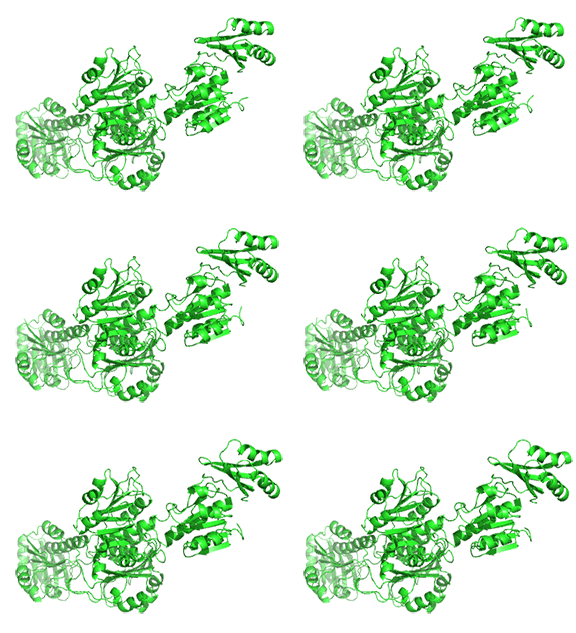
The functional enzyme is a tetramer (only the monomer is shown in the above figure). The rotation in the full tetramer aligns catalytic residues and closes off the active site.
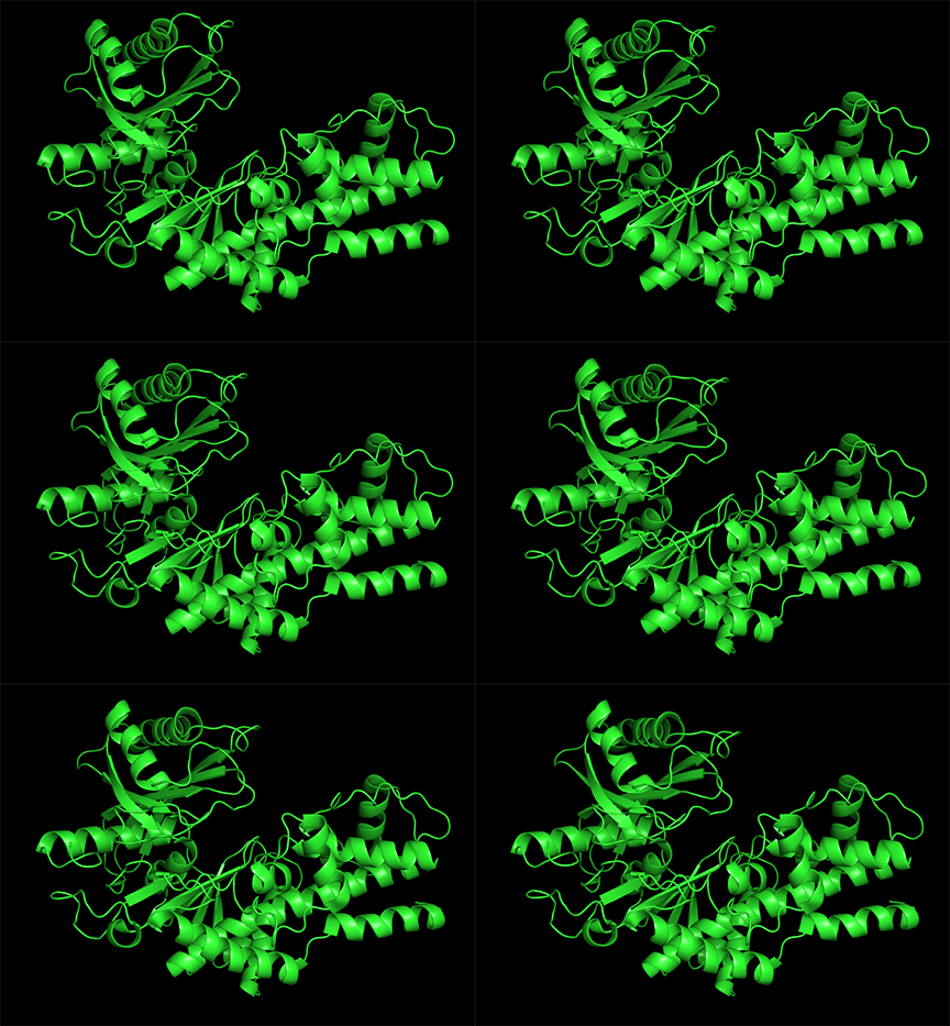
Conformational changes are ubiquitous in the binding of ligands to proteins. These changes also occur on substrate binding at the active site. It is important to recognize that substrate-induced changes in the active site are not allosteric since the conformational changes, no matter how big, are caused by binding at the active site, not at an allosteric site. A classic example of a large conformational change upon binding is the activation of hexokinase, the first enzyme in the glycolytic pathway, by glucose or glucose analogs. Figure \(\PageIndex{15}\) shows the large conformational change on binding a glucose analog to hexokinase (1hkg, no glucose; 2yhx, with glucose analog). The background is shown in black to remind readers that this is NOT an example of allosterism. The changes facilitate catalysis by excluding water, a competing substrate, from the active site after glucose and ATP bind (not shown). Upon binding the glucose analog in the active site, the site becomes sequestered ("jaw" clamping down on the binding) from solvent.
Allosterism by small molecules: Subtle electrostatic effects
Often, no large conformational change is evident in the protein. In those cases, subtle rearrangements of key residues in the active site or near it (which might promote access) may result from allosteric effector binding. A simple change in the electrostatic environment might account for the effect. This appears to be the case with chorismate mutase, an enzyme in the bacterial, fungal, and plant pathways for synthesizing the aromatic amino acids tyrosine and phenylalanine. The enzyme catalyzes the conversion of chorismate to prephenate, an intermediate in aromatic amino acid synthesis. An offshoot pathway takes chorismate to the last aromatic amino acid, tryptophan. Chorismate mutase is key in the metabolic decision pathway that leads to the production of tyrosine/phenylalanine or tryptophan. The enzyme is activated by tryptophan and inhibited by tyrosine. The aligned sequences of two crystal structures of the enzyme with the bound allosteric activator tryptophan (Trp 502 in pdb structure 1csm, showing a magenta glutamic acid 23) and with bound inhibitor (Tyr 300 in pdb structure 2csmA, showing cyan Glu 23) are shown in Figure \(\PageIndex{16}\):
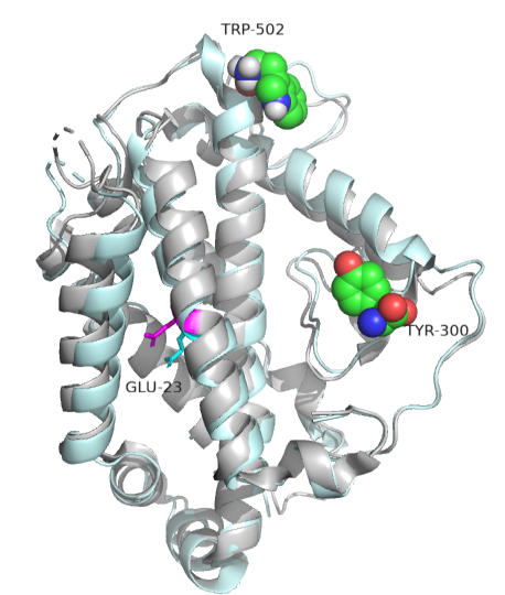
The protein's overall structure and active site show no significant changes. However, glutamic acid 23 shifts, which may underlie the observed allosteric effects. When active, glutamic acid 23 is buried in the active site pocket, but in the inhibited state, it swings into the binding site. Since chorismate is a charged dicarboxylic acid, Glu 23 probably repels it, inhibiting the enzyme's activity.
Allosterism by phosphorylation
We'll focus on one example of a large conformational change upon phosphorylation of a serine residue in glycogen phosphorylase (GP). This enzyme catalyzes the phosphorolysis of the acetal linkage between the terminal glucose of a glycogen polymer and the next glucose in the chain. This reaction is not a hydrolysis, in which water acts as a nucleophile to cleave acetal bonds. This reaction is key in metabolism because it cleaves a major energy-storage molecule, glycogen.
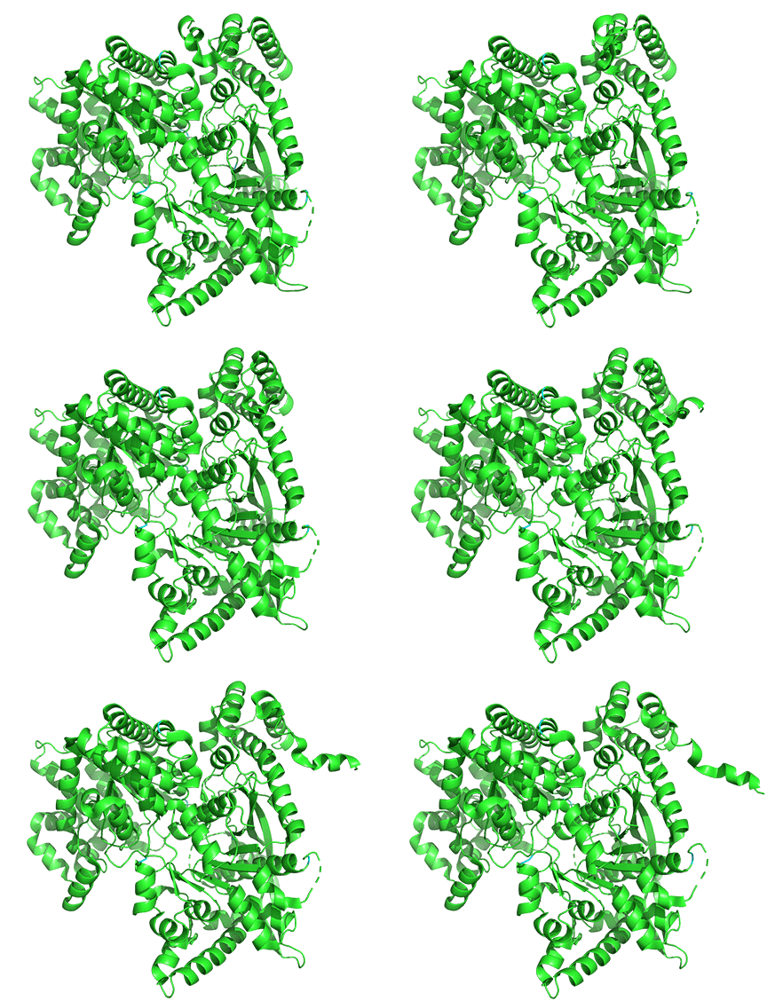
The enzyme is a multimer that exists in two major states: a T-state and an R-state glycogen phosphorylase (GP). We explored T and R states and allosterism in their interconversion when we discussed oxygen binding to hemoglobin in Chapter 5.3. Crystal structures of the T and R states bound to allosteric activators IMP and AMP are known. High AMP concentrations imply low ATP concentrations and the need to mobilize glycogen reserves. Phosphorylation of Serine 14 also promotes conversion to the R state, activating the enzyme. The allosteric activators AMP and IMP bind to a disordered loop (313-326) and change it to a form promoted by nucleotide binding. which is disordered in the free structure and adopts a conformation dictated mainly by the type of nucleotide that binds at this site. Figure \(\PageIndex{17}\) shows a monomer of the European rabbit glycogen phosphorylase in the R (active) state bound to AMP (3e3n).
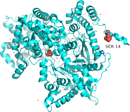
Only one chain of the active homodimer is shown. The helix containing the phospho-Ser 14 swings away from the rest of the protein.
Figure \(\PageIndex{18}\) shows the difference between the T (inactive) state of rabbit muscle glycogen phosphorylase (7P7D) and the R (active) state (3e3n, neither phospho-Ser 14 nor AMP shown). Note the huge conformational change elicited on phosphorylation of Ser 13.
Allosterism by small molecules: control of quaternary structure
The binding of small molecules at allosteric sites may promote or inhibit the formation of a protein's correct functional multimeric structure. One example is ATP phosphoribosyltransferase (APRT), which catalyzes the first step in synthesizing histidine. As we saw with phosphoglycerate dehydrogenase (PGDH), APRT has three domains. Domains I and II comprise the active site located between them, and domain III is a regulatory domain (the functional protein is a dimer). As with PGDH, APRT is allosterically inhibited by the pathway's end product, histidine. On histidine binding to the inactive form (1nh7), a large conformational change results (1nh8), as shown in Figure \(\PageIndex{19}\).
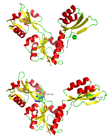
This change drives the formation of a hexamer (dimer)3, which closes off the active site and inactivates the enzyme. The enzyme ribonuclease reductase behaves similarly.
Allosteric by protein binding
Another protein can bind to an enzyme and activate it by promoting an allosteric change. An example is the binding of a regulatory protein, Cyclin A, to cyclin-dependent kinase 2 (CDK2). Cyclin binding induces a large conformational change that activates the enzyme by altering active-site geometry and making it more accessible. CDK enzymes are involved in cell-cycle progression. For greater control of the cell cycle, cyclin binding and CDK2 phosphorylation are required for activity. Upon binding cyclin, the active site becomes more accessible for substrate (ATP) binding. Next, the amino acid that must be phosphorylated for activation, Thr 16, becomes accessible. Figure \(\PageIndex{20}\) shows the conformational change in apo-CDK2 (1hc1) upon cyclin binding, which allows ATP binding (1fin).

Cyclin (the regulatory protein) is shown in gray. ATP is shown in spacefill. CDK2 is shown as a colored cartoon.
Allosterism by disulfide bonds
It should make sense that cleaving an intrachain or interchain disulfide, whose presence constrains a specific protein conformation, should significantly alter protein structure and function. One example is the protein botulinum neurotoxin type A, a neurotoxin and Zn protease. It cleaves proteins such as synaptobrevin, syntaxin, and SNAP-25 in the neurosynapse, which are required for neurotransmitter release.
It has a catalytic (Zn2+ peptidase/protease) domain and a translocation domain in a single protein chain and contains two disulfide bonds when synthesized (PDB code 3bta). Cleavage at a specific peptide bond splits the polypeptide into A and B chains, which remain connected by a single disulfide bond, with the A chain containing the catalytic domain. The B chain contains the translocation domain, which effectively still blocks the active site of the peptidase/protease domain. The disulfides are cleaved when the protein enters an endosome, which contains a more reducing environment. The separate A chain then dissociates from the inhibitory B domain and expresses protease activity. The active protein then enters the cytoplasm.
Figure \(\PageIndex{21}\) shows the domain structure and disulfide bonds of the unprocessed form. The N-terminal Zn proteinase domain (Peptidase_M27, amino acids 1-409) is followed by an intrachain disulfide between C429 and C 453.

The mature form is proteolyzed between amino acids 447 and 448 (within the sequences connected by the disulfide (429-453). The protein remains inactive until the disulfide bond is cleaved.
Figure \(\PageIndex{22}\) shows an interactive iCn3D model of botulinum neurotoxin type A (3bta).
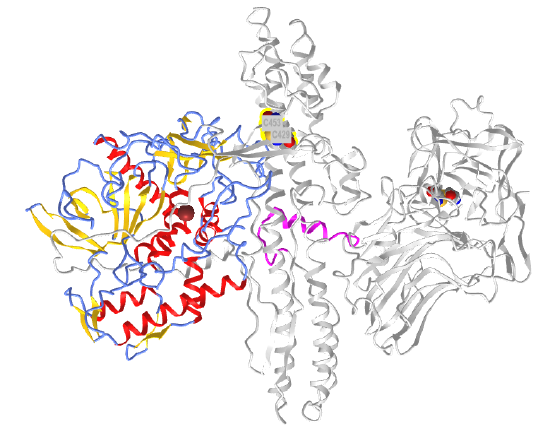
 Figure \(\PageIndex{22}\): Botulinum neurotoxin type A (3bta) (Copyright; author via source).
Click the image for a popup or use this external link: https://structure.ncbi.nlm.nih.gov/i...CfSsBH94jcPyb8
The N-terminal Zn peptidase/protease domain is shown in color, and the other regions of the protein are shown in gray. The two disulfide bonds are shown in spacefill, with CPK color. On cleavage of the disulfide bond between C429 and C453, the Zn protease domain, which is inhibited in the presence of the constraining disulfide bonds, is released and activated.
Allosterism by changes in protein flexibility and population shifts
Sometimes, the crystal structure shows little difference between the apo- and holo-forms of a protein that contains a separate allosteric binding site. One example is the enzyme dihydrodipicolinate synthase (DHDPS), which catalyzes the first step in the bacterial and plant lysine biosynthetic pathways. DHDPS catalyzes the synthesis of 4-hydroxy-2,3,4,5-tetrahydro-2-dipicolinic acid from aspartate-β-semialdehyde and pyruvate. One explanation for allosterism is that it occurs on time scales too short to be seen in crystal or NMR structures. Rather, some transient conformation might cause allosterism. Such changes are detectable in the molecular dynamics simulation of the protein in the presence and absence of the allosteric effector molecule.
This possibility suggests that the allosteric effector might change the distribution of possible transient conformational states within the population. If an allosteric effector bound one of the transient states in the population, that state would be "removed" from the ensemble of states, shifting the dynamic equilibrium among the populated states to produce more of the specific conformation that binds the allosteric effector. This sounds like the T to R state conversion in the MWC model of oxygen binding to hemoglobin and the allosterism discussed above with the enzyme glycogen phosphorylase, only on a much shorter time scale. This model is consistent with a conformational selection model, in which the allosteric molecule binds preferentially to a preexisting, transient conformational state.