

2.2.9: Enzyme Modulation
- Page ID
- 190476
\( \newcommand{\vecs}[1]{\overset { \scriptstyle \rightharpoonup} {\mathbf{#1}} } \)
\( \newcommand{\vecd}[1]{\overset{-\!-\!\rightharpoonup}{\vphantom{a}\smash {#1}}} \)
\( \newcommand{\dsum}{\displaystyle\sum\limits} \)
\( \newcommand{\dint}{\displaystyle\int\limits} \)
\( \newcommand{\dlim}{\displaystyle\lim\limits} \)
\( \newcommand{\id}{\mathrm{id}}\) \( \newcommand{\Span}{\mathrm{span}}\)
( \newcommand{\kernel}{\mathrm{null}\,}\) \( \newcommand{\range}{\mathrm{range}\,}\)
\( \newcommand{\RealPart}{\mathrm{Re}}\) \( \newcommand{\ImaginaryPart}{\mathrm{Im}}\)
\( \newcommand{\Argument}{\mathrm{Arg}}\) \( \newcommand{\norm}[1]{\| #1 \|}\)
\( \newcommand{\inner}[2]{\langle #1, #2 \rangle}\)
\( \newcommand{\Span}{\mathrm{span}}\)
\( \newcommand{\id}{\mathrm{id}}\)
\( \newcommand{\Span}{\mathrm{span}}\)
\( \newcommand{\kernel}{\mathrm{null}\,}\)
\( \newcommand{\range}{\mathrm{range}\,}\)
\( \newcommand{\RealPart}{\mathrm{Re}}\)
\( \newcommand{\ImaginaryPart}{\mathrm{Im}}\)
\( \newcommand{\Argument}{\mathrm{Arg}}\)
\( \newcommand{\norm}[1]{\| #1 \|}\)
\( \newcommand{\inner}[2]{\langle #1, #2 \rangle}\)
\( \newcommand{\Span}{\mathrm{span}}\) \( \newcommand{\AA}{\unicode[.8,0]{x212B}}\)
\( \newcommand{\vectorA}[1]{\vec{#1}} % arrow\)
\( \newcommand{\vectorAt}[1]{\vec{\text{#1}}} % arrow\)
\( \newcommand{\vectorB}[1]{\overset { \scriptstyle \rightharpoonup} {\mathbf{#1}} } \)
\( \newcommand{\vectorC}[1]{\textbf{#1}} \)
\( \newcommand{\vectorD}[1]{\overrightarrow{#1}} \)
\( \newcommand{\vectorDt}[1]{\overrightarrow{\text{#1}}} \)
\( \newcommand{\vectE}[1]{\overset{-\!-\!\rightharpoonup}{\vphantom{a}\smash{\mathbf {#1}}}} \)
\( \newcommand{\vecs}[1]{\overset { \scriptstyle \rightharpoonup} {\mathbf{#1}} } \)
\(\newcommand{\longvect}{\overrightarrow}\)
\( \newcommand{\vecd}[1]{\overset{-\!-\!\rightharpoonup}{\vphantom{a}\smash {#1}}} \)
\(\newcommand{\ket}[1]{\left| #1 \right>}\)
\(\newcommand{\bra}[1]{\left< #1 \right|}\)
\(\newcommand{\braket}[2]{\left< #1 \vphantom{#2} \right| \left. #2 \vphantom{#1} \right>}\)
\(\newcommand{\braopket}[3]{\left< #1 \vphantom{#2}\vphantom{#3} \right| #2 \vphantom{#1}\vphantom{#3} \left| #3 \vphantom{#1}\vphantom{#2} \right>}\)
\(\newcommand{\qmvec}[1]{\mathbf{\vec{#1}}}\)
\(\newcommand{\op}[1]{\hat{\mathbf{#1}}}\)
\(\newcommand{\expect}[1]{\langle #1 \rangle}\)
\(\newcommand{\dfn}[1]{\emph{\textbf{#1}}}\)
- Describe how temperature affects enzyme activity and explain why activity increases at lower temperatures but decreases sharply after denaturation.
- Describe how pH affects enzyme activity by altering ionization state, active site interactions, and protein structure.
- Compare the optimal pH and temperature ranges of enzymes and relate them to biological environment.
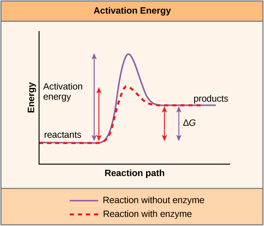
- Explain why changes in pH and temperature can alter enzyme activity without changing the overall free energy of reaction.
Enzymes are biological catalysts that increase reaction rate by lowering activation energy. Enzyme activity depends not only on substrate binding and catalysis, but also on the physical and chemical environment surrounding the enzyme. Two of the most important environmental factors that modulate enzyme activity are temperature and pH. Each enzyme functions best within a limited range of conditions, and activity falls outside that range because substrate binding, active site chemistry, or overall protein structure is disrupted.
Temperature Modulates Enzyme Activity
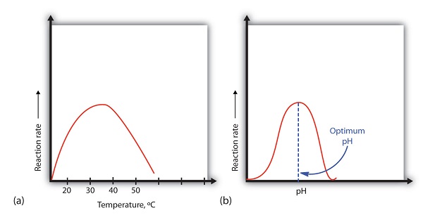
As temperature increases, molecules move more rapidly and collide more often. For this reason, the rate of an enzyme-catalyzed reaction usually increases as temperature rises. However, this trend holds only up to a point. Because most enzymes are proteins, excessive heat disrupts the noncovalent interactions that maintain protein structure. Once the enzyme begins to denature, the active site loses its functional shape and activity drops quickly.
The result is a characteristic curve: activity rises with temperature, reaches an optimum, and then declines sharply at higher temperatures. For many human enzymes, the optimum temperature is close to 37 °C, while enzymes from thermophilic organisms function best at much higher temperatures.

Low Temperature
Low temperature affects enzymes differently from high temperature. Cooling generally slows reaction rate because molecular motion decreases and productive collisions become less frequent. In most cases, this loss of activity is reversible because the enzyme remains folded and can regain activity when temperature returns to a favorable range.
High Temperature
High temperature initially increases reaction rate, but only within a limited range. Beyond the optimum temperature, thermal motion begins to disrupt the weak interactions that stabilize protein structure. As a result, activity declines rapidly as the enzyme loses the precise shape required for catalysis.
Temperature effects on enzyme activity also have biological and practical consequences. Refrigeration slows enzyme-catalyzed reactions in food and microbes. Boiling and autoclaving destroy enzymatic function by denaturing proteins. In contrast, thermophilic organisms produce enzymes that remain stable and active at temperatures that would inactivate most proteins.
Structural Basis of Protein Denaturation
Protein structure depends on many weak interactions between amino acid side chains, including hydrogen bonds, ionic interactions, and hydrophobic effects. Changes in temperature, pH, or exposure to chemicals can disrupt these interactions. When this occurs, the protein may lose its three-dimensional structure and become nonfunctional. This process is known as denaturation.
Denaturation does not break the covalent bonds that link amino acids together in the primary structure. Instead, it disrupts the secondary, tertiary, or quaternary structure of the protein. Because enzyme activity depends on the precise shape of the active site, even modest structural changes can greatly reduce catalytic activity.
Enzymes often operate only within narrow temperature and pH ranges because their structure must remain stable for catalysis to occur. For example, the digestive enzyme pepsin functions in the highly acidic environment of the stomach. At higher pH values, the structure of pepsin changes and the enzyme becomes inactive.

Reversibility of Denaturation
In some cases, denaturation can be reversible because the primary structure of the protein remains intact. If the denaturing conditions are removed, the protein may refold into its original conformation and regain its activity. However, severe heat or chemical damage often causes irreversible denaturation.
Cells contain helper proteins called chaperones or chaperonins that assist newly synthesized proteins in folding correctly. These proteins help prevent aggregation and provide an environment that favors proper folding.
pH Modulates Enzyme Activity
Enzymes are also highly sensitive to pH. Changes in pH alter the protonation state of ionizable groups in amino acid side chains and substrates. These changes can disrupt ionic interactions, alter hydrogen bonding, and change the charge pattern of the active site. Even when the overall protein remains folded, a shift in protonation of a single catalytic residue can greatly reduce activity.
Ionization and Catalytic Activity
Each enzyme therefore has an optimal pH at which its active site is in the proper ionization state for substrate binding and catalysis. Outside this range, activity declines because key functional groups no longer have the charge required for binding or catalysis. At extreme pH, the enzyme may also denature.
Optimal pH
Optimal pH often reflects the environment in which an enzyme normally functions. For example, salivary amylase functions near neutral pH, while pepsin is adapted to the acidic environment of the stomach. Other enzymes function best at more basic pH values. These differences reflect both the chemistry of the local environment and the ionization requirements of the active site.
Why pH and Temperature Matter at the Molecular Level
Temperature and pH influence enzyme activity in different ways, but both act through structure and chemistry. Temperature changes kinetic energy and collision frequency, while heat disrupts the weak interactions that stabilize protein structure. pH changes protonation state, which alters charge, binding interactions, and the catalytic properties of active site residues.
As a result, enzyme modulation by pH and temperature can affect both substrate binding and catalysis. A change in conditions may weaken substrate binding, reduce catalytic rate, or do both at once.
The influence of pH and temperature on enzyme activity appears across biology. Digestive enzymes function at pH values matched to their physiological location. Microbial growth rates shift as environmental temperature and pH change because different species have different enzyme optima. Food preservation methods such as refrigeration slow enzyme activity, while sterilization methods such as boiling or autoclaving destroy it. Thermophilic organisms rely on enzymes that remain stable at high temperature.
Summary
Enzyme activity is strongly modulated by temperature and pH. Rising temperature usually increases reaction rate at first because molecular collisions become more frequent, but excessive heat denatures the protein and causes activity to fall. Changes in pH alter the ionization state of amino acid side chains and substrates, which can disrupt substrate binding, catalytic chemistry, and protein stability. Because enzymes are adapted to the environments in which they function, each enzyme has characteristic pH and temperature optima. Understanding these effects helps explain enzyme behavior in physiology, microbiology, and biotechnology.
Attribution
This page was adapted from the following LibreTexts pages:
- Enzyme Activity, Chemistry LibreTexts, Page ID 16022.
- Enzymes, Principles of Biology, Biology LibreTexts, Page ID 64819.
- Factors Affecting Enzyme Activity, PGCC Microbiology, Biology LibreTexts, Page ID 93116.
- Influence of pH and temperature on enzyme activity, Biology LibreTexts, Page ID 131878.
- Changing the Shape of a Protein, Biology LibreTexts, Page ID 12701.