
4.2: Specialized Bacterial Staining Techniques
- Page ID
- 52243

Learning outcomes
- Describe the purpose of each of the stains listed: simple, Gram, Acid-Fast, Endospore, Capsule, Flagella, Spirochete.
- Recognize the cellular characteristics revealed by each of the stains listed
Simple Stains:
Commonly used dyes for simple stains: Crystal Violet, Methylene Blue, Safranin
- Uses one dye
- Used to provide color to otherwise transparent bacterial cells
- Can be used to determine cell size, morphology and arrangement
- All bacteria are the same color when stained with the single dye that is used
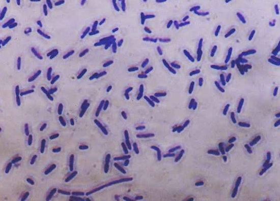
Image 1: Simple stain with crystal violet showing rod shaped bacteria. Note that all bacteria are the same color of the dye, crystal violet (purple), regardless of its cell wall composition. Image by Muntasir du, 2007 (https://commons.wikimedia.org/wiki/F...micrograph.jpg)
Gram Stain
Primary stain – crystal violet
Mordant – iodine; decolorizer- 95% Ethanol
Counterstain – Safranin
- Common differential stain
- Gram reaction (positive or negative) reflects cell wall properties
- Also used to determine cell size, morphology and arrangement

Image 2. Gram positive, rod, Bacillus subtilis. (Ann C. Smith, University of Maryland, College Park, MD)

Image 3: Gram stain showing Gram negative rods (pink) and Gram positive cocci (purple): https://commons.wikimedia.org/wiki/F...m_stain_01.jpg
Acid-Fast Stain
The acid-fast stain is a differential stain used to identify acid-fast organisms such as members of the genus Mycobacterium .
Acid-fast organisms are characterized by wax-like, nearly impermeable cell walls; they contain mycolic acid and large amounts of fatty acids, waxes, and complex lipids. Acid-fast organisms are highly resistant to disinfectants and dry conditions. (1)
Because the cell wall is so resistant to most compounds, acid-fast organisms require a special staining technique. The primary stain used in acid-fast staining, carbolfuchsin, is lipid-soluble and contains phenol, which helps the stain penetrate the cell wall. This is further assisted by the addition of heat. The smear is then rinsed with a very strong decolorizer, which strips the stain from all non-acid-fast cells but does not permeate the cell wall of acid-fast organisms. The decolorized non-acid-fast cells then take up the counterstain. (1)
Primary stain – Carbol fuchsin
Decolorizer – acid alcohol
Counterstain – Methylene blue
- A differential stain used to detect bacteria with mycolic acid cell walls (genera Mycobacterium and Nocardia)
- Developed to detect the bacterial species that causes tuberculosis
- Acid-fast organisms resist decolorization with acid-alcohol

Image 4. A mixed culture of Mycobacterium smegmatus (acid-fast, red/pink) and Staphylococcus epidermidis (non-acid-fast, light blue/purple). (Alan Schenkel, Peter Justice, and Erica Suchman, Colorado State University, Fort Collins, CO )
Endospore Stain
This stain is used to visualize bacterial endospores produced by members of the genera Bacillus and Clostridium. The nature of the spore makes it impervious to most ordinary stains and staining methods, but, once stained it strongly resists decolorization and counterstaining. In the Schaeffer-Fulton method, a primary stain with malachite green is forced into the endospore by steaming the bacterial emulsion. Malachite green is water soluble and has a low affinity for cellular material, so vegetative (actively dividing) cells may be decolorized with water. Vegetative cells are then counterstained with safranin. Microscopic examination of stained endospores will reveal their relative size and position in the cell, which are distinctive characteristics of each of the spore-forming species.
Primary stain - Malachite green
Counterstain - safranin
- Endospores resist staining with basic stains
- Endospores stain with malachite green; vegetative cells stain with safranin (red)

Image 5. Endospore stain of a Bacillus cereus culture using the Shaeffer-Fulton method and viewed at 1,000x total magnification under an oil immersion lens. Vegetative cells of B. cereus are in red; endospores are in green. Due to the age of the culture, endospores have been released from the cells. (Derek Weber and S. Finazzo, Broward Community College–Central campus, Davie, FL)
Capsule Stain (Negative staining)
Polysaccharide capsules and slime layers are generally difficult to stain selectively. Instead they are demonstrated by simply outlining them, staining the background and/or the cells without staining the capsule. This negative stain can be done several ways. The lab demonstration slide was prepared by staining the background and cells with basic fuchsin. Hence, the capsule appears as a clear zone or halo surrounding the red cell against a red background.
Uses an acidic stain: (Congo red or Nigrosin) and a basic stain: (crystal violet or safranin)
- Negative stains are neither heat-fixed nor rinsed
- The background of the slide is stained by acidic stains (capsule remains unstained)
- The cells within the capsule are stained with Basic stains
- Examples of encapsulated cells: Bacillus anthracis, Streptococcus pneumoniae, and Klebsiella pneumonia

Image 6: Encapsulated Enterobacter aerogenes stained with Anthony's capsule stain. (Gary E. Kaiser, The Community College of Baltimore County, Catonsville Campus, Baltimore, MD)
Flagella Stain
This stain coats the thin bacterial flagella with heavy metals or other compounds to make them visible in the light microscope. Once visible, the location and number of the flagella can be used diagnostically. The presence of flagella varies with cultural conditions, so a negative result is not proof of a cell's inability to produce flagella. The demonstration slide was prepared with a silver compound to coat the flagella and a red dye, basic fuchsin, to stain the cells.
Silver nitrate
- Used to see bacterial flagella that are too slender to be seen with other staining techniques
- Silver nitrate makes flagella appear larger than they are
- Can be used to determine arrangement of flagella for identification.
- Ex: Proteus vulgaris has peritrichous flagella

Image 7: Pseudomonas fluorescens stained with Presque Isle Cultures Flagella Stain. Arrows in the labeled view point to the flagella. (Jay Mellies and Introductory Biology students, Reed College, Portland, OR)

Image 8: (A) Schematic of E. coli flagellum. Basal body: Located at the base of the flagellum. The basal body, embedded in the cell wall and cell membrane, is the output device. It acts as an engine to provide energy for locomotion. The nearby switch determines the direction of rotation. Hook: This structure points directly away from the cell and has a sharp bend (about 90°) from which filaments protrude. Filament: This filiform structure protrudes from the bacterial cell. It is a hollow tube made of the protein flagellin. Its acts like a ship’s or plane’s propeller to move the bacterial cell. (B) Examples of bacterial flagellar arrangement. (C) Periplasmic flagella (flagella staining). The bacterial cell is stained red and the flagella are stained light red around the bacterial cell. From the Atlas of Oral Microbiology, edited by: Xuedong Zhou and Yuqing Li, 2015.
Spirochete stain
Silver nitrate
- Used to visualize slender spirochetes like Treponema pallidum

Image 9:Photomicrograph of skin biopsy showing secondary syphilis. Spirochete organisms are stained bright red by the Treponema pallidum immunohistochemical stain. 400x magnification by Jerad M. Gardner, MD. https://commons.wikimedia.org/wiki/F...high_power.jpg
References
1. Welcome to microbugz. Acid Fast Stain https://www.austincc.edu/microbugz/a...mplex%20lipids.