
4.3.4: Cyanobacteria
- Page ID
- 42488

Learning Objectives
- Describe the unique features of nonproteobacteria gram-negative bacteria
- Give an example of a nonproteobacteria bacterium in each category
- Describe the unique features of phototrophic bacteria
- Identify phototrophic bacteria
The majority of the gram-negative bacteria belong to the phylum Proteobacteria, discussed in the previous section. One of the most ecologically important Gram-negative phyla is the Cyanobacteria.
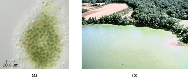
A large, diverse group of phototrophic bacteria compose the phylum Cyanobacteria; they get their blue-green color from the chlorophyll contained in their cells (Figure \(\PageIndex{5}\)). Species of this group perform oxygenic photosynthesis, producing megatons of gaseous oxygen. Scientists hypothesize that cyanobacteria played a critical role in the change of our planet’s anoxic atmosphere 1–2 billion years ago to the oxygen-rich environment we have today.3
Cyanobacteria have other remarkable properties. Amazingly adaptable, they thrive in many habitats, including marine and freshwater environments, soil, and even rocks. They can live at a wide range of temperatures, even in the extreme temperatures of the Antarctic. They can live as unicellular organisms or in colonies, and they can be filamentous, forming sheaths or biofilms. Many of them fix nitrogen, converting molecular nitrogen into nitrites and nitrates that other bacteria, plants, and animals can use. The reactions of nitrogen fixation occur in specialized cells called heterocysts.
Photosynthesis in Cyanobacteria is oxygenic, using the same type of chlorophyll a found in plants and algae as the primary photosynthetic pigment. Cyanobacteria also use phycocyanin and cyanophycin, two secondary photosynthetic pigments that give them their characteristic blue color. They are located in special organelles called phycobilisomes and in folds of the cellular membrane called thylakoids, which are remarkably similar to the photosynthetic apparatus of plants. Scientists hypothesize that plants originated from endosymbiosis of ancestral eukaryotic cells and ancestral photosynthetic bacteria.4 Cyanobacteria are also an interesting object of research in biochemistry,5 with studies investigating their potential as biosorbents6 and products of human nutrition.7
Unfortunately, cyanobacteria can sometimes have a negative impact on human health. Genera such as Microcystis can form harmful cyanobacterial blooms, forming dense mats on bodies of water and producing large quantities of toxins that can harm wildlife and humans. These toxins have been implicated in tumors of the liver and diseases of the nervous system in animals and humans.8
Summary
- Gram-negative nonproteobacteria include the taxa spirochetes; the Cytophaga, Fusobacterium, Bacteroides group; Planctomycetes; and many representatives of phototrophic bacteria.
- Spirochetes are motile, spiral bacteria with a long, narrow body; they are difficult or impossible to culture.
- Several genera of spirochetes contain human pathogens that cause such diseases as syphilis and Lyme disease.
- Cytophaga, Fusobacterium, and Bacteroides are classified together as a phylum called the CFB group. They are rod-shaped anaerobic organoheterotrophs and avid fermenters. Cytophaga are aquatic bacteria with the gliding motility. Fusobacteria inhabit the human mouth and may cause severe infectious diseases. Bacteroides are present in vast numbers in the human gut, most of them being mutualistic but some are pathogenic.
- Planctomycetes are aquatic bacteria that reproduce by budding; they may form large colonies, and develop a holdfast.
- Phototrophic bacteria are not a taxon but, rather, a group categorized by their ability to use the energy of sunlight. They include Proteobacteria and nonproteobacteria, as well as sulfur and nonsulfur bacteria colored purple or green.
- Sulfur bacteria perform anoxygenic photosynthesis, using sulfur compounds as donors of electrons, whereas nonsulfur bacteria use organic compounds (succinate, malate) as donors of electrons.
- Some phototrophic bacteria are able to fix nitrogen, providing the usable forms of nitrogen to other organisms.
- Cyanobacteria are oxygen-producing bacteria thought to have played a critical role in the forming of the earth’s atmosphere.
Footnotes
- 1 R.C. Fuller et al. “Carbon Metabolism in Chromatium.” Journal of Biological Chemistry 236 (1961):2140–2149.
- 2 T.T. Selao et al. “Comparative Proteomic Studies in Rhodospirillum rubrum Grown Under Different Nitrogen Conditions.” Journal of Proteome Research 7 no. 8 (2008):3267–3275.
- 3 A. De los Rios et al. “Ultrastructural and Genetic Characteristics of Endolithic Cyanobacterial Biofilms Colonizing Antarctic Granite Rocks.” FEMS Microbiology Ecology 59 no. 2 (2007):386–395.
- 4 T. Cavalier-Smith. “Membrane Heredity and Early Chloroplast Evolution.” Trends in Plant Science 5 no. 4 (2000):174–182.
- 5 S. Zhang, D.A. Bryant. “The Tricarboxylic Acid Cycle in Cyanobacteria.” Science 334 no. 6062 (2011):1551–1553.
- 6 A. Cain et al. “Cyanobacteria as a Biosorbent for Mercuric Ion.” Bioresource Technology 99 no. 14 (2008):6578–6586.
- 7 C.S. Ku et al. “Edible Blue-Green Algae Reduce the Production of Pro-Inflammatory Cytokines by Inhibiting NF-κB Pathway in Macrophages and Splenocytes.” Biochimica et Biophysica Acta 1830 no. 4 (2013):2981–2988.
- 8 I. Stewart et al. Cyanobacterial Poisoning in Livestock, Wild Mammals and Birds – an Overview. Advances in Experimental Medicine and Biology 619 (2008):613–637.
Contributor
Nina Parker, (Shenandoah University), Mark Schneegurt (Wichita State University), Anh-Hue Thi Tu (Georgia Southwestern State University), Philip Lister (Central New Mexico Community College), and Brian M. Forster (Saint Joseph’s University) with many contributing authors. Original content via Openstax (CC BY 4.0; Access for free at https://openstax.org/books/microbiology/pages/1-introduction)