
8.4: Virus Replication
- Page ID
- 35710
What you’ll learn to do: Identify different viruses and how they replicate
While viruses technically aren’t living things (they don’t have cells), they still have DNA or RNA. Despite being “nonliving,” viruses play an important role in evolutionary pressures on all living things, so it is important to study them.
Viruses are diverse entities. They vary in their structure, their replication methods, and in their target hosts. Nearly all forms of life—from bacteria and archaea to eukaryotes such as plants, animals, and fungi—have viruses that infect them. While most biological diversity can be understood through evolutionary history, such as how species have adapted to conditions and environments, much about virus origins and evolution remains unknown.
In this section, we’ll learn how viruses reproduce. As we do, you can compare viral replication to DNA replication in living things. We will finish by looking at other nonliving infectious agents.
- Understand the different types of viral infections, based on the host cell
- Discuss the basics of virus structure
- Describe prions and their basic properties
- Define viroids and their targets of infection
Viral Infectious Cycles
As you’ve learned, viruses are often very specific as to which hosts and which cells within the host they will infect. This feature of a virus makes it specific to one or a few species of life on Earth. On the other hand, so many different types of viruses exist on Earth that nearly every living organism has its own set of viruses that tries to infect its cells. Even the smallest and simplest of cells, prokaryotic bacteria, may be attacked by specific types of viruses. Viruses that target bacteria are known as bacteriophages.
A bacteriophage has both lytic and lysogenic cycles. In the lytic cycle, the phage replicates and lyses the host cell. In the lysogenic cycle, phage DNA is incorporated into the host genome, where it is passed on to subsequent generations. When the phage DNA is incorporated into the host cell genome, it is called a prophage. Environmental stressors such as starvation or exposure to toxic chemicals may cause the prophage to excise and enter the lytic cycle.
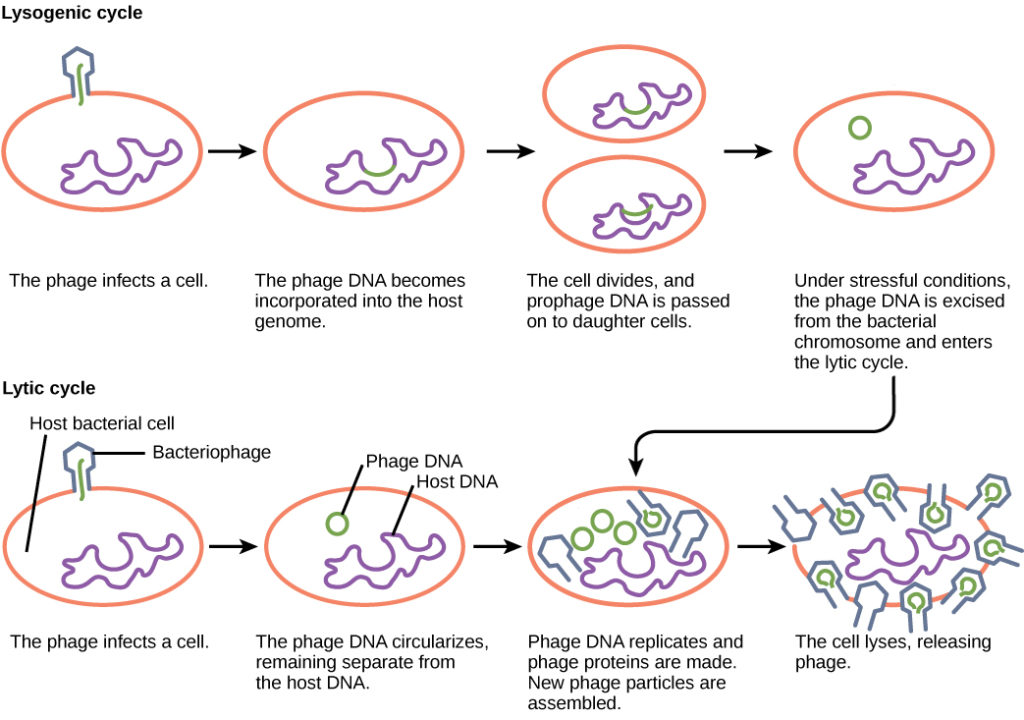
Which of the following statements is false?
- In the lytic cycle, new phage are produced and released into the environment.
- In the lysogenic cycle, phage DNA is incorporated into the host genome.
- An environmental stressor can cause the phage to initiate the lysogenic cycle.
- Cell lysis only occurs in the lytic cycle.
- Show Answer
-
Statement c is false.
Animal Viruses
Animal viruses, unlike the viruses of plants and bacteria, do not have to penetrate a cell wall to gain access to the host cell. Non-enveloped or “naked” animal viruses may enter cells in two different ways. As a protein in the viral capsid binds to its receptor on the host cell, the virus may be taken inside the cell via a vesicle during the normal cell process of receptor-mediated endocytosis. An alternative method of cell penetration used by non-enveloped viruses is for capsid proteins to undergo shape changes after binding to the receptor, creating channels in the host cell membrane. The viral genome is then “injected” into the host cell through these channels in a manner analogous to that used by many bacteriophages. Enveloped viruses also have two ways of entering cells after binding to their receptors: receptor-mediated endocytosis, or fusion. Many enveloped viruses enter the cell by receptor-mediated endocytosis in a fashion similar to some non-enveloped viruses. On the other hand, fusion only occurs with enveloped virions. These viruses, which include HIV among others, use special fusion proteins in their envelopes to cause the envelope to fuse with the plasma membrane of the cell, thus releasing the genome and capsid of the virus into the cell cytoplasm.
After making their proteins and copying their genomes, animal viruses complete the assembly of new virions and exit the cell. As we have already discussed using the example of HIV, enveloped animal viruses may bud from the cell membrane as they assemble themselves, taking a piece of the cell’s plasma membrane in the process. On the other hand, non-enveloped viral progeny, such as rhinoviruses, accumulate in infected cells until there is a signal for lysis or apoptosis, and all virions are released together.
Animal viruses are associated with a variety of human diseases. Some of them follow the classic pattern of acute disease, where symptoms get increasingly worse for a short period followed by the elimination of the virus from the body by the immune system and eventual recovery from the infection. Examples of acute viral diseases are the common cold and influenza. Other viruses cause long-term chronic infections, such as the virus causing hepatitis C, whereas others, like herpes simplex virus, only cause intermittent symptoms. Still other viruses, such as human herpesviruses 6 and 7, which in some cases can cause the minor childhood disease roseola, often successfully cause productive infections without causing any symptoms at all in the host, and thus we say these patients have an asymptomatic infection.
In hepatitis C infections, the virus grows and reproduces in liver cells, causing low levels of liver damage. The damage is so low that infected individuals are often unaware that they are infected, and many infections are detected only by routine blood work on patients with risk factors such as intravenous drug use. On the other hand, since many of the symptoms of viral diseases are caused by immune responses, a lack of symptoms is an indication of a weak immune response to the virus. This allows for the virus to escape elimination by the immune system and persist in individuals for years, all the while producing low levels of progeny virions in what is known as a chronic viral disease. Chronic infection of the liver by this virus leads to a much greater chance of developing liver cancer, sometimes as much as 30 years after the initial infection.
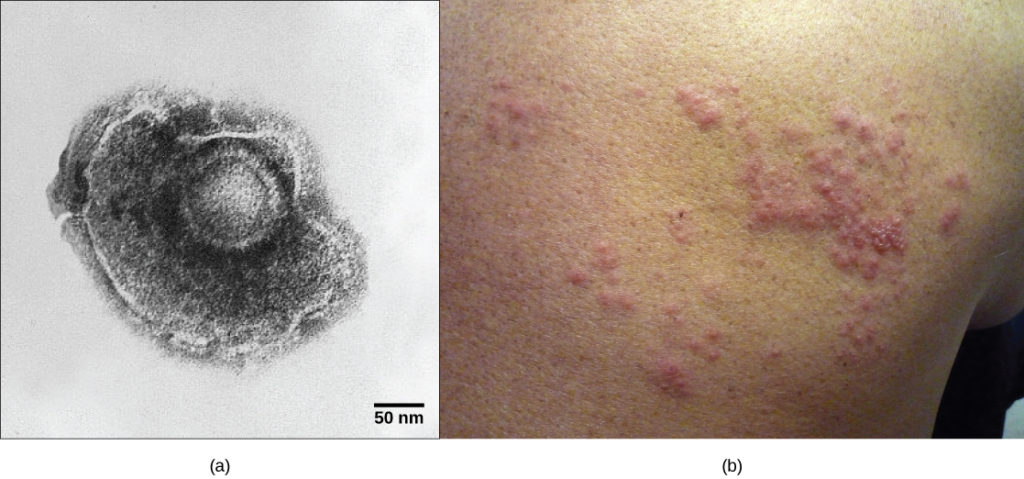
As already discussed, herpes simplex virus can remain in a state of latency in nervous tissue for months, even years. As the virus “hides” in the tissue and makes few if any viral proteins, there is nothing for the immune response to act against, and immunity to the virus slowly declines. Under certain conditions, including various types of physical and psychological stress, the latent herpes simplex virus may be reactivated and undergo a lytic replication cycle in the skin, causing the lesions associated with the disease. Once virions are produced in the skin and viral proteins are synthesized, the immune response is again stimulated and resolves the skin lesions in a few days by destroying viruses in the skin. As a result of this type of replicative cycle, appearances of cold sores and genital herpes outbreaks only occur intermittently, even though the viruses remain in the nervous tissue for life. Latent infections are common with other herpesviruses as well, including the varicella-zoster virus that causes chickenpox. After having a chickenpox infection in childhood, the varicella-zoster virus can remain latent for many years and reactivate in adults to cause the painful condition known as “shingles” (Figure 2).

Some animal-infecting viruses, including the hepatitis C virus discussed above, are known as oncogenic viruses: They have the ability to cause cancer. These viruses interfere with the normal regulation of the host cell cycle either by either introducing genes that stimulate unregulated cell growth (oncogenes) or by interfering with the expression of genes that inhibit cell growth. Oncogenic viruses can be either DNA or RNA viruses.
Cancers known to be associated with viral infections include cervical cancer caused by human papillomavirus (HPV) , liver cancer caused by hepatitis B virus, T-cell leukemia, and several types of lymphoma.
HPV, or human papillomavirus (as seen in Figure 3), has a naked icosahedral capsid visible in this transmission electron micrograph and a double-stranded DNA genome that is incorporated into the host DNA. The virus, which is sexually transmitted, is oncogenic and can lead to cervical cancer.
Visit the interactive animations showing the various stages of the replicative cycles of animal viruses and click on the flash animation links.
Plant Viruses
Plant viruses, like other viruses, contain a core of either DNA or RNA. You have already learned about one of these, the tobacco mosaic virus. As plants have a cell wall to protect their cells, these viruses do not use receptor-mediated endocytosis to enter host cells as is seen with animal viruses. For many plant viruses to be transferred from plant to plant, damage to some of the plants’ cells must occur to allow the virus to enter a new host. This damage is often caused by weather, insects, animals, fire, or human activities like farming or landscaping. Additionally, plant offspring may inherit viral diseases from parent plants. Plant viruses can be transmitted by a variety of vectors, through contact with an infected plant’s sap, by living organisms such as insects and nematodes, and through pollen. When plants viruses are transferred between different plants, this is known as horizontal transmission, and when they are inherited from a parent, this is called vertical transmission.
Symptoms of viral diseases vary according to the virus and its host (see the table below). One common symptom is hyperplasia, the abnormal proliferation of cells that causes the appearance of plant tumors known as galls. Other viruses induce hypoplasia, or decreased cell growth, in the leaves of plants, causing thin, yellow areas to appear. Still other viruses affect the plant by directly killing plant cells, a process known as cell necrosis. Other symptoms of plant viruses include malformed leaves, black streaks on the stems of the plants, altered growth of stems, leaves, or fruits, and ring spots, which are circular or linear areas of discoloration found in a leaf.
| Table 1. Some Common Symptoms of Plant Viral Diseases | |
|---|---|
| Symptom | Appears as |
| Hyperplasia | Galls (tumors) |
| Hypoplasia | Thinned, yellow splotches on leaves |
| Cell necrosis | Dead, blackened stems, leaves, or fruit |
| Abnormal growth patterns | Malformed stems, leaves, or fruit |
| Discoloration | Yellow, red, or black lines, or rings in stems, leaves, or fruit |
Plant viruses can seriously disrupt crop growth and development, significantly affecting our food supply. They are responsible for poor crop quality and quantity globally, and can bring about huge economic losses annually. Others viruses may damage plants used in landscaping. Some viruses that infect agricultural food plants include the name of the plant they infect, such as tomato spotted wilt virus, bean common mosaic virus, and cucumber mosaic virus. In plants used for landscaping, two of the most common viruses are peony ring spot and rose mosaic virus. There are far too many plant viruses to discuss each in detail, but symptoms of bean common mosaic virus result in lowered bean production and stunted, unproductive plants. In the ornamental rose, the rose mosaic disease causes wavy yellow lines and colored splotches on the leaves of the plant.
Viral Morphology
Viruses are acellular, meaning they are biological entities that do not have a cellular structure. They therefore lack most of the components of cells, such as organelles, ribosomes, and the plasma membrane. Viruses are sometimes called virions: a virion is a ‘complete’ virus free in the environment (not in a host). A virion consists of at least a nucleic acid core and an outer protein coating or capsid; sometimes a virus will have an outer envelope made of protein and phospholipid membranes derived from the host cell. Viruses may also contain additional proteins, such as enzymes. The most obvious difference between members of viral families is their morphology, which is quite diverse. An interesting feature of viral complexity is that the complexity of the host does not correlate with the complexity of the virion. Some of the most complex virion structures are observed in bacteriophages, viruses that infect the simplest living organisms, bacteria.
Types of Nucleic Acid
Unlike nearly all living organisms that use DNA as their genetic material, viruses may use either DNA or RNA as theirs. The virus core contains the genome or total genetic content of the virus. Viral genomes tend to be small, containing only those genes that encode proteins that the virus cannot get from the host cell. This genetic material may be single- or double-stranded. It may also be linear or circular.
DNA viruses cause human diseases, such as chickenpox, hepatitis B, and some venereal diseases, like herpes and genital warts. Human diseases caused by RNA viruses include hepatitis C, measles, and rabies.
Morphology

Viruses come in many shapes and sizes, but these are consistent and distinct for each viral family. All virions have a nucleic acid genome covered by a protective layer of proteins, called a capsid. The capsid is made up of protein subunits called capsomeres. Some viral capsids are simple polyhedral “spheres,” whereas others are quite complex in structure.
Many viruses use some sort of glycoprotein to attach to their host cells via molecules on the cell called viral receptors (Figure 4).
Among the most complex virions known, the T4 bacteriophage, which infects the Escherichia coli bacterium, has a tail structure that the virus uses to attach to host cells and a head structure that houses its DNA.
Overall, the shape of the virion and the presence or absence of an envelope tell us little about what disease the virus may cause or what species it might infect, but they are still useful means to begin viral classification (Figure 5).

Which of the following statements about virus structure is true?
- All viruses are encased in a viral membrane.
- The capsomere is made up of small protein subunits called capsids.
- DNA is the genetic material in all viruses.
- Glycoproteins help the virus attach to the host cell.
- Show Answer
-
Statement d is true.
Prions and Viroids
Prions and viroids are pathogens (agents with the ability to cause disease) that have simpler structures than viruses but, in the case of prions, still can produce deadly diseases.
Prions
Prions, so-called because they are proteinaceous, are infectious particles—smaller than viruses—that contain no nucleic acids (neither DNA nor RNA).
Fatal neurodegenerative diseases, such as kuru in humans and bovine spongiform encephalopathy (BSE) in cattle (commonly known as “mad cow disease”), were shown to be transmitted by prions. The disease was spread by the consumption of meat, nervous tissue, or internal organs from infected individuals, usually by members of the same species. Individuals with kuru and BSE show symptoms of loss of motor control and unusual behaviors, such as uncontrolled bursts of laughter with kuru, followed by death. These symptoms are due to lesions in the brain tissue.
The cause of spongiform encephalopathies, such as kuru and BSE, is an infectious structural variant of a normal cellular protein called PrP (prion protein). It is this variant that constitutes the prion particle. PrP exists in two forms, PrPc, the normal form of the protein, and PrPsc, the infectious form. Once introduced into the body, the PrPsc contained within the prion binds to PrPc and converts it to PrPsc. This leads to an exponential increase of the PrPsc protein, which aggregates. PrPsc is folded abnormally, and the resulting conformation (shape) is directly responsible for the lesions seen in the brains of infected cattle. Thus, although not fully accepted among scientists, the prion seems likely to be an entirely new form of infectious agent, the first one found whose transmission is not reliant upon genes made of DNA or RNA.
Viroids

Viroids are plant pathogens: small, single-stranded, circular RNA particles that are much simpler than a virus. They do not have a capsid or outer envelope, but like viruses can reproduce only within a host cell. Viroids do not, however, manufacture any proteins, and they only produce a single, specific RNA molecule. Human diseases caused by viroids have yet to be identified.
Viroids are known to infect plants and are responsible for crop failures and the loss of millions of dollars in agricultural revenue each year. Some of the plants they infect include potatoes, cucumbers, tomatoes, chrysanthemums, avocados, and coconut palms. For example, the potato spindle tuber viroid (PSTVd), which typically spreads when infected knives cut healthy potatoes in preparation for planting, can affect potatoes and tomatoes. The symptoms of PSTVd can be seen in Figure 6.
Prions are infectious agents that consist of protein, but no DNA or RNA, and seem to produce their deadly effects by duplicating their shapes and accumulating in tissues. They are thought to contribute to several progressive brain disorders, including mad cow disease and Creutzfeldt-Jakob disease. Viroids are single-stranded RNA pathogens that infect plants. Their presence can have a severe impact on the agriculture industry.
Check Your Understanding
Answer the question(s) below to see how well you understand the topics covered in the previous section. This short quiz does not count toward your grade in the class, and you can retake it an unlimited number of times.
Use this quiz to check your understanding and decide whether to (1) study the previous section further or (2) move on to the next section.
Contributors and Attributions
- Introduction to Virus Replication. Authored by: Shelli Carter and Lumen Learning. Provided by: Lumen Learning. License: CC BY: Attribution
- Biology. Provided by: OpenStax CNX. Located at: http://cnx.org/contents/185cbf87-c72e-48f5-b51e-f14f21b5eabd@10.8. License: CC BY: Attribution. License Terms: http://cnx.org/contents/b3c1e1d2-839...9a8aafbdd@9.25