
7.3: The Cell Cycle
- Page ID
- 35699
What you’ll learn to do: Identify the stages of the cell cycle, by picture and by description of major milestones
The cell cycle is an ordered series of events involving cell growth and cell division that produces two new daughter cells. Cells on the path to cell division proceed through a series of precisely timed and carefully regulated stages of growth, DNA replication, and division that produces two identical (clone) cells. The cell cycle has two major phases: interphase and the mitotic phase (Figure 1). During interphase, the cell grows and DNA is replicated. During the mitotic phase, the replicated DNA and cytoplasmic contents are separated, and the cell divides.

- Identify the characteristics and sub-phases of interphase
- Identify the characteristics and stages of mitosis
- Identify the characteristics of cytokinesis
Interphase
Stages of Interphase
During interphase, the cell undergoes normal growth processes while also preparing for cell division. It is the longest phase of the cell cycle, cell spends approximately 90% of its time in this phase. In order for a cell to move from interphase into the mitotic phase, many internal and external conditions must be met.
The three stages of interphase are called G1, S, and G2.
G1 Phase (First Gap)
The first stage of interphase is called the G1 phase (first gap) where the cell is quite active at the biochemical level. The cell is accumulating the building blocks of chromosomal DNA and the associated proteins as well as accumulating sufficient energy reserves to complete the task of replicating each chromosome in the nucleus.
S Phase (Synthesis of DNA)
In the S phase, DNA replication results in the formation of identical pairs of DNA molecules—sister chromatids—that are firmly attached to the centromeric region.
G2 Phase (Second Gap)
In the G2 phase, the cell replenishes its energy stores and synthesizes proteins necessary for chromosome manipulation. Some cell organelles are duplicated, and the cytoskeleton is dismantled to provide resources for the mitotic phase.
Mitosis
The mitotic phase (also known as M phase) is a multistep process during which the duplicated chromosomes are aligned, separated, and move into two new, identical daughter cells. The first portion of the mitotic phase is called karyokinesis, or nuclear division. The second portion of the mitotic phase, called cytokinesis, is the physical separation of the cytoplasmic components into the two daughter cells.
Karyokinesis (Mitosis)
Karyokinesis, also known as mitosis, is divided into a series of phases—prophase, metaphase, anaphase, and telophase—that result in the division of the cell (Figure 2).
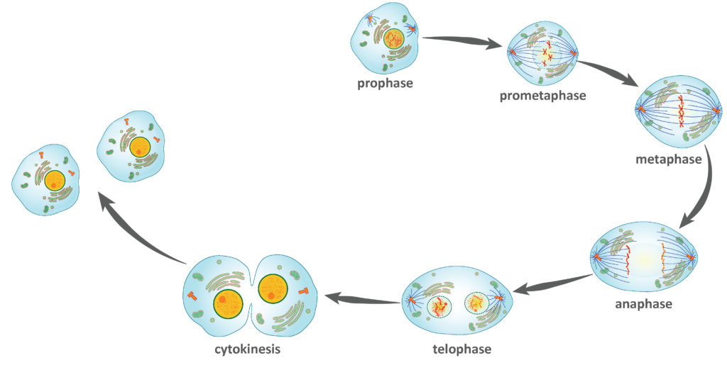
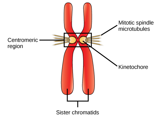
During prophase, the “first phase,” the nuclear envelope starts to dissociate into small vesicles, and the membranous organelles fragment and disperse toward the periphery of the cell. The nucleolus disappears . The centrosomes begin to move to opposite poles of the cell. Microtubules that will form the mitotic spindle extend between the centrosomes, pushing them farther apart as the microtubule fibers lengthen. The sister chromatids begin to coil more tightly and become visible under a light microscope. Each sister chromatid develops a protein structure called a kinetochore in the centromeric region (Figure 3). The proteins of the kinetochore attract and bind mitotic spindle microtubules.
During prometaphase, the nuclear envelope is fully broken down and chromosomes are attached to microtubules from both poles of the mitotic spindle, which begin to move them toward the middle of the cell.
During metaphase, all the chromosomes are aligned in a plane called the metaphase plate, or the equatorial plane, midway between the two poles of the cell. At this time, the chromosomes are maximally condensed.
During anaphase, the sister chromatids separate at the centromere. Each chromatid, now called a chromosome, is pulled rapidly toward the centrosome to which its microtubule is attached. The cell becomes visibly elongated (oval shaped) as the polar microtubules slide against each other at the metaphase plate where they overlap.
During telophase, the chromosomes reach the opposite poles and begin to decondense (unravel), relaxing into a chromatin configuration. Nuclear envelopes form around the chromosomes, and nucleosomes appear within the nuclear area.
The activity below will walk you through mitosis—providing you with the chance to review the different steps of the process and how they work together.
Click here for a text-only version of the activity.
Cytokinesis
Cytokinesis is the second main stage of the mitotic phase during which cell division is completed via the physical separation of the cytoplasmic components into two daughter cells. Division is not complete until the cell components have been apportioned and completely separated into the two daughter cells. Although the stages of mitosis are similar for most eukaryotes, the process of cytokinesis is quite different for eukaryotes that have cell walls, such as plant cells.
In cells such as animal cells that lack cell walls, cytokinesis follows the onset of anaphase. A contractile ring composed of actin filaments forms just inside the plasma membrane at the former metaphase plate. The actin filaments pull the equator of the cell inward, forming a fissure. This fissure, or “crack,” is called the cleavage furrow. The furrow deepens as the actin ring contracts, and eventually the membrane is cleaved in two (Figure 4).
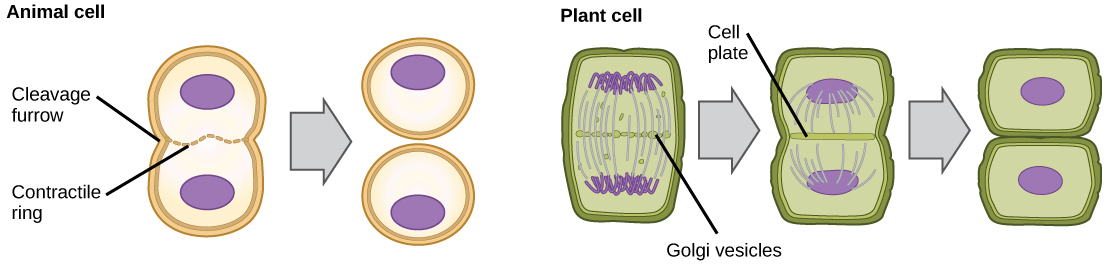
In plant cells, a new cell wall must form between the daughter cells. During interphase, the Golgi apparatus accumulates enzymes, structural proteins, and glucose molecules prior to breaking into vesicles and dispersing throughout the dividing cell. During telophase, these Golgi vesicles are transported on microtubules to form a phragmoplast (a vesicular structure) at the metaphase plate. There, the vesicles fuse and coalesce from the center toward the cell walls; this structure is called a cell plate. As more vesicles fuse, the cell plate enlarges until it merges with the cell walls at the periphery of the cell. Enzymes use the glucose that has accumulated between the membrane layers to build a new cell wall. The Golgi membranes become parts of the plasma membrane on either side of the new cell wall (Figure 4).
The Complete Cell Cycle
Remember, mitosis is the process of cell division, but it’s just a portion of the full cell cycle. Figure 5 shows approximately how long a cell spends in each stage of the cell cycle:
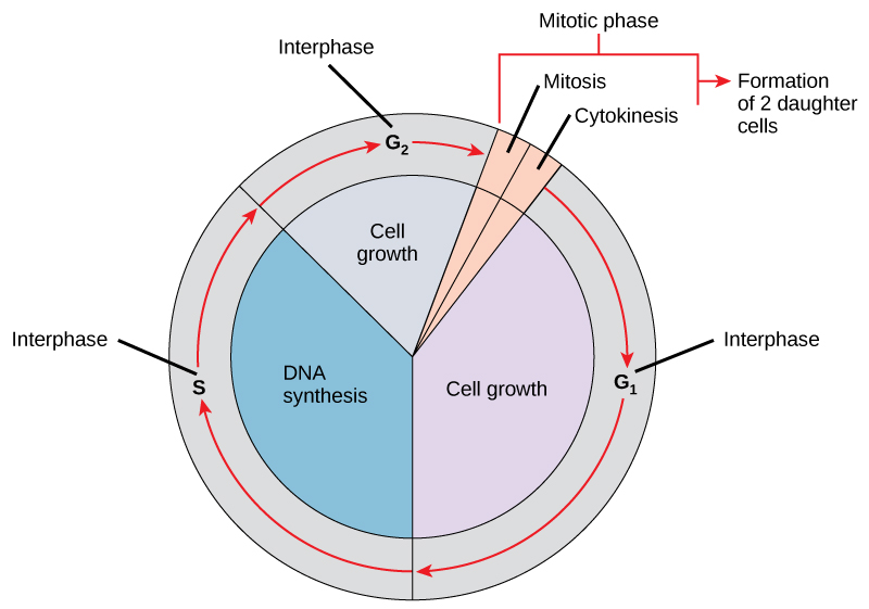
As you can see, cells spend most of their time in interphase.
This video reviews all the steps of mitosis; seeing it all together is a great review at this stage.
Check Your Understanding
Answer the question(s) below to see how well you understand the topics covered in the previous section. This short quiz does not count toward your grade in the class, and you can retake it an unlimited number of times.
Use this quiz to check your understanding and decide whether to (1) study the previous section further or (2) move on to the next section.
Contributors and Attributions
- Introduction to the Cell Cycle. Authored by: Shelli Carter and Lumen Learning. Provided by: Lumen Learning. License: CC BY: Attribution
- Biology. Provided by: OpenStax CNX. Located at: http://cnx.org/contents/185cbf87-c72e-48f5-b51e-f14f21b5eabd@10.8. License: CC BY: Attribution. License Terms: Download for free at http://cnx.org/contents/185cbf87-c72...f21b5eabd@10.8
- Mitosis diagram. Authored by: Marek Kultys. Located at: https://commons.wikimedia.org/wiki/File:Mitosis_diagram.jpg. License: CC BY-SA: Attribution-ShareAlike
- Mitosis. Provided by: Lumen Learning. Located at: www.oppia.org/explore/q4NBr1JG7Qrt. License: CC BY-SA: Attribution-ShareAlike
- Mitosis: Splitting Up Is Complicated: Crash Course Biology #12. Authored by: CrashCourse. Located at: https://youtu.be/L0k-enzoeOM. Project: Crash Course Biology. License: All Rights Reserved. License Terms: Standard YouTube License
- Modification of Mitosis cells sequence. Authored by: LadyofHats. Located at: https://commons.wikimedia.org/wiki/File:Mitosis_cells_sequence.svg. License: Public Domain: No Known Copyright