
Lab 10 Nervous System Anatomy Part 2: Functional Anatomy of the Peripheral Nerves and Sense Organs
- Page ID
- 59138
Objectives:
At the end of this lab, you will be able to…
- Correctly name the cranial nerves and indicate general functions
- Correctly and independently perform simple dissections
- Use correct anatomical terminology when discussing structures of the nervous system
- Correctly identify the various structures of the eye and ear
Pre-Lab Exercise:
After reading through the lab activities prior to lab, complete the following before you start your lab
1. The mapping of the peripheral nervous system that indicates sensation based on nerve roots is called , while for motor control is called the .
2. The nerves that will control the upper extremity originate in the plexus while the nerves controlling the lower extremity will originate in the , , and the plexuses.
3. The receptor in the eye responsible for vision is the while the is found in the ear and is responsible for hearing and the for the sense of movement.
4. Color the images for use as a reference for identifying the models and dissected specimens.
Materials:
- Nervous System Model
- Sagittal Head
- Eye Model
- Ear Model
- Stickers
- Felt Pens
- Colored Pencils
Activity 1: Dermatome and Myotomes
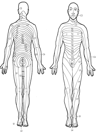
The myotomes and dermatomes provide the clinician with a reference map for the origin of the nerves that control movement or are responsible for conveying somatosensation. The myotome is the map that indicates the region of control of muscles for the spinal nerve and originates during fetal development as motor neurons (nerves) exit the spinal cord and innervates regions of non-differentiated skeletal muscle tissue (referred to as somites). Dermatomes are areas of sensation of the body that relate to the nerve root that forms the peripheral nerves that synapse with the receptors in that region. The dermatomes also form during fetal development and mirror the mapping that occurs with the myotomes. Using the dermatome and myotomes allow for map of origin of peripheral nerves through the body should peripheral nerve issues arise for the person.
Procedures:
1. Check your lab group for completion of the color the map of the myotome and dermatome.
2. Select a “team leader” and using the colored images as reference have members of the group take turns tracing a dermatome or myotomes themselves and then move to the next activity.
|
Peripheral Nervous System based on Myotome and Dermatome CN V: Trigeminal N C2 Cervical Nerve 2 C3 Cervical Nerve 3 C4 Cervical Nerve 4 C5 Cervical nerve 5 C6 Cervical Nerve 6 C7 Cervical Nerve 7 C8 Cervical Nerve 8 T1 Thoracic Nerve 1 T2 Thoracic Nerve 2 T3 Thoracic Nerve 3 T4 Thoracic Nerve 4 T5 Thoracic Nerve 5 T6 Thoracic Nerve 6 T7 Thoracic Nerve 7 T8 Thoracic Nerve 8 T9 Thoracic Nerve 9 T10 Thoracic Nerve 10 T11 Thoracic Nerve 11 T12 Thoracic Nerve 12 L1 Lumbar Nerve 1 L2 Lumbar Nerve 2 L3 Lumbar Nerve 3 L4 Lumbar Nerve 4 L5 Lumbar Nerve 5 S1 Sacral Nerve 1 S2 Sacral Nerve 2 S3 Sacral Nerve 3
Color each structure with a different color for use in identification. |
Peripheral nerves:
The somatic peripheral system will function via two primary divisions or plexus, the Brachial Plexus and Lumbar/Sacral Plexus. The plexus all for the innervation of the extremities of the body through the convergence and divergence of the spinal nerves. While the plexus are convergences and divergence so spinal nerves, the means by which they develop are not identical. The peripheral nerves of the extremities will have their origin within each of these plexuses. In which the brachial plexus will provide the peripheral nerves to the upper extremities while the lumbar and sacral plexus will provide the peripheral nerves the lower extremities. These peripheral nerves will allow for the transmission of somatic sensation and proprioception from periphery to the spinal cord and central nervous system. While they will be involved with conveying the motor response for reflexive behavior along with cognitive control of the skeletal muscle that allows for spinal reflex, fine and gross motor control of the limb (or limb segment). Clinically, we are also able to trace the origins of motor issues to spinal nerve segment of involvement or to what extent afferent neurons are impacting the ability to convey the sensory signal based on where within the limb issues might be arising.
Activity 2: Nerves from the Brachial Plexus
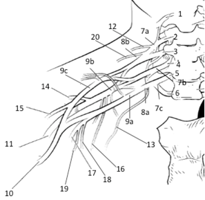
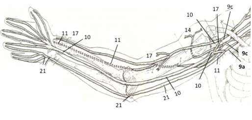
The brachial plexus is built through the convergence of spinal nerves forming 3 plexus trunks (Superior, Middle, Inferior) that then converge and alter their orientation forming 2 plexus divisions (Anterior, Posterior) all within a short distance of the inferior cervical vertebrae. As the nerves move lateral toward the scapula, the division once again converge and diverge forming 3 plexus cords (Lateral, Medial, Posterior) that they diverge within the lateral scapular region forming the peripheral nerves of the upper extremities.
|
Peripheral Nervous System for the Upper Extremity 1 C4 Nerve Root 2 C5 Nerve Root 3 C6 Nerve Root 4 C7 Nerve Root 5 C8 Nerve Root 6 T1 Nerve Root 7a Superior Trunk 7b Middle Trunk 7c Inferior Trunk 8a Anterior Division 8b Posterior Division 9a Medial Cord 9b Posterior Cord 9c Lateral Cord 10 Median Nerve 11 Radial Nerve 12 Dorsal Scapular Nerve 13 Long Thoracic Nerve 14 Axillary Nerve 15 Subscapular Nerve 16 Lateral Pectoral Nerve 17 Musculocutaneous Nerve 18 Medial Brachial Cutaneous Nerve 19 Medial Antebrachial Cutaneous Nerve 20 Suprascapular Nerve 21 Ulnar Nerve
Color each structure with a different color for use in identification.
|
Procedures:
1. Obtain nervous system models, stickers, felt pens
2. On the stickers, write the regions of the plexus and major nerves of the upper extremity
Superior Trunk, Middle Trunk, Inferior Trunk, Anterior Division, Posterior Division, Lateral Chord, Medial Chord, Posterior Chord, Radial N, Ulnar N, Musculocutaneous N
3. Select a “team leader” and using the colored images as reference have members of the group take turns labeling the model with the correct stickers.
4. Have your instructor check your work and then move to the next activity.
Activity 3: Nerves from the Lumbar, Lumbosacral and Sacral Plexus
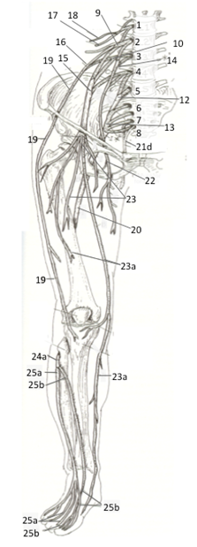
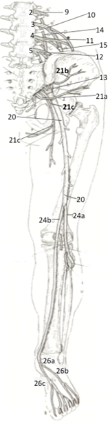
The lumbar and sacral plexus form 3 plexus trunks from the spinal nerves converging onto each other (Lumbar, Lumbosacral, Sacral) that will the converge and diverge in order to form into 2 divisions (Anterior or Posterior) near the sacroiliac region of the pelvis. These divisions will then converge and diverge within the posterior aspect of the pelvic bowl forming the peripheral nerves of the lower extremities.
|
Peripheral Nervous System for the Lower Extremity 1 L1 Nerve Root 2 L2 Nerve Root 3 L3 Nerve Root 4 L4 Nerve Root 5 L5 Nerve Root 6 S1 Nerve Root 7 S2 Nerve Root 8 S3 Nerve Root 9 Lumbar Plexus 10 Lumbosacral Plexus 11 Posterior Division 12 Lumbosacral Plexus 13 Sacral Plexus 14 Posterior Division 15 Anterior Division 16 Anterior (Lateral) Division 17 Ilioinguinal Nerve 18 Iliohypogastric Nerve 19 Lateral Femoral Cutaneous Nerve 20 Sciatic Nerve 21a Inferior Gluteal Nerve 21b Superior Gluteal Nerve 21c Posterior Femoral Cutaneous Nerve 21d Pudendal Nerve 22 Nerve to Obturator 23 Femoral Nerve 23a Saphenous Nerve 24a Fibular Nerve 24b Tibial Nerve 25a Superficial Fibular Nerve 25b Deep Fibular Nerve 26a Plantar Nerve 26b Lateral Plantar 26c Medial Plantar
Color each structure with a different color for use in identification. |
Procedures:
1. Obtain nervous system models, stickers, felt pens
2. On the stickers, write the regions of the plexus and major nerves of the lower extremity
Lumbar Plexus, Lumbosacral Plexus, Sacral Plexus, Anterior Divisions, Posterior Divisions, Sciatic N, Femoral N, Tibial N, Peroneal (Fibular) N
3. Select a “team leader” and using the colored images as reference have members of the group take turns labeling the model with the correct stickers.
4. Have your instructor check your work and then move to the next activity.
Activity 4: Sense Organs
Tongue and Nose:
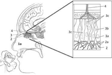
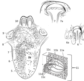
Gustatory complexes are the receptors are located throughout the mouth and tongue and not in specialized regions that has been previously noted (due to a mistranslation and misconception of a classical study). The receptor itself is comprised of separate cells that have ligand-gated receptors within the cilia projections. The cells cluster these cilia projection along folds within the pit to form a microvilli projection along the inner surface of a pit (gustatory receptor). These will respond to chemicals that get trapped within the pit that will then link with a chemical class ligand receptor that leads to an action potential being transmitted to one of three cranial nerves, CN VII, IX and X, depending on the location that stimulation is occurring within the tongue. The olfactory epithelium is tissue lining the superior aspect of the nasal opening that has chemoreceptors embedded into the tissue. These receptors appear to be sensitive to distinct classes of chemicals and not individual chemicals. The olfactory receptors are bipolar neuron with cilia called olfactory hairs that project into the nasal concha. These will respond to chemical stimulation of a particular volatile chemical (odorant molecule) via a ligand gated channel leading the action potential being transmitted to CN I via passage through the Cribriform plate of the Ethmoid bone.
|
Structures of the nose and yongue related to olfactory and gustatory reception 1a Nasal Cavity 1b Tongue 2 Olfactory Epithelium 2a Olfactory Receptors 3 Neural 3a Olfactory Cells 3b Bipolar Cells 3c Support Cells 4 Olfactory Nerve (CN I) 5 Filiform Papillae 6 Fungiform Papillae 6a Taste bud 7 Circumvallate Papillae 7a Taste Bud 8 Lingual Tonsils 9 Palatine Tonsils 10 Epiglottis 11 Taste Bud 11a Receptors 11b Gustatory Cells 11c Support Cells 11d Nerve
Color each structure with a different color for use in identification. |
Eye:
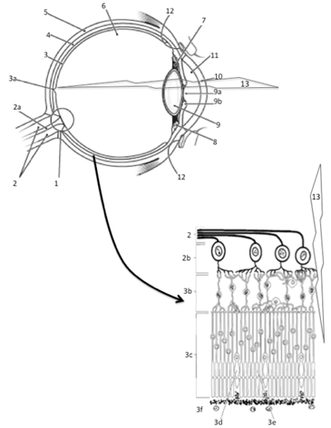
Vision is the sense that provides for reception of light energy from the environment. We perceive light energy within a spectrum of wavelength that is deemed the visible light spectrum of electromagnetic radiation. The receptor organ for vision is the retina of eye, where the structure of the eye is meant to focus light onto the fovea centralis based on the principle that light will always travel in straight lines and will bend when it passes through a medium whose density changes (such as going from air through the cornea and lens or the fluids that fill the eye, aqueous and vitreous humor). The receptors for vision are found in retina. To receive this sense, the retina will utilize 2 distinct types of photoreceptive cells that are stimulated (or inhibited) by distinct wavelengths of light. The receptors are arranged in radiations of density with the cone cells being in greatest density near the fovea and lessen as move towards the periphery of the retina (away from the fovea), while rods are greatest in density at the periphery and least at the fovea. Cone cells are in effect 3 distinct photoreceptors that exhibit a threshold based on location within retina and wavelength of light that cause “excitation”. Where the short wave-cell, aka Blue Cone, has a peak response at 437 nm of light wavelength with range of 400-550 nm. The medium wave-cell, aka Green Cone, has a peak response at 530 nm of light wavelength with range of 400-650 nm. The long wave-cell, aka Red Cone, has a peak response at 564 nm of light wavelength with rang from 450-700 nm. The rods on the other hand are generalized photoreceptors that while having a peak response at 490 nm of light will respond with the entire spectrum of visible light.
|
Structures of the eye related to vision: 1 Optic Disk 2 Optic Nerve (CN II) 2a Blood Vessels 2b Ganglion Cells 3 Retina 3a Fovea Centralis 3b Bipolar Cells 3c Photoreceptors 3d Cone Cells 3e Rod Cells 3f Pigment Layer 4 Choroid Plexus 5 Sclera 6 Posterior Chamber 7 Suspensory Ligaments 8 Ciliary Muscles 9 Lens 9a Pupil (Iris) 9b Pupil (Radial/Circular Muscles) 10 Cornea 11 Anterior Chamber 12 Scleral Sinus 13 Direction of Light Travel Color each structure with a different color for use in identification. |
Ear (Hearing and Vestibular Organs)
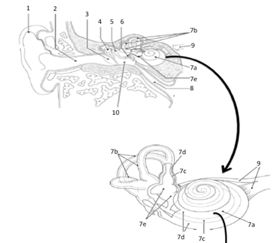
The structure that is responsible for the auditory sense and response is the ear and the cochlea. The anatomy of the ear is formed in such a way as to funnel sound waves into the external auditory canal and onto the tympanic membrane. The tympanic membrane then transforms the energy wave from a sinusoidal to a compressive wave that can then transfer that energy onto the bones of the middle ear (auditory Ossicles). The energy transfer causes the Ossicles to vibrate against each other that will then provide the mechanisms to transform the compressive wave into a sinusoidal wave to transfer the same energy wave into the cochlea through the oval window. The cochlea itself is a “snail” coiled boney labyrinth within the temporal bone of the cranium. Within the cochlea are fluid filled membranous cavities (the tympanies). It is within tympanies that the receptor organ with the cochlea is located. This receptor organ, the Organ of Corti, is comprised on two membranes and a stereoscopic organization of mechanoreceptors (hair cells).
Vestibular sense provides input as to the direction of head (and body) movement relative to the force of gravity. To perceive this sense, it will utilize receptors found in the semi-circular canals that are surrounding the cochlea of the inner ear, stimulate the neurons found in CN VIII (vestibular branch). Overall, vestibular sensation gives the person the ability to sense this movement is based on the acceleration taking place to give the “sense” of movement and speed of movement of the body in space without the need for visual or proprioceptive information.
|
Structures of the ear related to hearing and vestibular response: 1 Pina of Ear (Auricle) 2 External Auditory Canal 3 Tympanic Membrane 4 Malleus 5 Incus 6 Stapes 7a Cochlea 7b Semicircular Canals 7c Endolymph (Scala Vestibuli) 7d Labyrinth (Scala Tympani) 7e Vestibule (Saccule and Utricle) 8 Auditory (Eustachian) Tube 9 CN VIII (Vestibulocochlear)
Color each structure with a different color for use in identification. |
Procedures:
1. Obtain ear, eye and sagittal head models, stickers, felt pens
2. Write the names of the anatomical structures of the ear and eye that you are responsible for knowing on to the stickers
Eye: Fovea Centralis, Macula Lacteal, Ciliary Body, Suspensory Ligaments, Lens, Cornea, Retina, Pupil, Sclera
Ear: Auricle (Pina of Ear), Cochlea, Tympanic Membrane, Ossicles, Anterior Semicircular Canal, Lateral Semicircular Canal, Posterior Semicircular Canal
3. Select a “team leader” and using the colored images as reference have members of the group take turns labeling the model with the correct stickers.
a. As you label, indicate the function of the structure that you are labeling
4. Have your instructor check your work and then move to the next activity.