
5.5: Skeletal Muscle Functions and Structures
- Page ID
- 103466
Turn your eyes—a tiny movement, considering the conspicuously large and strong external eye muscles that control eyeball movements. These muscles have been called the strongest muscles in the human body relative to the work they do. However, the external eye muscles actually do a surprising amount of work. Eye movements occur almost constantly during waking hours, especially when we are scanning faces or reading. Eye muscles are also exercised nightly during the phase of sleep called rapid eye movement sleep. External eye muscles can move the eyes because they are made mainly of muscle tissue.

Skeletal Muscle Functions
The best-known feature of skeletal muscle is its ability to contract and cause movement. Skeletal muscles act not only to produce movement but also to stop movement, such as resisting gravity to maintain posture. Small, constant adjustments of the skeletal muscles are needed to hold a body upright or balanced in any position. Muscles also prevent excess movement of the bones and joints, maintaining skeletal stability and preventing skeletal structure damage or deformation. Joints can become misaligned or dislocated entirely by pulling on the associated bones; muscles work to keep joints stable. Skeletal muscles are located throughout the body at the openings of internal tracts to control the movement of various substances. These muscles allow functions, such as swallowing, urination, and defecation, to be under voluntary control. Skeletal muscles also protect internal organs (particularly abdominal and pelvic organs) by acting as an external barrier or shield to external trauma and by supporting the weight of the organs.
Skeletal muscles contribute to the maintenance of homeostasis in the body by generating heat. Muscle contraction requires energy, and when ATP is broken down, heat is produced. This heat is very noticeable during exercise, when sustained muscle movement causes body temperature to rise, and in cases of extreme cold, when shivering produces random skeletal muscle contractions to generate heat.
Major Skeletal Muscles
Skeletal muscle is muscle tissue attached to bones by tendons, which are bundles of collagen fibers. Whether you are moving your eyes or running a marathon, you are using skeletal muscles. Contractions of skeletal muscles are voluntary or under the conscious control of the central nervous system via the somatic nervous system. Skeletal muscle tissue is the most common type of muscle tissue in the human body. By weight, an average adult male is about 42 percent skeletal muscles, and the average adult female is about 36 percent skeletal muscles.
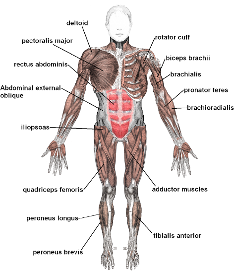
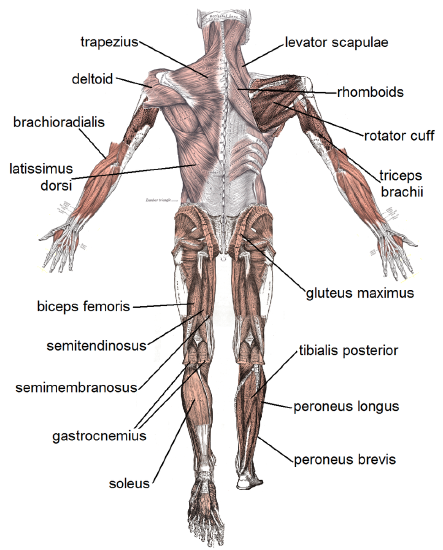
Some of the major skeletal muscles in the human body are labeled in Figures \(\PageIndex{3}\) and Figure \(\PageIndex{4}\) and listed in Table \(\PageIndex{1}\).
| Muscles visible in Figure \(\PageIndex{3}\) | Muscles visible in Figure \(\PageIndex{4}\) |
|---|---|
| rotator cuff (multiple muscles are part of this group) | levator scapulae |
| biceps brachii | rhomboids |
| brachialis | rotator cuff |
| pronator teres | triceps brachii |
| brachioradialis | gluteus maximus |
| adductor muscles | tibialis posterior |
| tibialis anterior | peroneus longus |
| deltoid | peroneus brevis |
| pectoralis major | trapezius |
| rectus abdominis | deltoid |
| abdominal external oblique | brachioradialis |
| iliopsoas | latissimus dorsi |
| quadriceps femoris | biceps femoris |
| peroneus longus | semitendinosus |
| peroneus bravis | semimembranousus |
| gastrocnemius | |
| soleus |
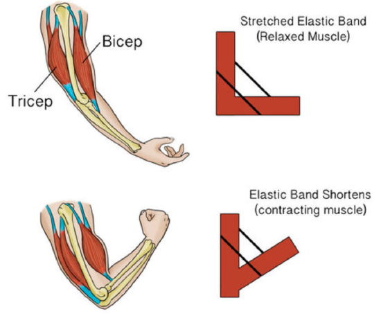
Skeletal Muscle Pairs
To move bones in opposite directions, skeletal muscles often consist of muscle pairs that work in opposition to one another. For example, when the biceps muscle (on the anterior of the upper arm) contracts, it can cause the elbow joint to flex or bend the arm (flexion), as shown in Figure \(\PageIndex{5}\). When the triceps muscle (on the posterior of the upper arm) contracts, it can cause the elbow to extend or straighten the arm (extension). The biceps and triceps muscles are examples of a muscle pair where the muscles work in opposition to each other.
In another example, to flex the knee joint a set of muscles called the hamstrings (n the posterior of the thigh) is activated. However, to extend the knee a group of four muscles called the quadriceps femoris (on the anterior of the thigh) are activated.
Skeletal Muscle Structure
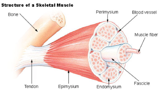
Each skeletal muscle is an organ that consists of various integrated tissues. These tissues include the skeletal muscle fibers, blood vessels, nerve fibers, and connective tissue. Each skeletal muscle has three layers of connective tissue that enclose it and provide structure to the muscle as a whole, and also compartmentalize the muscle fibers within the muscle. Each muscle is wrapped in a sheath of dense, irregular connective tissue called the epimysium, which allows a muscle to contract and move powerfully while maintaining its structural integrity. The epimysium also separates muscle from other tissues and organs in the area, allowing the muscle to move independently.
Each skeletal muscle consists of hundreds — or even thousands — of skeletal muscle fibers, which are long, string-like cells. As shown in Figure \(\PageIndex{6}\), skeletal muscle fibers are individually wrapped in connective tissue called endomysium. The skeletal muscle fibers are bundled together in units called muscle fascicles, surrounded by sheaths of connective tissue called perimysium. Each fascicle contains between ten and 100 (or even more!) skeletal muscle fibers. Fascicles, in turn, are bundled together to form individual skeletal muscles, which are wrapped in connective tissue called epimysium. The connective tissues in skeletal muscles have a variety of functions. They support and protect muscle fibers, allowing them to withstand contraction forces by distributing the forces applied to the muscle. They also provide pathways for nerves and blood vessels to reach the muscles. Also, the epimysium anchors the muscles to tendons.
The same bundles-within-bundles structure is replicated within each muscle fiber. As shown in Figure \(\PageIndex{7}\), a muscle fiber consists of a bundle of myofibrils, which are themselves bundles of protein filaments. These protein filaments consist of thin filaments of the protein actin, anchored to structures called Z discs — and thick filaments of the protein myosin. The filaments are arranged together within a myofibril in repeating units called sarcomeres, which run from one Z disc to the next. The sarcomere is the basic functional unit of skeletal (and cardiac) muscles. It contracts as actin and myosin filaments slide over one another. Skeletal muscle tissue is said to be striated because it appears striped. It has this appearance because of the regular, alternating A (dark) and I (light) bands of filaments arranged in sarcomeres inside the muscle fibers. Other components of a skeletal muscle fiber include multiple nuclei and mitochondria.

Slow- and Fast-Twitch Skeletal Muscle Fibers
Skeletal muscle fibers can be divided into two types, called slow-twitch (or type I) muscle fibers and fast-twitch (or type II) muscle fibers.
- Slow-twitch muscle fibers are dense with capillaries and rich in mitochondria and myoglobin, a protein that stores oxygen until needed for muscle activity. Relative to fast-twitch fibers, slow-twitch fibers can carry more oxygen and sustain aerobic (oxygen-using) activity. Slow-twitch fibers can contract for long periods of time, but not with very much force. They are relied upon primarily in endurance events, such as distance running or cycling.
- Fast-twitch muscle fibers contain fewer capillaries and mitochondria and less myoglobin. This type of muscle fiber can contract rapidly and powerfully, but it fatigues very quickly. Fast-twitch fibers can sustain only short, anaerobic (non-oxygen-using) bursts of activity. Relative to slow-twitch fibers, fast-twitch fibers contribute more to muscle strength and have a greater potential for increasing mass. They are relied upon primarily in short, strenuous events, such as sprinting or weight lifting.
Proportions of fiber types vary considerably from muscle to muscle and from person to person. Individuals may be genetically predisposed to have a larger percentage of one type of muscle fiber than the other. Generally, an individual who has more slow-twitch fibers is better suited for activities requiring endurance. In contrast, an individual who has more fast-twitch fibers is better suited for activities requiring short bursts of power.
Review
1. Where is the skeletal muscle found, and what is its general function?
2. Why do many skeletal muscles work in pairs?
3. Describe the structure of a skeletal muscle.
4. Relate muscle fiber structure to the functional units of muscles.
5. Why is skeletal muscle tissue striated?
6. Compare and contrast slow-twitch and fast-twitch skeletal muscle fibers.
7. Arrange the following units within a skeletal muscle in order, from smallest to largest: fascicle; sarcomere; muscle fiber; myofibril
8. Give one example of connective tissue that is found in muscles. Describe one of its functions.
9. True or False: skeletal muscle fibers are cells with multiple nuclei.
Attributions
- Eyes by Nappy; public domain
- Muscle tissue by Mdunning13, CC BY 3.0 via Wikimedia Commons
- Muscles anterior labeled by Häggström, Mikael (2014). "Medical gallery of Mikael Häggström 2014". WikiJournal of Medicine 1 (2). DOI:10.15347/wjm/2014.008. ISSN 2002-4436. Public Domain. via Wikimedia Commons
- Muscles posterior labeled by Häggström, Mikael (2014). "Medical gallery of Mikael Häggström 2014". WikiJournal of Medicine 1 (2). DOI:10.15347/wjm/2014.008. ISSN 2002-4436. Public Domain. via Wikimedia Commons
- Muscle movement by CK-12 licensed CC BY-NC 3.0
- Muscle structure by National Cancer Institute, public domain via Wikimedia Commons
- Muscle fibers by OpenStax, CC BY 4.0 via Wikimedia Commons
- Text adapted from Human Biology by CK-12 licensed CC BY-NC 3.0