
5.3: Types of Muscle Tissue
- Page ID
- 103459

What is Muscle Tissue?
.svg?revision=1&size=bestfit&width=481&height=361)
Muscle tissue is a soft tissue that makes up most of the tissues in the muscles of the human muscular system. Other tissues in muscles are connective tissues, such as tendons that attach skeletal muscles to bones and sheaths of connective tissues that cover or line muscle tissues. Only muscle tissue per se, however, has cells with the ability to contract.
There are three major types of muscle tissues in the human body: skeletal, smooth, and cardiac muscle tissues. Figure \(\PageIndex{2}\) shows how the three types of muscle tissues appear under a microscope. When you read about each type below, you will learn why the three types appear as they do.
All three muscle tissues have some properties in common; they all exhibit a quality called excitability as their plasma membranes can change their electrical states (from polarized to depolarized) and send an electrical wave called an action potential along the entire length of the membrane. While the nervous system can influence the excitability of cardiac and smooth muscle to some degree, skeletal muscle completely depends on signaling from the nervous system to work properly. On the other hand, both cardiac muscle and smooth muscle can respond to other stimuli, such as hormones and local stimuli.
Differences among the three muscle types include the microscopic organization of their contractile proteins—actin and myosin. The actin and myosin proteins are arranged very regularly in the cytoplasm of individual muscle cells (referred to as fibers) in both skeletal muscle and cardiac muscle, which creates a pattern, or stripes, called striations. The striations are visible with a light microscope under high magnification
Skeletal Muscle
Skeletal muscle has a distinctive striped pattern, called striations. The striations result form actin and myosin proteins arranged very regularly in the cytoplasm of individual muscle cells (fibers), and are visible with a light microscope under high magnification. Skeletal muscle fibers (cells) are multinucleated structures that compose the skeletal muscle.
The next sections of this chapter will go into more detail about the structure and function of skeletal muscle tissue and skeletal muscles.
Smooth Muscle
Smooth muscle is muscle tissue in the walls of internal organs and other internal structures such as blood vessels. When smooth muscles contract, they help the organs and vessels carry out their functions. When smooth muscles in the stomach wall contract, they squeeze the food inside the stomach, helping to mix and churn the food and break it into smaller pieces. This is an important part of digestion. Contractions of smooth muscles are involuntary, so they are not under conscious control. Instead, they are controlled by the autonomic nervous system, hormones, neurotransmitters, and other physiological factors.
Structure of Smooth Muscle
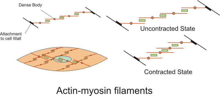
The cells that make up smooth muscle are generally called myocytes. Unlike the muscle fibers of striated muscle tissue, the myocytes of smooth muscle tissue do not have their filaments arranged in sarcomeres. Therefore, smooth tissue is not striated. However, the myocytes of smooth muscle contain myofibrils, which contain bundles of myosin and actin filaments. The filaments cause contractions when they slide over each other, as shown in Figure \(\PageIndex{8}\).
Functions of Smooth Muscle
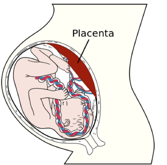
Unlike striated muscle, smooth muscle can sustain very long-term contractions. Smooth muscle can also stretch and still maintain its contractile function, which striated muscle cannot. An extracellular matrix secreted by myocytes enhances the elasticity of smooth muscle. The matrix consists of elastin, collagen, and other stretchy fibers. The ability to stretch and still contract is an important attribute of smooth muscle in organs such as the stomach and uterus (Figure \(\PageIndex{9}\)), both of which must stretch considerably as they perform their normal functions.
The following list indicates where many smooth muscles are found, along with some of their specific functions.
- Walls of the gastrointestinal tract (such as the esophagus, stomach, and intestines), moving food through the tract by peristalsis.
- Walls of air passages of the respiratory tract (such as the bronchi), controlling the diameter of the passages and the volume of air that can pass through them
- Walls of organs of the male and female reproductive tracts; in the uterus, for example, pushing a baby out of the uterus and into the birth canal
- Walls of the urinary system structures, including the urinary bladder, allow the bladder to expand so it can hold more urine and then contract as urine is released.
- Walls of blood vessels, controlling the diameter of the vessels and thereby affecting blood flow and blood pressure
- Walls of lymphatic vessels, squeezing the fluid called lymph through the vessels.
- Iris of the eyes, controlling the size of the pupils and thereby the amount of light entering the eyes
- Arrector pili in the skin, raising hairs in hair follicles in the dermis.
Cardiac Muscle

Cardiac muscle is found only in the wall of the heart. It is also called myocardium. As shown in Figure \(\PageIndex{10}\), the myocardium is enclosed within connective tissues, including the endocardium on the inside of the heart and pericardium on the outside of the heart. When cardiac muscle contracts, the heart beats and pumps blood. Contractions of cardiac muscle are involuntary, like those of smooth muscles. They are controlled by electrical impulses from specialized cardiac muscle cells in the heart muscle area called the sinoatrial node.
Like skeletal muscle, cardiac muscle is striated because its filaments are arranged in sarcomeres inside the muscle fibers. However, in cardiac muscle, the myofibrils are branched at irregular angles rather than arranged in parallel rows (as they are in skeletal muscle). This explains why cardiac and skeletal muscle tissues look different from one another.
The cells of cardiac muscle tissue are arranged in interconnected networks. This arrangement allows rapid transmission of electrical impulses, which stimulate virtually simultaneous contractions of the cells. This enables the cells to coordinate contractions of the heart muscle.
The heart is the muscle that performs the greatest amount of physical work in a lifetime. Although the heart's power output is much less than the maximum power output of some other muscles in the human body, the heart does its work continuously over an entire lifetime without rest. The cardiac muscle contains many mitochondria, which produce ATP for energy and help the heart resist fatigue.
Review
1. What is muscle tissue?
2. Where is the skeletal muscle found, and what is its general function?
3. Why is skeletal muscle tissue striated?
4. Where is the smooth muscle found? What controls the contraction of smooth muscle?
5. Compare and contrast smooth muscle and striated muscle (such as skeletal muscle).
6. Where is the cardiac muscle found? What controls its contractions?
7. Both cardiac and skeletal muscle tissues are striated, but they look different from one another. Why?
8. The heart muscle is smaller and less powerful than some other muscles in the body. Why is the heart the muscle that performs the greatest amount of physical work in a lifetime? How does the heart resist fatigue?
9. True or False: skeletal muscle fibers are cells with multiple nuclei.
Explore More
You can learn more about the three types of muscle tissues by watching this Khan Academy video:
Attributions
- Actin-myosin filament by Boumphreyfr, CC BY 3.0 via Wikimedia Commons
- Placenta by Gray38, public domain via Wikimedia Commons
- Heart Wall by OpenStax College, CC BY 3.0 via Wikimedia Commons
- Text adapted from Human Biology by CK-12 licensed CC BY-NC 3.0