4.2: Introduction to the Skeletal System
- Last updated
- Save as PDF
- Page ID
- 103186
Skull and Cross-Bones
The skull and cross-bones symbol has been used for a very long time to represent death, perhaps because after death and decomposition, bones are all that remain. Many people think of bones as being dead, dry, and brittle. These adjectives may correctly describe the bones of a preserved skeleton, but the bones of a living human being are very much alive. Living bones are also strong and flexible. Bones are the major organs of the skeletal system.
_insignia%252C_1980.png?revision=1)
The skeletal system is the organ system that provides an internal framework for the human body. Why do you need a skeletal system? Try to imagine what you would look like without it. You would be a soft, wobbly pile of skin containing muscles and internal organs but no bones. You might look something like a very large slug. Not that you would be able to see yourself — folds of skin would droop down over your eyes and block your vision because of your lack of skull bones. You could push the skin out of the way if you could only move your arms, but you need bones for that as well!
Components of the Skeletal System
In adults, the skeletal system includes 206 bones, many of which are shown in Figure \(\PageIndex{2}\). Bones are organs made of dense connective tissues, mainly the tough protein collagen. Bones contain blood vessels, nerves, and other tissues. Bones are hard and rigid due to deposits of calcium and other mineral salts within their living tissues. Locations, where two or more bones meet, are called joints. Many joints allow bones to move like levers. For example, your elbow is a joint that allows you to bend and straighten your arm.
Besides bones, the skeletal system includes cartilage and ligaments.
- Cartilage is a type of dense connective tissue, made of tough protein fibers. It is strong but flexible and very smooth. It covers the ends of bones at joints, providing a smooth surface for bones to move over.
- Ligaments are bands of fibrous connective tissue that hold bones together. They keep the bones of the skeleton in place.
Axial and Appendicular Skeletons
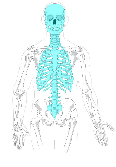
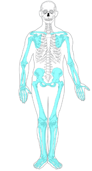
The skeleton is traditionally divided into two major parts: the axial skeleton and the appendicular skeleton, both of which are pictured in Figure \(\PageIndex{3}\).
- The axial skeleton forms the axis of the body. It includes the skull, vertebral column (spine), and rib cage. The bones of the axial skeleton, along with ligaments and muscles, allow the human body to maintain its upright posture. The axial skeleton also transmits weight from the head, trunk, and upper extremities down the back to the lower extremities. In addition, the bones protect the brain and organs in the chest.
- The appendicular skeleton forms the appendages and their attachments to the axial skeleton. It includes the bones of the arms and legs, hands and feet, and shoulder and pelvic girdles. The bones of the appendicular skeleton make possible locomotion and other movements of the appendages. They also protect the major organs of digestion, excretion, and reproduction.


Functions of the Skeletal System
The skeletal system has many different functions that are necessary for human survival. Some of the functions, such as supporting the body, are relatively obvious. Other functions are less obvious but no less important. For example, three tiny bones (hammer, anvil, and stirrup) inside the middle ear transfer sound waves into the inner ear.
Support, Shape, and Protection
The skeleton supports the body and gives it shape. Without the rigid bones of the skeletal system, the human body would be just a bag of soft tissues, as described above. The bones of the skeleton are very hard and provide protection to the delicate tissues of internal organs. For example, the skull encloses and protects the soft tissues of the brain, and the vertebral column protects the nervous tissues of the spinal cord. The vertebral column, ribs, and sternum (breast bone) protect the heart, lungs, and major blood vessels. Providing protection to these latter internal organs requires the bones to be able to expand and contract. The ribs and the cartilage that connects them to the sternum and vertebrae are capable of small shifts that allow breathing and other internal organ movements.
Movement
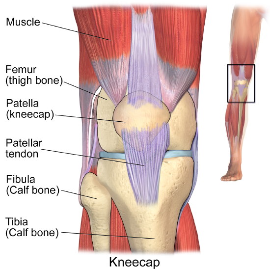
The bones of the skeleton provide attachment surfaces for skeletal muscles. When the muscles contract, they pull on and move the bones. The figure below, for example, shows the muscles attached to the bones at the knee. They help stabilize the joint and allow the leg to bend at the knee. The bones at joints act like levers moving at a fulcrum point, and the muscles attached to the bones apply the force needed for movement.

Hematopoiesis
Hematopoiesis is the process in which blood cells are produced. This process occurs in a tissue called red marrow, which is found inside some bones, including the pelvis, ribs, and vertebrae. Red marrow synthesizes red blood cells, white blood cells, and platelets. Billions of these blood cells are produced inside the bones every day.
Mineral Storage and Homeostasis
Another function of the skeletal system is storing minerals, especially calcium and phosphorus. This storage function is related to the role of bones in maintaining mineral homeostasis. Just the right levels of calcium and other minerals are needed in the blood for the normal functioning of the body. When mineral levels in the blood are too high, bones absorb some of the minerals and store them as mineral salts, which is why bones are so hard. When blood levels of minerals are too low, bones release some of the minerals back into the blood. Bone minerals are alkaline (basic), so their release into the blood buffers the blood against excessive acidity (low pH), whereas their absorption back into bones buffers the blood against excessive alkalinity (high pH). In this way, bones help maintain acid-base homeostasis in the blood.
Another way bones help to maintain homeostasis is by acting as an endocrine organ. One endocrine hormone secreted by bone cells is osteocalcin, which helps regulate blood glucose and fat deposition. It increases insulin secretion and also the sensitivity of cells to insulin. In addition, it boosts the number of insulin-producing cells and reduces fat stores.
Review
- What is the skeletal system? How many bones are there in the adult skeleton?
- Describe the composition of bones.
- Besides bones, what other organs are included in the skeletal system?
- Identify the two major divisions of the skeleton.
- List several functions of the skeletal system.
- Discuss sexual dimorphism in the human skeleton.
- Bones, cartilage, and ligaments are all made of types of ____________ tissue.
- True or False. Bones contain living tissue and can affect processes in other parts of the body.
- True or False. Bone cells contract to pull on muscles in order to initiate a movement.
- If a person has a problem with blood cell production, what type of bone tissue is most likely involved? Explain your answer.
- Are the pelvic girdles part of the axial or appendicular skeleton?
- What are three forms of homeostasis that the skeletal system regulates? Briefly explain how each one is regulated by the skeletal system.
- What do you think would happen to us if we did not have ligaments? Explain your answer.
- a. Define a joint in the skeletal system.
b. How is cartilage related to joints?
c. Identify one joint in the human body and describe its function.
Explore More
Attributions
- Fighter squadron 84 by US Navy, public domain via Wikimedia Commons
- Human skeleton front by LadyofHats Mariana Ruiz Villarreal, public domain via Wikimedia Commons
- Axial skeleton by LadyofHats Mariana Ruiz Villarreal, public domain via Wikimedia Commons
- Appendicular skeleton by LadyofHats Mariana Ruiz Villarreal, public domain via Wikimedia Commons
- Knee anatomy by Blausen.com staff (2014). "Medical gallery of Blausen Medical 2014". WikiJournal of Medicine 1 (2). DOI:10.15347/wjm/2014.010. ISSN 2002-4436. CC BY 3.0 via Wikimedia Commons
- Text adapted from Human Biology by CK-12 licensed CC BY-NC 3.0


