
16.2: Structure and Function of the Respiratory System
- Page ID
- 22562

Why can you “see your breath” on a cold day? The air you exhale through your nose and mouth is warm, like the inside of your body. Exhaled air also contains a lot of water vapor because it passes over moist surfaces from the lungs to the nose or mouth. The water vapor in your breath cools suddenly when it reaches the much colder outside air. This causes the water vapor to condense into a fog of tiny droplets of liquid water. You release water vapor and other gases from your body through the process of respiration.
.jpg?revision=1&size=bestfit&width=399&height=241)
What is Respiration?
Respiration is the life-sustaining process in which gases are exchanged between the body and the outside atmosphere. Specifically, oxygen moves from the outside air into the body; and water vapor, carbon dioxide, and other waste gases move from inside the body into the outside air. Respiration is carried out mainly by the respiratory system. It is important to note that respiration by the respiratory system is not the same process as cellular respiration that occurs inside cells, although the two processes are closely connected. Cellular respiration is the metabolic process in which cells obtain energy, usually by “burning” glucose in the presence of oxygen. When cellular respiration is aerobic, it uses oxygen and releases carbon dioxide as a waste product. Respiration by the respiratory system supplies the oxygen needed by cells for aerobic cellular respiration and removes the carbon dioxide produced by cells during cellular respiration.
Respiration by the respiratory system actually involves two subsidiary processes. One process is ventilation or breathing. This is the physical process of conducting air to and from the lungs. The other process is gas exchange. This is the biochemical process in which oxygen diffuses out of the air and into the blood while carbon dioxide and other waste gases diffuse out of the blood and into the air. All of the organs of the respiratory system are involved in breathing, but only the lungs are involved in gas exchange.
Respiratory Organs
The organs of the respiratory system form a continuous system of passages called the respiratory tract, through which air flows into and out of the body. The respiratory tract has two major divisions: the upper respiratory tract and the lower respiratory tract. The organs in each division are shown in Figure \(\PageIndex{2}\). In addition to these organs, certain muscles of the thorax (the body cavity that fills the chest) are also involved in respiration by enabling breathing. Most important is a large muscle called the diaphragm, which lies below the lungs and separates the thorax from the abdomen. Smaller muscles between the ribs also play a role in breathing. You can learn more about breathing muscles in the concept of Breathing.
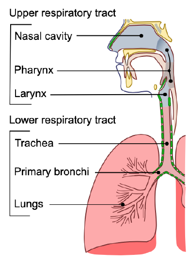
Upper Respiratory Tract
All of the organs and other structures of the upper respiratory tract are involved in the conduction or the movement of air into and out of the body. Upper respiratory tract organs provide a route for air to move between the outside atmosphere and the lungs. They also clean, humidity, and warm the incoming air. However, no gas exchange occurs in these organs.
Nasal Cavity
The nasal cavity is a large, air-filled space in the skull above and behind the nose in the middle of the face. It is a continuation of the two nostrils. As inhaled air flows through the nasal cavity, it is warmed and humidified. Hairs in the nose help trap larger foreign particles in the air before they go deeper into the respiratory tract. In addition to its respiratory functions, the nasal cavity also contains chemoreceptors that are needed for the sense of smell and that contribute importantly to the sense of taste.
Pharynx
The pharynx is a tube-like structure that connects the nasal cavity and the back of the mouth to other structures lower in the throat, including the larynx. The pharynx has dual functions: both air and food (or other swallowed substances) pass through it, so it is part of both the respiratory and digestive systems. Air passes from the nasal cavity through the pharynx to the larynx (as well as in the opposite direction). Food passes from the mouth through the pharynx to the esophagus.
Larynx
The larynx connects the pharynx and trachea and helps to conduct air through the respiratory tract. The larynx is also called the voice box because it contains the vocal cords, which vibrate when air flows over them, thereby producing sound. You can see the vocal cords in the larynx in Figure \(\PageIndex{3}\). Certain muscles in the larynx move the vocal cords apart to allow breathing. Other muscles in the larynx move the vocal cords together to allow the production of vocal sounds. The latter muscles also control the pitch of sounds and help control their volume.
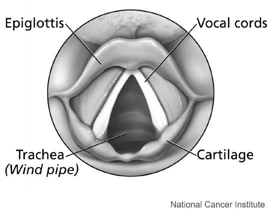
A very important function of the larynx is protecting the trachea from aspirated food. When swallowing occurs, the backward motion of the tongue forces a flap called the epiglottis to close over the entrance to the larynx. You can see the epiglottis in Figure \(\PageIndex{3}\). This prevents swallowed material from entering the larynx and moving deeper into the respiratory tract. If swallowed material does start to enter the larynx, it irritates the larynx and stimulates a strong cough reflex. This generally expels the material out of the larynx and into the throat.
Lower Respiratory Tract
The trachea and other passages of the lower respiratory tract conduct air between the upper respiratory tract and the lungs. These passages form an inverted tree-like shape (Figure \(\PageIndex{4}\)), with repeated branching as they move deeper into the lungs. All told, there are an astonishing 1,500 miles of airways conducting air through the human respiratory tract! It is only in the lungs, however, that gas exchange occurs between the air and the bloodstream.
Trachea
The trachea, or windpipe, is the widest passageway in the respiratory tract. It is about 2.5 cm (1 in.) wide and 10-15 cm (4-6 in.) long. It is formed by rings of cartilage, which make it relatively strong and resilient. The trachea connects the larynx to the lungs for the passage of air through the respiratory tract. The trachea branches at the bottom to form two bronchial tubes.
Bronchi and Bronchioles
There are two main bronchial tubes, or bronchi (singular, bronchus), called the right and left bronchi. The bronchi carry air between the trachea and lungs. Each bronchus branches into smaller, secondary bronchi; and secondary bronchi branch into still smaller tertiary bronchi. The smallest bronchi branch into very small tubules called bronchioles. The tiniest bronchioles end in alveolar ducts, which terminate in clusters of minuscule air sacs, called alveoli (singular, alveolus), in the lungs.
Lungs

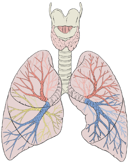
The lungs are the largest organs of the respiratory tract. They are suspended within the pleural cavity of the thorax. In Figure \(\PageIndex{5}\), you can see that each of the two lungs is divided into sections. These are called lobes, and they are separated from each other by connective tissues. The right lung is larger and contains three lobes. The left lung is smaller and contains only two lobes. The smaller left lung allows room for the heart, which is just left of the center of the chest.
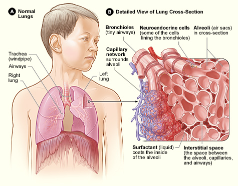
Lung tissue consists mainly of alveoli (Figure \(\PageIndex{6}\)). These tiny air sacs are the functional units of the lungs where gas exchange takes place. The two lungs may contain as many as 700 million alveoli, providing a huge total surface area for gas exchange to take place. In fact, alveoli in the two lungs provide as much surface area as half a tennis court! Each time you breathe in, the alveoli fill with air, making the lungs expand. Oxygen in the air inside the alveoli is absorbed by the blood in the mesh-like network of tiny capillaries that surrounds each alveolus. The blood in these capillaries also releases carbon dioxide into the air inside the alveoli. Each time you breathe out, air leaves the alveoli and rushes into the outside atmosphere, carrying waste gases with it.
The lungs receive blood from two major sources. They receive deoxygenated blood from the heart. This blood absorbs oxygen in the lungs and carries it back to the heart to be pumped to cells throughout the body. The lungs also receive oxygenated blood from the heart that provides oxygen to the cells of the lungs for cellular respiration.
Protecting the Respiratory System
You may be able to survive for weeks without food and for days without water, but you can survive without oxygen for only a matter of minutes except under exceptional circumstances. Therefore, protecting the respiratory system is vital. That’s why making sure a patient has an open airway is the first step in treating many medical emergencies. Fortunately, the respiratory system is well protected by the ribcage of the skeletal system. However, the extensive surface area of the respiratory system is directly exposed to the outside world and all its potential dangers in inhaled air. Therefore, it should come as no surprise that the respiratory system has a variety of ways to protect itself from harmful substances such as dust and pathogens in the air.
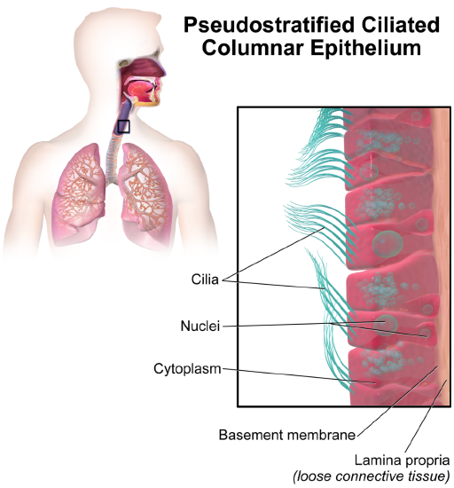
The main way the respiratory system protects itself is called the mucociliary escalator. From the nose through the bronchi, the respiratory tract is covered in the epithelium that contains mucus-secreting goblet cells. The mucus traps particles and pathogens in the incoming air. The epithelium of the respiratory tract is also covered with tiny cell projections called cilia (singular, cilium), as shown in Figure \(\PageIndex{7}\). The cilia constantly move in a sweeping motion upward toward the throat, moving the mucus and trapped particles and pathogens away from the lungs and toward the outside of the body.
What happens to the material that moves up the mucociliary escalator to the throat? It is generally removed from the respiratory tract by clearing the throat or coughing. Coughing is a largely involuntary response of the respiratory system that occurs when nerves lining the airways are irritated. The response causes air to be expelled forcefully from the trachea, helping to remove mucus and any debris it contains (called phlegm) from the upper respiratory tract to the mouth. The phlegm may spit out (expectorated), or it may be swallowed and destroyed by stomach acids.
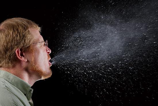
Sneezing is a similar involuntary response that occurs when nerves lining the nasal passage are irritated. It results in forceful expulsion of air from the mouth, which sprays millions of tiny droplets of mucus and other debris out of the mouth and into the air, as shown in Figure \(\PageIndex{8}\). This explains why it is so important to sneeze into a sleeve rather than the air to help prevent the transmission of respiratory pathogens.
How the Respiratory System Works with Other Organ Systems
The amount of oxygen and carbon dioxide in the blood must be maintained within a limited range for the survival of the organism. Cells cannot survive for long without oxygen, and if there is too much carbon dioxide in the blood, the blood becomes dangerously acidic (pH is too low). Conversely, if there is too little carbon dioxide in the blood, the blood becomes too basic (pH is too high). The respiratory system works hand-in-hand with the nervous and cardiovascular systems to maintain homeostasis in blood gases and pH.
It is the level of carbon dioxide rather than the level of oxygen that is most closely monitored to maintain blood gas and pH homeostasis. The level of carbon dioxide in the blood is detected by cells in the brain, which speed up or slow down the rate of breathing through the autonomic nervous system as needed to bring the carbon dioxide level within the normal range. Faster breathing lowers the carbon dioxide level (and raises the oxygen level and pH); slower breathing has the opposite effects. In this way, the levels of carbon dioxide and oxygen, as well as pH, are maintained within normal limits.
The respiratory system also works closely with the cardiovascular system to maintain homeostasis. The respiratory system exchanges gases between the blood and the outside air, but it needs the cardiovascular system to carry them to and from body cells. Oxygen is absorbed by the blood in the lungs and then transported through a vast network of blood vessels to cells throughout the body where it is needed for aerobic cellular respiration. The same system absorbs carbon dioxide from cells and carries it to the respiratory system for removal from the body.
Choking is the mechanical obstruction of the flow of air from the atmosphere into the lungs. It prevents breathing and may be partial or complete. Partial choking allows some though inadequate airflow into the lung—prolonged or complete choking results in asphyxia, or suffocation, which is potentially fatal.
Obstruction of the airway typically occurs in the pharynx or trachea. Young children are more prone to choking than are older people, in part because they often put small objects in their mouths and do not appreciate the risk of choking that they pose. Young children may choke on small toys or parts of toys or on household objects in addition to food. Foods that can adapt their shape to that of the pharynx, such as bananas and marshmallows, are especially dangerous and may cause choking in adults as well as children.
How can you tell if a loved one is choking? The person cannot speak or cry out or has great difficulty doing so. Breathing, if possible, is labored, producing gasping or wheezing. The person may desperately clutch at his or her throat or mouth. If breathing is not soon restored, the person’s face will start to turn blue from lack of oxygen. This will be followed by unconsciousness if oxygen deprivation continues beyond a few minutes.
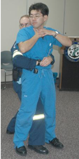
If an infant is choking, turning the baby upside down and slapping on the back may dislodge the obstructing object. To help an older person who is choking, first, encourage the person to cough. Give them a few hardback slaps to help force the lodged object out of the airway. If these steps fail, perform the Heimlich maneuver on the person. You can easily find instructional videos online to learn how to do it. If the Heimlich maneuver also fails, call for emergency medical care immediately.
Review
- What is respiration, as carried out by the respiratory system? Name the two subsidiary processes it involves.
- Describe the respiratory tract.
- Identify the organs of the upper respiratory tract, and state their functions.
- List the organs of the lower respiratory tract. Which organs are involved only in conduction?
- Where does gas exchange take place?
- How does the respiratory system protect itself from potentially harmful substances in the air?
- Explain how the rate of breathing is controlled.
- Why does the respiratory system need the cardiovascular system to help it perform its main function of gas exchange?
- Place the following organs or structures of the respiratory system in order of when they are encountered by air entering the body — from earliest to latest.
trachea; nasal cavity; alveoli; bronchioles; larynx; bronchi; pharynx
- Which organ is part of both the digestive and respiratory systems?
A. Larynx
B. Trachea
C. Pharynx
D. Bronchus
- Describe two ways in which the body prevents food from entering the lungs.
- True or False. The lungs receive some oxygenated blood.
- True or False. Gas exchange occurs in both the upper and lower respiratory tracts.
- Coughing can expel ___________ from the body.
A. mucus
B. food particles
C. phlegm
D. All of the above
- What is the relationship between respiration and cellular respiration?
Explore More
Attributions
- Snowboarders breath on a cold day by Alain Wong via Unsplash License
- Conducting Passages by Lord Akryl, Jmarchn, public domain via Wikimedia Commons
- Larynx by Alan Hoofring, National Cancer Institute, public domain via Wikimedia Commons
- Lung Diagram by Patrick J. Lynch; CC BY 2.5 via Wikimedia Commons
- Lung Structure by National Heart Lung and Blood Institute, public domain via Wikimedia Commons
- Alveoli by helix84 licensed CC BY 2.5, via Wikimedia Commons
- Ciliated Epithelium by Blausen.com staff (2014). "Medical gallery of Blausen Medical 2014". WikiJournal of Medicine 1 (2). DOI:10.15347/wjm/2014.010. ISSN 2002-4436. licensed CC BY 3.0 via Wikimedia Commons
- Sneeze by James Gathany, CDC, public domain via Wikimedia Commons
- Abdominal Thrusts by Amanda M. Woodhead, public domain via Wikimedia Commons
- Text adapted from Human Biology by CK-12 licensed CC BY-NC 3.0