Biology LibreTexts

19.2.3: Cleavage

  • Page ID
    75307
  • \( \newcommand{\vecs}[1]{\overset { \scriptstyle \rightharpoonup} {\mathbf{#1}} } \)

    \( \newcommand{\vecd}[1]{\overset{-\!-\!\rightharpoonup}{\vphantom{a}\smash {#1}}} \)

    \( \newcommand{\id}{\mathrm{id}}\) \( \newcommand{\Span}{\mathrm{span}}\)

    ( \newcommand{\kernel}{\mathrm{null}\,}\) \( \newcommand{\range}{\mathrm{range}\,}\)

    \( \newcommand{\RealPart}{\mathrm{Re}}\) \( \newcommand{\ImaginaryPart}{\mathrm{Im}}\)

    \( \newcommand{\Argument}{\mathrm{Arg}}\) \( \newcommand{\norm}[1]{\| #1 \|}\)

    \( \newcommand{\inner}[2]{\langle #1, #2 \rangle}\)

    \( \newcommand{\Span}{\mathrm{span}}\)

    \( \newcommand{\id}{\mathrm{id}}\)

    \( \newcommand{\Span}{\mathrm{span}}\)

    \( \newcommand{\kernel}{\mathrm{null}\,}\)

    \( \newcommand{\range}{\mathrm{range}\,}\)

    \( \newcommand{\RealPart}{\mathrm{Re}}\)

    \( \newcommand{\ImaginaryPart}{\mathrm{Im}}\)

    \( \newcommand{\Argument}{\mathrm{Arg}}\)

    \( \newcommand{\norm}[1]{\| #1 \|}\)

    \( \newcommand{\inner}[2]{\langle #1, #2 \rangle}\)

    \( \newcommand{\Span}{\mathrm{span}}\) \( \newcommand{\AA}{\unicode[.8,0]{x212B}}\)

    \( \newcommand{\vectorA}[1]{\vec{#1}}      % arrow\)

    \( \newcommand{\vectorAt}[1]{\vec{\text{#1}}}      % arrow\)

    \( \newcommand{\vectorB}[1]{\overset { \scriptstyle \rightharpoonup} {\mathbf{#1}} } \)

    \( \newcommand{\vectorC}[1]{\textbf{#1}} \)

    \( \newcommand{\vectorD}[1]{\overrightarrow{#1}} \)

    \( \newcommand{\vectorDt}[1]{\overrightarrow{\text{#1}}} \)

    \( \newcommand{\vectE}[1]{\overset{-\!-\!\rightharpoonup}{\vphantom{a}\smash{\mathbf {#1}}}} \)

    \( \newcommand{\vecs}[1]{\overset { \scriptstyle \rightharpoonup} {\mathbf{#1}} } \)

    \( \newcommand{\vecd}[1]{\overset{-\!-\!\rightharpoonup}{\vphantom{a}\smash {#1}}} \)

    \(\newcommand{\avec}{\mathbf a}\) \(\newcommand{\bvec}{\mathbf b}\) \(\newcommand{\cvec}{\mathbf c}\) \(\newcommand{\dvec}{\mathbf d}\) \(\newcommand{\dtil}{\widetilde{\mathbf d}}\) \(\newcommand{\evec}{\mathbf e}\) \(\newcommand{\fvec}{\mathbf f}\) \(\newcommand{\nvec}{\mathbf n}\) \(\newcommand{\pvec}{\mathbf p}\) \(\newcommand{\qvec}{\mathbf q}\) \(\newcommand{\svec}{\mathbf s}\) \(\newcommand{\tvec}{\mathbf t}\) \(\newcommand{\uvec}{\mathbf u}\) \(\newcommand{\vvec}{\mathbf v}\) \(\newcommand{\wvec}{\mathbf w}\) \(\newcommand{\xvec}{\mathbf x}\) \(\newcommand{\yvec}{\mathbf y}\) \(\newcommand{\zvec}{\mathbf z}\) \(\newcommand{\rvec}{\mathbf r}\) \(\newcommand{\mvec}{\mathbf m}\) \(\newcommand{\zerovec}{\mathbf 0}\) \(\newcommand{\onevec}{\mathbf 1}\) \(\newcommand{\real}{\mathbb R}\) \(\newcommand{\twovec}[2]{\left[\begin{array}{r}#1 \\ #2 \end{array}\right]}\) \(\newcommand{\ctwovec}[2]{\left[\begin{array}{c}#1 \\ #2 \end{array}\right]}\) \(\newcommand{\threevec}[3]{\left[\begin{array}{r}#1 \\ #2 \\ #3 \end{array}\right]}\) \(\newcommand{\cthreevec}[3]{\left[\begin{array}{c}#1 \\ #2 \\ #3 \end{array}\right]}\) \(\newcommand{\fourvec}[4]{\left[\begin{array}{r}#1 \\ #2 \\ #3 \\ #4 \end{array}\right]}\) \(\newcommand{\cfourvec}[4]{\left[\begin{array}{c}#1 \\ #2 \\ #3 \\ #4 \end{array}\right]}\) \(\newcommand{\fivevec}[5]{\left[\begin{array}{r}#1 \\ #2 \\ #3 \\ #4 \\ #5 \\ \end{array}\right]}\) \(\newcommand{\cfivevec}[5]{\left[\begin{array}{c}#1 \\ #2 \\ #3 \\ #4 \\ #5 \\ \end{array}\right]}\) \(\newcommand{\mattwo}[4]{\left[\begin{array}{rr}#1 \amp #2 \\ #3 \amp #4 \\ \end{array}\right]}\) \(\newcommand{\laspan}[1]{\text{Span}\{#1\}}\) \(\newcommand{\bcal}{\cal B}\) \(\newcommand{\ccal}{\cal C}\) \(\newcommand{\scal}{\cal S}\) \(\newcommand{\wcal}{\cal W}\) \(\newcommand{\ecal}{\cal E}\) \(\newcommand{\coords}[2]{\left\{#1\right\}_{#2}}\) \(\newcommand{\gray}[1]{\color{gray}{#1}}\) \(\newcommand{\lgray}[1]{\color{lightgray}{#1}}\) \(\newcommand{\rank}{\operatorname{rank}}\) \(\newcommand{\row}{\text{Row}}\) \(\newcommand{\col}{\text{Col}}\) \(\renewcommand{\row}{\text{Row}}\) \(\newcommand{\nul}{\text{Nul}}\) \(\newcommand{\var}{\text{Var}}\) \(\newcommand{\corr}{\text{corr}}\) \(\newcommand{\len}[1]{\left|#1\right|}\) \(\newcommand{\bbar}{\overline{\bvec}}\) \(\newcommand{\bhat}{\widehat{\bvec}}\) \(\newcommand{\bperp}{\bvec^\perp}\) \(\newcommand{\xhat}{\widehat{\xvec}}\) \(\newcommand{\vhat}{\widehat{\vvec}}\) \(\newcommand{\uhat}{\widehat{\uvec}}\) \(\newcommand{\what}{\widehat{\wvec}}\) \(\newcommand{\Sighat}{\widehat{\Sigma}}\) \(\newcommand{\lt}{<}\) \(\newcommand{\gt}{>}\) \(\newcommand{\amp}{&}\) \(\definecolor{fillinmathshade}{gray}{0.9}\)

    Cleavage refers to the early cell divisions that occur as a fertilized egg begins to develop into an embryo.

    Holoblastic Cleavage

    In eggs that contain no (mammals) or only moderate amounts (frog) of yolk, cytokinesis divides the cells completely. The figure shows the results of the first two cleavages in the frog embryo.

    Two illustrations of cell stages. The left depicts a 2-cell stage, and the right a 4-cell stage, with dividing lines visible. Red text below each indicates the respective stage.
    Figure 14.3.1: First two cleavages in a frog embryo

    Meroblastic Cleavage

    In eggs that contain a large amount of yolk, cytokinesis does not divide the egg completely.

    Diagram showing bird egg development stages: yolk with disk of cytoplasm, 2-cell stage, 4-cell stage, and late cleavage with multiple cells.
    Figure 14.3.2: Bird cleavage

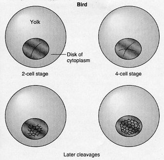
    The hen's egg consists of just a tiny patch of cytoplasm resting on the surface of a large ball of yolk (the "white" of the egg is noncellular accessory protein). When the first cleavages occur in the hen's egg, the cleavage furrows do not continue down through the mass of yolk. Therefore, each of the cells produced in the earliest stages is bound on the top and on the sides by a plasma membrane, but the bottom of the cell is in direct contact with yolk.

    Close-up black and white image of a morula, an early-stage embryo, showing tightly packed spherical cells on a round structure in a cellular formation.
    Figure 14.3.3: Zebrafish cleavage

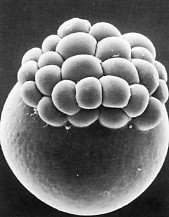
    This type of meroblastic cleavage is also found in the eggs of fish, reptiles, and 4 species of mammals — the monotremes. This photo, courtesy of H. W. Beames and Richard G. Kessel, shows the zebrafish (Danio) embryo at the 32-cell stage. Note that the cleavage furrows have not continued down through the yolk of the egg.

    Insects use a different type of meroblastic cleavage.

    Illustration of an egg diagram showing three stages: yolk, fertilized egg with small dots, and blastoderm. Each stage is labeled, depicting progression from yolk to blastoderm.
    Figure 14.3.4: Insect cleavage

    The yolk of the eggs of insects is concentrated in the center of the egg. The daughter nuclei produced by mitosis of the zygote nucleus remain suspended within the single egg compartment. After several thousand nuclei have been produced, they migrate to the cytoplasm-rich margin of the egg. Only then does a plasma membrane form around each one.

    What does cleavage accomplish in the development of the organism? First, it provides a stockpile of cells out of which the embryo will be constructed. Second, cleavage establishes a normal relationship between the nucleus and the volume of cytoplasm it regulates (and which in turn regulates it). Even small eggs are enormous when compared with other kinds of cells. The volume of the frog egg is about 1.6 million times larger than that of a normal frog cell. But it, too, contains only a single nucleus. During cleavage, thousands of new nuclei are produced by mitosis all of which finally end up in a cell of normal dimensions. The frog blastula, with its thousands of cells is no larger than the original fertilized egg.


    This page titled 19.2.3: Cleavage is shared under a CC BY 3.0 license and was authored, remixed, and/or curated by John W. Kimball via source content that was edited to the style and standards of the LibreTexts platform.