Biology LibreTexts

5.3: The Plasma Membrane and The Cytoplasm

  • Page ID
    68561
  • \( \newcommand{\vecs}[1]{\overset { \scriptstyle \rightharpoonup} {\mathbf{#1}} } \) \( \newcommand{\vecd}[1]{\overset{-\!-\!\rightharpoonup}{\vphantom{a}\smash {#1}}} \)\(\newcommand{\id}{\mathrm{id}}\) \( \newcommand{\Span}{\mathrm{span}}\) \( \newcommand{\kernel}{\mathrm{null}\,}\) \( \newcommand{\range}{\mathrm{range}\,}\) \( \newcommand{\RealPart}{\mathrm{Re}}\) \( \newcommand{\ImaginaryPart}{\mathrm{Im}}\) \( \newcommand{\Argument}{\mathrm{Arg}}\) \( \newcommand{\norm}[1]{\| #1 \|}\) \( \newcommand{\inner}[2]{\langle #1, #2 \rangle}\) \( \newcommand{\Span}{\mathrm{span}}\) \(\newcommand{\id}{\mathrm{id}}\) \( \newcommand{\Span}{\mathrm{span}}\) \( \newcommand{\kernel}{\mathrm{null}\,}\) \( \newcommand{\range}{\mathrm{range}\,}\) \( \newcommand{\RealPart}{\mathrm{Re}}\) \( \newcommand{\ImaginaryPart}{\mathrm{Im}}\) \( \newcommand{\Argument}{\mathrm{Arg}}\) \( \newcommand{\norm}[1]{\| #1 \|}\) \( \newcommand{\inner}[2]{\langle #1, #2 \rangle}\) \( \newcommand{\Span}{\mathrm{span}}\)\(\newcommand{\AA}{\unicode[.8,0]{x212B}}\)

    At this point, it should be clear that eukaryotic cells have a more complex structure than do prokaryotic cells. Organelles allow for various functions to occur in the cell at the same time. Before discussing the functions of organelles within a eukaryotic cell, let us first examine two important components of all cells (prokaryotic and eukaryotic): the plasma membrane and the cytoplasm.

    An illustration of a prokaryotic cell. The fluid inside the cell is called the cytoplasm. Ribosomes, depicted as small circles, float in the cytoplasm. The cytoplasm is encased by a plasma membrane, which in turn is encased by a cell wall. A capsule surrounds the cell wall.
    Figure \(\PageIndex{1}\): A prokaryotic cell. The cytoplasm is not labeled, but is the light blue area inside the cell membrane. The ribosome label is pointing to one of the small brown dots representing the ribosome.
    typical animal cell
    Figure \(\PageIndex{2}\): This figure shows a typical animal cell.
    typical plant cell
    Figure \(\PageIndex{3}\): This figure shows a typical plant cell.

    The Plasma Membrane

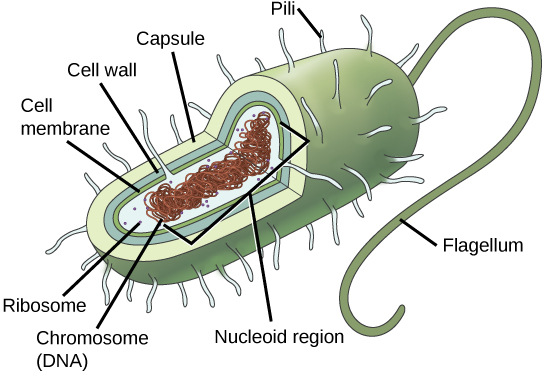
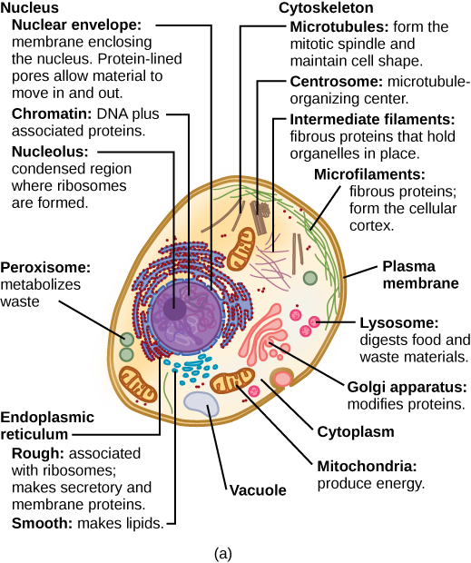
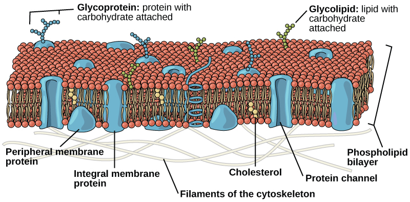
    Like prokaryotes, eukaryotic cells have a plasma membrane (Find it in Figures \(\PageIndex{1}\)-\(\PageIndex{3}\), then look at the detailed structure in Figure \(\PageIndex{4}\)) made up of a phospholipid bilayer with embedded proteins that separates the internal contents of the cell from its surrounding environment. A phospholipid is a lipid molecule composed of two fatty acid chains, a glycerol backbone, and a phosphate group. The plasma membrane regulates the passage of some substances, such as organic molecules, ions, and water, preventing the passage of some to maintain internal conditions, while actively bringing in or removing others. Other compounds move passively across the membrane.

    The plasma membrane is composed of a phospholipid bilayer. In the bilayer, the two long hydrophobic tails of phospholipids face toward the center, and the hydrophilic head group faces the exterior. Integral membrane proteins and protein channels span the entire bilayer. Protein channels have a pore in the middle. Peripheral membrane proteins sit on the surface of the phospholipids and are associated with the head groups. On the exterior side of the membrane, carbohydrates are attached to certain proteins and lipids. Filaments of the cytoskeleton line the interior of the membrane.
    Figure \(\PageIndex{4}\): The plasma membrane is a phospholipid bilayer with embedded proteins. There are other components, such as cholesterol and carbohydrates, which can be found in the membrane in addition to phospholipids and protein.

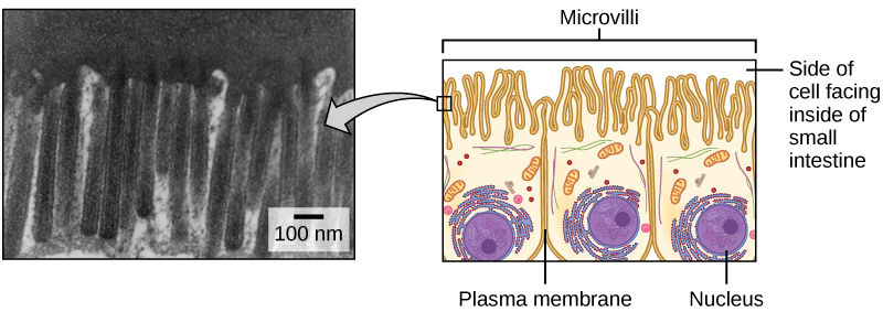
    The plasma membranes of cells that specialize in absorption are folded into fingerlike projections called microvilli (singular = microvillus). This folding increases the surface area of the plasma membrane. Such cells are typically found lining the small intestine, the organ that absorbs nutrients from digested food (Figure \(\PageIndex{5}\)). This is an excellent example of form matching the function of a structure.

    electron micrograph and cartoon of microvilli
    Figure \(\PageIndex{5}\): Microvilli, shown here as they appear on cells lining the small intestine, increase the surface area available for absorption. These microvilli are only found on the area of the plasma membrane that faces the cavity from which substances will be absorbed. (credit “micrograph”: modification of work by Louisa Howard)

    The Cytoplasm

    The cytoplasm comprises the contents of a cell between the plasma membrane and the nuclear envelope (a structure to be discussed shortly). It is made up of organelles suspended in the gel-like cytosol, the cytoskeleton, and various chemicals (Find it in Figures \(\PageIndex{1}\)-\(\PageIndex{3}\)). Even though the cytoplasm consists of 70 to 80 percent water, it has a semi-solid consistency, which comes from the proteins within it. However, proteins are not the only organic molecules found in the cytoplasm. Glucose and other simple sugars, polysaccharides, amino acids, nucleic acids, fatty acids, and derivatives of glycerol are found there too. Ions of sodium, potassium, calcium, and many other elements are also dissolved in the cytoplasm. Many metabolic reactions, including protein synthesis, take place in the cytoplasm. Take note that the cytoplasm is not “empty” or “filler” – it is a vitally important component of cells that allows chemical reactions to take place!

    References

    Unless otherwise noted, images on this page are licensed under CC-BY 4.0 by OpenStax.

    Text adapted from: OpenStax, Concepts of Biology. OpenStax CNX. May 18, 2016 http://cnx.org/contents/b3c1e1d2-839...9a8aafbdd@9.10


    5.3: The Plasma Membrane and The Cytoplasm is shared under a CC BY license and was authored, remixed, and/or curated by LibreTexts.

    • Was this article helpful?