
39.4: Systems of Gas Exchange - Mammalian Systems and Protective Mechanisms
- Page ID
- 14021

The mammalian respiratory system equilibrates air to the body, protects against foreign materials, and allows for gas exchange.
- Explain how air passes from the outside environment to the lungs, protecting them from particulate matter
Key Points
- The air that moves from the external environment into the body must pass through the nasal cavity where it is warmed, humidified, and surveyed for particulates.
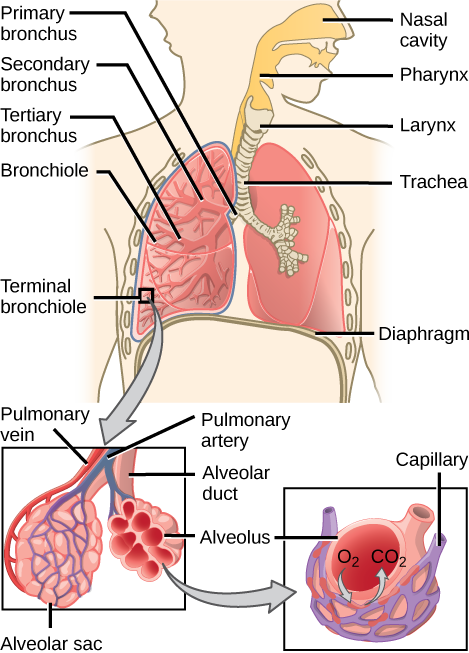
- As air moves out of the nasal cavity, it moves into the pharynx, larynx, trachea, the primary bronchi (right and left lung), secondary and tertiary bronchi, bronchioles, terminal then respiratory bronchioles, alveolar ducts then alveolar sacs where gas exchange occurs with the capillaries.
- Components in the respiratory system allow for protection from foreign material; these include mucus production in the lungs and cilia in the bronchi and bronchioles to move matter out of the system.
- Components in the respiratory system that allow for protection from foreign material and include mucus production in the lungs and cilia in the bronchi and bronchioles.
Key Terms
- alveolus: a small air sac in the lungs, where oxygen and carbon dioxide are exchanged with the blood
- bifurcate: to divide or fork into two channels or branches
- bronchus: either of two airways, which are primary branches of the trachea, leading directly into the lungs
Mammalian Respiratory System
In mammals, pulmonary ventilation occurs via inhalation when air enters the body through the nasal cavity. Air passes through the nasal cavity and is warmed to body temperature and humidified. The respiratory tract is coated with mucus that is high in water to seal the tissues from direct contact with air. As air crosses the surfaces of the mucous membranes, it picks up water. This equilibrates the air to the body, reducing damage that cold, dry air can cause. Particulates in the air are also removed in the nasal passages. These processes are all protective mechanisms that prevent damage to the trachea and lungs.
From the nasal cavity, air passes through the pharynx and the larynx to the trachea. The function of the trachea is to funnel the inhaled air to the lungs and the exhaled air out of the body. The human trachea, a cylinder about 10-12cm long, 2cm in diameter found in front of the esophagus, extends from the larynx into the chest cavity. It is made of incomplete rings of hyaline cartilage and smooth muscle that divides into the two primary bronchi at the midthorax. The trachea is lined with mucus-producing goblet cells and ciliated epithelia that propel foreign particles trapped in the mucus toward the pharynx. The cartilage provides strength and support to the trachea to keep the passage open. The smooth muscle can contract, causing a decrease in the trachea’s diameter, which propels expired air upwards from the lungs at a great force. The forced exhalation helps expel mucus when we cough.
Lungs: Bronchi and Alveoli
The end of the trachea bifurcates to the right and left lungs, which are not identical. The larger right lung has three lobes, while the smaller left lung has two lobes. The muscular diaphragm, which facilitates breathing, is inferior to the lungs, marking the end of the thoracic cavity.
As air enters the lungs, it is diverted through bronchi beginning with the two primary bronchi. Each bronchus divides into secondary, then into tertiary bronchi, which further divide to create smaller diameter bronchioles that split and spread through the lung. The bronchi are made of cartilage and smooth muscle; at the bronchioles, the cartilage is replaced with elastic fibers. Bronchi are innervated by nerves of both the parasympathetic and sympathetic nervous systems that control muscle contraction or relaxation, respectively. In humans, bronchioles with a diameter smaller than 0.5 mm are the respiratory bronchioles. Since they lack cartilage, they rely on inhaled air to support their shape. As the passageways decrease in diameter, the relative amount of smooth muscle increases.
The terminal bronchioles then subdivide into respiratory bronchioles which subdivide into alveolar ducts. Numerous alveoli (sing. alveolus) and alveolar sacs surround the alveolar ducts. The alveolar ducts are attached to the end of each bronchiole; each duct ends in approximately 100 alveolar sacs. Each sac contains 20-30 alveoli that are 200-300 microns in diameter. Alveoli are made of thin-walled, parenchymal cells that are in direct contact with capillaries of the circulatory system. This ensures that oxygen will diffuse from alveoli into the blood and that carbon dioxide produced by cells as a waste product will diffuse from the blood into alveoli to be exhaled. The anatomical arrangement of capillaries and alveoli emphasizes the relationship of the respiratory and circulatory systems. As there are so many alveoli (around 300 million per lung) within each alveolar sac and so many sacs at the end of each alveolar duct, the lungs have a sponge-like consistency. This organization produces a very large surface area that is available for gas exchange.
Protective Mechanisms
The air that organisms breathe contains particulate matter such as dust, dirt, viral particles, and bacteria that can damage the lungs. The respiratory system has protective mechanisms to avoid damage. In the nasal cavity, hairs and mucus trap small particles, viruses, bacteria, dust, and dirt to prevent entry. If particulates make it beyond the nose or enter via the mouth, the bronchi and bronchioles contain several protective devices. The lungs produce mucus that traps particulates. The bronchi and bronchioles contain cilia, small hair-like projections that line the walls of the bronchi and bronchioles. These cilia move mucus and particles out of the bronchi and bronchioles back up to the throat where it is swallowed and eliminated via the esophagus.
In humans, tar and other substances in cigarette smoke destroy or paralyze the cilia, making the removal of particles more difficult. In addition, smoking causes the lungs to produce more mucus, which the damaged cilia are unable to move. This causes a persistent cough, as the lungs try to rid themselves of particulate matter, making smokers more susceptible to respiratory ailments.
Contributions and Attributions
- OpenStax College, Biology. October 17, 2013. Provided by: OpenStax CNX. Located at: http://cnx.org/content/m44792/latest...ol11448/latest. License: CC BY: Attribution
- OpenStax College, Biology. October 23, 2013. Provided by: OpenStax CNX. Located at: http://cnx.org/content/m44790/latest...ol11448/latest. License: CC BY: Attribution
- Boundless. Provided by: Boundless Learning. Located at: www.boundless.com//biology/de...tion/diffusion. License: CC BY-SA: Attribution-ShareAlike
- Boundless. Provided by: Boundless Learning. Located at: www.boundless.com//biology/de...n/deoxygenated. License: CC BY-SA: Attribution-ShareAlike
- aerobic. Provided by: Wiktionary. Located at: http://en.wiktionary.org/wiki/aerobic. License: CC BY-SA: Attribution-ShareAlike
- OpenStax College, Systems of Gas Exchange. October 17, 2013. Provided by: OpenStax CNX. Located at: http://cnx.org/content/m44792/latest...e_39_01_02.jpg. License: CC BY: Attribution
- OpenStax College, Biology. October 17, 2013. Provided by: OpenStax CNX. Located at: http://cnx.org/content/m44792/latest...ol11448/latest. License: CC BY: Attribution
- OpenStax College, Biology. October 23, 2013. Provided by: OpenStax CNX. Located at: http://cnx.org/content/m44792/latest...ol11448/latest. License: CC BY: Attribution
- gill. Provided by: Wiktionary. Located at: en.wiktionary.org/wiki/gill. License: CC BY-SA: Attribution-ShareAlike
- coelom. Provided by: Wiktionary. Located at: en.wiktionary.org/wiki/coelom. License: CC BY-SA: Attribution-ShareAlike
- spiracle. Provided by: Wiktionary. Located at: en.wiktionary.org/wiki/spiracle. License: CC BY-SA: Attribution-ShareAlike
- OpenStax College, Systems of Gas Exchange. October 17, 2013. Provided by: OpenStax CNX. Located at: http://cnx.org/content/m44792/latest...e_39_01_02.jpg. License: CC BY: Attribution
- OpenStax College, Systems of Gas Exchange. October 17, 2013. Provided by: OpenStax CNX. Located at: http://cnx.org/content/m44792/latest...e_39_01_04.jpg. License: CC BY: Attribution
- OpenStax College, Systems of Gas Exchange. October 17, 2013. Provided by: OpenStax CNX. Located at: http://cnx.org/content/m44792/latest...e_39_01_03.jpg. License: CC BY: Attribution
- OpenStax College, Systems of Gas Exchange. October 17, 2013. Provided by: OpenStax CNX. Located at: http://cnx.org/content/m44792/latest...e_39_01_05.jpg. License: CC BY: Attribution
- Breathing. Provided by: OpenStax CNX. Located at: http://cnx.org/contents/GFy_h8cu@9.87:E4i-YQIZ@5/Breathing. License: CC BY-SA: Attribution-ShareAlike
- Bird Anatomy. Provided by: Wikipedia. Located at: en.Wikipedia.org/wiki/Bird_anatomy%23Respiratory_system. License: CC BY-SA: Attribution-ShareAlike
- Respiratory System. Provided by: Wikipedia. Located at: en.Wikipedia.org/wiki/Respiratory_system. License: CC BY-SA: Attribution-ShareAlike
- Gill. Provided by: Wiktionary. Located at: en.wiktionary.org/wiki/gill. License: CC BY-SA: Attribution-ShareAlike
- OpenStax College, Systems of Gas Exchange. October 17, 2013. Provided by: OpenStax CNX. Located at: http://cnx.org/content/m44792/latest/Figure_39_01_02.jpg. License: CC BY: Attribution
- OpenStax College, Systems of Gas Exchange. October 17, 2013. Provided by: OpenStax CNX. Located at: http://cnx.org/content/m44792/latest/Figure_39_01_04.jpg. License: CC BY: Attribution
- OpenStax College, Systems of Gas Exchange. October 17, 2013. Provided by: OpenStax CNX. Located at: http://cnx.org/content/m44792/latest/Figure_39_01_03.jpg. License: CC BY: Attribution
- OpenStax College, Systems of Gas Exchange. October 17, 2013. Provided by: OpenStax CNX. Located at: http://cnx.org/content/m44792/latest...e_39_01_05.jpg. License: CC BY: Attribution
- BirdRespiration.svg.png. Provided by: Wikipedia. Located at: en.Wikipedia.org/wiki/Bird_a...espiration.svg. License: Public Domain: No Known Copyright
- OpenStax College, Biology. October 17, 2013. Provided by: OpenStax CNX. Located at: http://cnx.org/content/m44792/latest...ol11448/latest. License: CC BY: Attribution
- OpenStax College, Biology. December 3, 2013. Provided by: OpenStax CNX. Located at: http://cnx.org/content/m44792/latest...ol11448/latest. License: CC BY: Attribution
- bronchus. Provided by: Wiktionary. Located at: en.wiktionary.org/wiki/bronchus. License: CC BY-SA: Attribution-ShareAlike
- bifurcate. Provided by: Wiktionary. Located at: en.wiktionary.org/wiki/bifurcate. License: CC BY-SA: Attribution-ShareAlike
- alveolus. Provided by: Wiktionary. Located at: en.wiktionary.org/wiki/alveolus. License: CC BY-SA: Attribution-ShareAlike
- OpenStax College, Systems of Gas Exchange. October 17, 2013. Provided by: OpenStax CNX. Located at: http://cnx.org/content/m44792/latest/Figure_39_01_02.jpg. License: CC BY: Attribution
- OpenStax College, Systems of Gas Exchange. October 17, 2013. Provided by: OpenStax CNX. Located at: http://cnx.org/content/m44792/latest/Figure_39_01_04.jpg. License: CC BY: Attribution
- OpenStax College, Systems of Gas Exchange. October 17, 2013. Provided by: OpenStax CNX. Located at: http://cnx.org/content/m44792/latest/Figure_39_01_03.jpg. License: CC BY: Attribution
- OpenStax College, Systems of Gas Exchange. October 17, 2013. Provided by: OpenStax CNX. Located at: http://cnx.org/content/m44792/latest/Figure_39_01_05.jpg. License: CC BY: Attribution
- BirdRespiration.svg.png. Provided by: Wikipedia. Located at: en.Wikipedia.org/wiki/Bird_anatomy%23/media/File:BirdRespiration.svg. License: Public Domain: No Known Copyright
- OpenStax College, Systems of Gas Exchange. October 17, 2013. Provided by: OpenStax CNX. Located at: http://cnx.org/content/m44792/latest...e_39_01_08.jpg. License: CC BY: Attribution
- OpenStax College, Systems of Gas Exchange. October 17, 2013. Provided by: OpenStax CNX. Located at: http://cnx.org/content/m44792/latest...e_39_01_10.jpg. License: CC BY: Attribution
- OpenStax College, Systems of Gas Exchange. October 17, 2013. Provided by: OpenStax CNX. Located at: http://cnx.org/content/m44792/latest...e_39_01_09.jpg. License: CC BY: Attribution
- OpenStax College, Systems of Gas Exchange. October 17, 2013. Provided by: OpenStax CNX. Located at: http://cnx.org/content/m44792/latest...e_39_01_07.jpg. License: CC BY: Attribution
- OpenStax College, Systems of Gas Exchange. October 17, 2013. Provided by: OpenStax CNX. Located at: http://cnx.org/content/m44792/latest...e_39_01_06.png. License: CC BY: Attribution