
35.4: How Neurons Communicate - Nerve Impulse Transmission within a Neuron- Resting Potential
- Page ID
- 13868

- Explain the formation of the resting potential in neurons
Nerve Impulse Transmission within a Neuron
For the nervous system to function, neurons must be able to send and receive signals. These signals are possible because each neuron has a charged cellular membrane (a voltage difference between the inside and the outside). The charge of this membrane can change in response to neurotransmitter molecules released from other neurons and environmental stimuli. Any voltage is a difference in electric potential between two points; for example, the separation of positive and negative electric charges on opposite sides of a resistive barrier. To understand how neurons communicate, one must first understand the basis of charged membranes and the baseline or ‘resting’ membrane charge.
Neuronal Charged Membranes
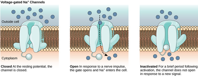
The lipid bilayer membrane that surrounds a neuron is impermeable to charged molecules or ions. To enter or exit the neuron, ions must pass through special proteins called ion channels that span the membrane. Ion channels have different configurations: open, closed, and inactive. Some ion channels need to be activated in order to open and allow ions to pass into or out of the cell. These ion channels are sensitive to the environment and can change their shape accordingly. Ion channels that change their structure in response to voltage changes are called voltage-gated ion channels. Voltage-gated ion channels regulate the relative concentrations of different ions inside and outside the cell. The difference in total charge between the inside and outside of the cell is called the membrane potential.
Resting Membrane Potential
For quiescent cells, the relatively-static membrane potential is known as the resting membrane potential. The resting membrane potential is at equilibrium since it relies on the constant expenditure of energy for its maintenance. It is dominated by the ionic species in the system that has the greatest conductance across the membrane. For most cells, this is potassium. As potassium is also the ion with the most-negative equilibrium potential, usually the resting potential can be no more negative than the potassium equilibrium potential.
A neuron at rest is negatively charged because the inside of a cell is approximately 70 millivolts more negative than the outside (−70 mV); this number varies by neuron type and by species. This voltage is called the resting membrane potential and is caused by differences in the concentrations of ions inside and outside the cell. If the membrane were equally permeable to all ions, each type of ion would flow across the membrane and the system would reach equilibrium. Because ions cannot simply cross the membrane at will, there are different concentrations of several ions inside and outside the cell. The difference in the number of positively-charged potassium ions (K+) inside and outside the cell dominates the resting membrane potential. When the membrane is at rest, K+ ions accumulate inside the cell due to a net movement with the concentration gradient. The negative resting membrane potential is created and maintained by increasing the concentration of cations outside the cell (in the extracellular fluid) relative to inside the cell (in the cytoplasm). The negative charge within the cell is created by the cell membrane being more permeable to K+ movement than Na+movement.

In neurons, potassium ions (K+) are maintained at high concentrations within the cell, while sodium ions (Na+) are maintained at high concentrations outside of the cell. The cell possesses potassium and sodium leakage channels that allow the two cations to diffuse down their concentration gradient. However, the neurons have far more potassium leakage channels than sodium leakage channels. Therefore, potassium diffuses out of the cell at a much faster rate than sodium leaks in. More cations leaving the cell than entering it causes the interior of the cell to be negatively charged relative to the outside of the cell. The actions of the sodium-potassium pump help to maintain the resting potential, once it is established. Recall that sodium-potassium pumps bring two K+ ions into the cell while removing three Na+ ions per ATP consumed. As more cations are expelled from the cell than are taken in, the inside of the cell remains negatively charged relative to the extracellular fluid.
Key Points
- When the neuronal membrane is at rest, the resting potential is negative due to the accumulation of more sodium ions outside the cell than potassium ions inside the cell.
- Potassium ions diffuse out of the cell at a much faster rate than sodium ions diffuse into the cell because neurons have many more potassium leakage channels than sodium leakage channels.
- Sodium-potassium pumps move two potassium ions inside the cell as three sodium ions are pumped out to maintain the negatively-charged membrane inside the cell; this helps maintain the resting potential.
Key Terms
- ion channel: a protein complex or single protein that penetrates a cell membrane and catalyzes the passage of specific ions through that membrane
- membrane potential: the difference in electrical potential across the enclosing membrane of a cell
- resting potential: the nearly latent membrane potential of inactive cells