
28.1B: Morphology of Sponges
- Page ID
- 13709

Instead of true tissues or organs, sponges have specialized cells that are in charge of important bodily functions and processes.
- Explain the various cell forms and bodily functions of sponges
Key Points
- Although sponges do not have organized tissue, they depend on specialized cells, such as choanocytes, porocytes, amoebocytes, and pinacocytes, for specialized functions within their bodies.
- The mesohyl acts as a type of endoskeleton, helping to maintain the tubular shape of sponges.
- Porocytes control the amount of water that enters pores into the spongocoel, while choanocytes, which are flagellated cells, aid the movement of water through the sponge, thereby helping the sponge to trap and ingest food particles.
- Amoebocytes carry out several special functions: they deliver nutrients from choanocytes to other cells, give rise to eggs for sexual reproduction, deliver phagocytized sperm from choanocytes to eggs, and can transform into other cell types.
- Collencytes, lophocytes, sclerocytes, and spongocytes are examples of cells that are derived from amoebocytes; these cells manage other vital functions in the body of sponges.
Key Terms
- choanocyte: any of the cells in sponges that contain a flagellum and are used to control the movement of water
- spongocoel: the large, central cavity of sponges
- osculum: an opening in a sponge from which water is expelled
- mesohyl: the gelatinous matrix within a sponge
Morphology of Sponges
The morphology of the simplest sponges takes the shape of a cylinder with a large central cavity, the spongocoel, occupying the inside of the cylinder. Water can enter into the spongocoel from numerous pores in the body wall. Water entering the spongocoel is extruded via a large, common opening called the osculum. However, sponges exhibit a range of diversity in body forms, including variations in the size of the spongocoel, the number of osculi, and where the cells that filter food from the water are located.
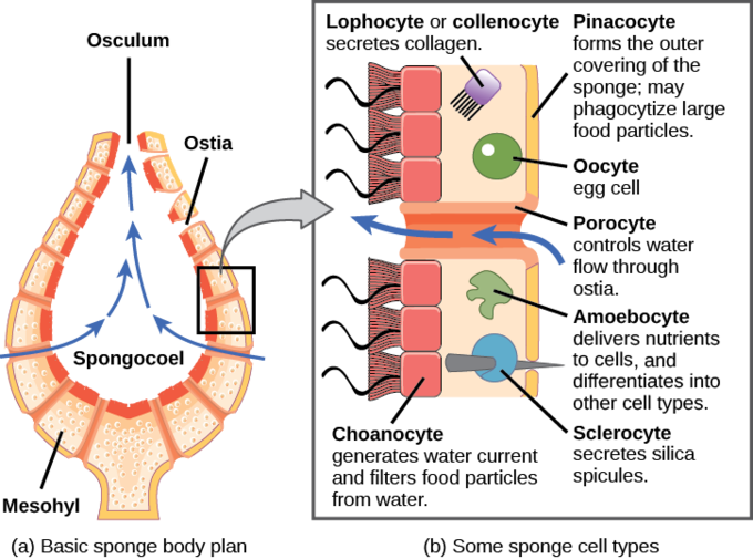
While sponges (excluding the Hexactinellids) do not exhibit tissue-layer organization, they do have different cell types that perform distinct functions. Pinacocytes, which are epithelial-like cells, form the outermost layer of sponges, enclosing a jelly-like substance called mesohyl. Mesohyl is an extracellular matrix consisting of a collagen -like gel with suspended cells that perform various functions. The gel-like consistency of mesohyl acts as an endoskeleton, maintaining the tubular morphology of sponges. In addition to the osculum, sponges have multiple pores called ostia on their bodies that allow water to enter the sponge. In some sponges, ostia are formed by porocytes: single, tube-shaped cells that act as valves to regulate the flow of water into the spongocoel. In other sponges, ostia are formed by folds in the body wall of the sponge.
Choanocytes (“collar cells”) are present at various locations, depending on the type of sponge; however, they always line the inner portions of some space through which water flows: the spongocoel in simple sponges; canals within the body wall in more complex sponges; and chambers scattered throughout the body in the most complex sponges. Whereas pinacocytes line the outside of the sponge, choanocytes tend to line certain inner portions of the sponge body that surround the mesohyl. The structure of a choanocyte is critical to its function, which is to generate a water current through the sponge and to trap and ingest food particles by phagocytosis. Note that there is a similarity in appearance between the sponge choanocyte and choanoflagellates (Protista). This similarity suggests that sponges and choanoflagellates are closely related and probably share a recent, common ancestry.
The cell body is embedded in mesohyl. It contains all organelles required for normal cell function, but protruding into the “open space” inside of the sponge is a mesh-like collar composed of microvilli with a single flagellum in the center of the column. The cumulative effect of the flagella from all choanocytes aids the movement of water through the sponge: drawing water into the sponge through the numerous ostia, into the spaces lined by choanocytes, and eventually out through the osculum (or osculi). Meanwhile, food particles, including waterborne bacteria and algae, are trapped by the sieve-like collar of the choanocytes, slide down into the body of the cell, are ingested by phagocytosis, and become encased in a food vacuole. Finally, choanocytes will differentiate into sperm for sexual reproduction; they will become dislodged from the mesohyl, leaving the sponge with expelled water through the osculum.
The second crucial cells in sponges are called amoebocytes (or archaeocytes), named for the fact that they move throughout the mesohyl in an amoeba-like fashion. Amoebocytes have a variety of functions: delivering nutrients from choanocytes to other cells within the sponge; giving rise to eggs for sexual reproduction (which remain in the mesohyl); delivering phagocytized sperm from choanocytes to eggs; and differentiating into more-specific cell types. Some of these more-specific cell types include collencytes and lophocytes, which produce the collagen-like protein to maintain the mesohyl; sclerocytes, which produce spicules in some sponges; and spongocytes, which produce the protein spongin in the majority of sponges. These cells produce collagen to maintain the consistency of the mesohyl.