
3.11: The Cytoskeleton
- Page ID
- 3988

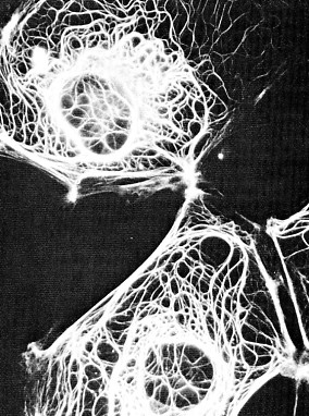
Cells contain elaborate arrays of protein fibers that serve such functions as establishing cell shape, providing mechanical strength, and locomotion. These fibers participate in chromosome separation in mitosis and meiosis and intracellular transport of organelles. The cytoskeleton is made up of three kinds of protein filaments: Actin filaments (also called microfilaments), Intermediate filaments, and Microtubules.
Actin Filaments
Monomers of the protein actin polymerize to form long, thin fibers. These are about 8 nm in diameter and, being the thinnest of the cytoskeletal filaments, are also called microfilaments (in skeletal muscle fibers they are called "thin" filaments). Some functions of actin filaments are:
- form a band just beneath the plasma membrane that
- provides mechanical strength to the cell
- links transmembrane proteins (e.g., cell surface receptors) to cytoplasmic proteins
- pinches dividing animal cells apart during cytokinesis
- generate cytoplasmic streaming in some cells
- generate locomotion in cells such as white blood cells and the amoeba
- interact with myosin ("thick") filaments in skeletal muscle fibers to provide the force of muscular contraction
Intermediate Filaments
These cytoplasmic fibers average 10 nm in diameter (and thus are "intermediate" in size between actin filaments (8 nm) and microtubules (25 nm) (as well as of the thick filaments of skeletal muscle fibers). There are several types of intermediate filament, each constructed from one or more proteins characteristic of it.
- keratins are found in epithelial cells and also form hair and nails;
- nuclear lamins form a meshwork that stabilizes the inner membrane of the nuclear envelope;
- neurofilaments strengthen the long axons of neurons;
- vimentins provide mechanical strength to muscle (and other) cells.
Despite their chemical diversity, intermediate filaments play similar roles in the cell: providing a supporting framework within the cell. For example, the nucleus in epithelial cells is held within the cell by a basketlike network of intermediate filaments made of keratins.
Different kinds of epithelia use different keratins to build their intermediate filaments. Over 20 different kinds of keratins have been found, although each kind of epithelial cell may use no more than 2 of them. Up to 85% of the dry weight of squamous epithelial cells can consist of keratins.
Microtubules
Microtubules are straight, hollow cylinders whose wall is made up of a ring of 13 "protofilaments" and have a diameter of about 25 nm. They are variable in length but can grow 1000 times as long as they are wide. They are built by the assembly of dimers of alpha tubulin and beta tubulin. Microtubules are found in both animal and plant cells. In plant cells, microtubules are created at many sites scattered through the cell. In animal cells, the microtubules originate at the centrosome. The attached end is called the minus end; the other end is the plus end.
Microtubules grow at the plus end by the polymerization of tubulin dimers (powered by the hydrolysis of GTP), and shrink by the release of tubulin dimers (depolymerization) at the same end. They participate in a wide variety of cell activities. Most involve motion. The motion is provided by protein "motors" that use the energy of ATP to move along the microtubule.
Microtubule motors
There are two major groups of microtubule motors kinesins (most of these move toward the plus end of the microtubules) and dyneins (which move toward the minus end)
- The rapid transport of organelles, like vesicles and mitochondria, along the axons of neurons takes place along microtubules with their plus ends pointed toward the end of the axon. The motors are kinesins.
- The migration of chromosomes in mitosis and meiosis takes place on microtubules that make up the spindle fibers. Both kinesins and dyneins are used as motors
Cilia and Flagella
Cilia and flagella are built from arrays of microtubules.