
24.1B: Fungi Cell Structure and Function
- Page ID
- 13598

Fungi are unicellular or multicellular thick-cell-walled heterotroph decomposers that eat decaying matter and make tangles of filaments.
- Describe the physical structures associated with fungi
Key Points
- Fungal cell walls are rigid and contain complex polysaccharides called chitin (adds structural strength) and glucans.
- Ergosterol is the steroid molecule in the cell membranes that replaces the cholesterol found in animal cell membranes.
- Fungi can be unicellular, multicellular, or dimorphic, which is when the fungi is unicellular or multicellular depending on environmental conditions.
- Fungi in the morphological vegetative stage consist of a tangle of slender, thread-like hyphae, whereas the reproductive stage is usually more obvious.
- Fungi like to be in a moist and slightly acidic environment; they can grow with or without light or oxygen.
- Fungi are saprophyte heterotrophs in that they use dead or decomposing organic matter as a source of carbon.
Key Terms
- glucan: any polysaccharide that is a polymer of glucose
- ergosterol: the functional equivalent of cholesterol found in cell membranes of fungi and some protists, as well as, the steroid precursor of vitamin D2
- mycelium: the vegetative part of any fungus, consisting of a mass of branching, threadlike hyphae, often underground
- hypha: a long, branching, filamentous structure of a fungus that is the main mode of vegetative growth
- septum: cell wall division between hyphae of a fungus
- thallus: vegetative body of a fungus
- saprophyte: any organism that lives on dead organic matter, as certain fungi and bacteria
- chitin: a complex polysaccharide, a polymer of N-acetylglucosamine, found in the exoskeletons of arthropods and in the cell walls of fungi; thought to be responsible for some forms of asthma in humans
Cell Structure and Function
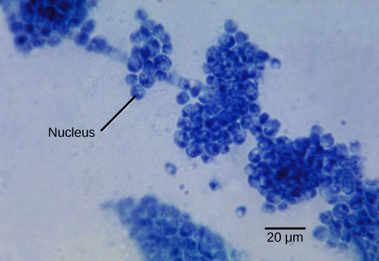
Fungi are eukaryotes and have a complex cellular organization. As eukaryotes, fungal cells contain a membrane-bound nucleus where the DNA is wrapped around histone proteins. A few types of fungi have structures comparable to bacterial plasmids (loops of DNA). Fungal cells also contain mitochondria and a complex system of internal membranes, including the endoplasmic reticulum and Golgi apparatus.
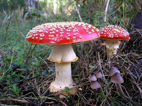
Unlike plant cells, fungal cells do not have chloroplasts or chlorophyll. Many fungi display bright colors arising from other cellular pigments, ranging from red to green to black. The poisonous Amanita muscaria (fly agaric) is recognizable by its bright red cap with white patches. Pigments in fungi are associated with the cell wall. They play a protective role against ultraviolet radiation and can be toxic.
The rigid layers of fungal cell walls contain complex polysaccharides called chitin and glucans. Chitin, also found in the exoskeleton of insects, gives structural strength to the cell walls of fungi. The wall protects the cell from desiccation and predators. Fungi have plasma membranes similar to other eukaryotes, except that the structure is stabilized by ergosterol: a steroid molecule that replaces the cholesterol found in animal cell membranes. Most members of the kingdom Fungi are nonmotile.
Growth
The vegetative body of a fungus is a unicellular or multicellular thallus. Dimorphic fungi can change from the unicellular to multicellular state depending on environmental conditions. Unicellular fungi are generally referred to as yeasts. Saccharomyces cerevisiae (baker’s yeast) and Candida species (the agents of thrush, a common fungal infection) are examples of unicellular fungi.
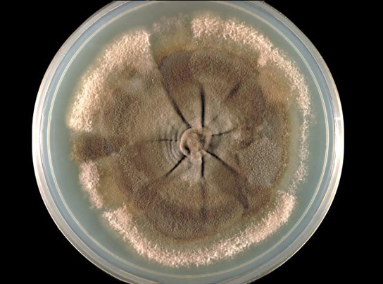
Most fungi are multicellular organisms. They display two distinct morphological stages: the vegetative and reproductive. The vegetative stage consists of a tangle of slender thread-like structures called hyphae (singular, hypha ), whereas the reproductive stage can be more conspicuous. The mass of hyphae is a mycelium. It can grow on a surface, in soil or decaying material, in a liquid, or even on living tissue. Although individual hyphae must be observed under a microscope, the mycelium of a fungus can be very large, with some species truly being “the fungus humongous.” The giant Armillaria solidipes (honey mushroom) is considered the largest organism on Earth, spreading across more than 2,000 acres of underground soil in eastern Oregon; it is estimated to be at least 2,400 years old.
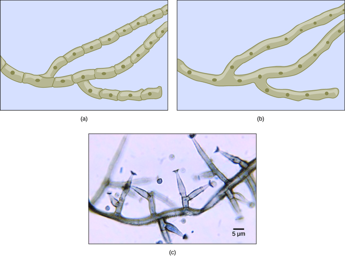
Most fungal hyphae are divided into separate cells by endwalls called septa (singular, septum) ( a, c). In most phyla of fungi, tiny holes in the septa allow for the rapid flow of nutrients and small molecules from cell to cell along the hypha. They are described as perforated septa. The hyphae in bread molds (which belong to the Phylum Zygomycota) are not separated by septa. Instead, they are formed by large cells containing many nuclei, an arrangement described as coenocytic hyphae ( b). Fungi thrive in environments that are moist and slightly acidic; they can grow with or without light.
Nutrition
Like animals, fungi are heterotrophs: they use complex organic compounds as a source of carbon, rather than fix carbon dioxide from the atmosphere as do some bacteria and most plants. In addition, fungi do not fix nitrogen from the atmosphere. Like animals, they must obtain it from their diet. However, unlike most animals, which ingest food and then digest it internally in specialized organs, fungi perform these steps in the reverse order: digestion precedes ingestion. First, exoenzymes are transported out of the hyphae, where they process nutrients in the environment. Then, the smaller molecules produced by this external digestion are absorbed through the large surface area of the mycelium. As with animal cells, the polysaccharide of storage is glycogen rather than the starch found in plants.
Fungi are mostly saprobes (saprophyte is an equivalent term): organisms that derive nutrients from decaying organic matter. They obtain their nutrients from dead or decomposing organic matter, mainly plant material. Fungal exoenzymes are able to break down insoluble polysaccharides, such as the cellulose and lignin of dead wood, into readily-absorbable glucose molecules. The carbon, nitrogen, and other elements are thus released into the environment. Because of their varied metabolic pathways, fungi fulfill an important ecological role and are being investigated as potential tools in bioremediation.
Some fungi are parasitic, infecting either plants or animals. Smut and Dutch elm disease affect plants, whereas athlete’s foot and candidiasis (thrush) are medically important fungal infections in humans.