
10.2B: The Mitotic Phase and the G0 Phase
- Page ID
- 13236

- Describe the events that occur at the different stages of mitosis
The Mitotic Phase
The mitotic phase is a multistep process during which the duplicated chromosomes are aligned, separated, and move into two new, identical daughter cells. The first portion of the mitotic phase is called karyokinesis or nuclear division. The second portion of the mitotic phase, called cytokinesis, is the physical separation of the cytoplasmic components into the two daughter cells.
Karyokinesis (Mitosis)
Karyokinesis, also known as mitosis, is divided into a series of phases (prophase, prometaphase, metaphase, anaphase, and telophase) that result in the division of the cell nucleus.

During prophase, the “first phase,” the nuclear envelope starts to dissociate into small vesicles. The membranous organelles (such as the Golgi apparatus and endoplasmic reticulum) fragment and disperse toward the periphery of the cell. The nucleolus disappears and the centrosomes begin to move to opposite poles of the cell. Microtubules that will eventually form the mitotic spindle extend between the centrosomes, pushing them farther apart as the microtubule fibers lengthen. The sister chromatids begin to coil more tightly with the aid of condensin proteins and become visible under a light microscope.
During prometaphase, the “first change phase,” many processes that began in prophase continue to advance. The remnants of the nuclear envelope fragment. The mitotic spindle continues to develop as more microtubules assemble and stretch across the length of the former nuclear area. Chromosomes become more condensed and discrete. Each sister chromatid develops a protein structure called a kinetochore in the centromeric region. The proteins of the kinetochore attract and bind mitotic spindle microtubules.
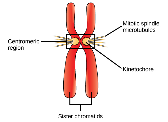
During metaphase, the “change phase,” all the chromosomes are aligned on a plane called the metaphase plate, or the equatorial plane, midway between the two poles of the cell. The sister chromatids are still tightly attached to each other by cohesin proteins. At this time, the chromosomes are maximally condensed.
During anaphase, the “upward phase,” the cohesin proteins degrade, and the sister chromatids separate at the centromere. Each chromatid, now called a chromosome, is pulled rapidly toward the centrosome to which its microtubule is attached. The cell becomes visibly elongated (oval shaped) as the polar microtubules slide against each other at the metaphase plate where they overlap.
During telophase, the “distance phase,” the chromosomes reach the opposite poles and begin to decondense (unravel), relaxing into a chromatin configuration. The mitotic spindles are depolymerized into tubulin monomers that will be used to assemble cytoskeletal components for each daughter cell. Nuclear envelopes form around the chromosomes and nucleosomes appear within the nuclear area.
Cytokinesis
Cytokinesis, or “cell motion,” is the second main stage of the mitotic phase during which cell division is completed via the physical separation of the cytoplasmic components into two daughter cells. Division is not complete until the cell components have been apportioned and completely separated into the two daughter cells. Although the stages of mitosis are similar for most eukaryotes, the process of cytokinesis is quite different for eukaryotes that have cell walls, such as plant cells.
In cells such as animal cells, which lack cell walls, cytokinesis follows the onset of anaphase. A contractile ring composed of actin filaments forms just inside the plasma membrane at the former metaphase plate. The actin filaments pull the equator of the cell inward, forming a fissure. This fissure or “crack” is called the cleavage furrow. The furrow deepens as the actin ring contracts; eventually the membrane is cleaved in two.

In plant cells, a new cell wall must form between the daughter cells. During interphase, the Golgi apparatus accumulates enzymes, structural proteins, and glucose molecules prior to breaking into vesicles and dispersing throughout the dividing cell. During telophase, these Golgi vesicles are transported on microtubules to form a phragmoplast (a vesicular structure) at the metaphase plate. There, the vesicles fuse and coalesce from the center toward the cell walls; this structure is called a cell plate. As more vesicles fuse, the cell plate enlarges until it merges with the cell walls at the periphery of the cell. Enzymes use the glucose that has accumulated between the membrane layers to build a new cell wall. The Golgi membranes become parts of the plasma membrane on either side of the new cell wall.
G0 Phase
Not all cells adhere to the classic cell cycle pattern in which a newly-formed daughter cell immediately enters the preparatory phases of interphase, closely followed by the mitotic phase. Cells in G0 phase are not actively preparing to divide. The cell is in a quiescent (inactive) stage that occurs when cells exit the cell cycle. Some cells enter G0 temporarily until an external signal triggers the onset of G1. Other cells that never or rarely divide, such as mature cardiac muscle and nerve cells, remain in G0 permanently.
Key Points
- During prophase, the nucleus disappears, spindle fibers form, and DNA condenses into chromosomes ( sister chromatids ).
- During metaphase, the sister chromatids align along the equator of the cell by attaching their centromeres to the spindle fibers.
- During anaphase, sister chromatids are separated at the centromere and are pulled towards opposite poles of the cell by the mitotic spindle.
- During telophase, chromosomes arrive at opposite poles and unwind into thin strands of DNA, the spindle fibers disappear, and the nuclear membrane reappears.
- Cytokinesis is the actual splitting of the cell membrane; animal cells pinch apart, while plant cells form a cell plate that becomes the new cell wall.
- Cells enter the G0 (inactive) phase after they exit the cell cycle when they are not actively preparing to divide; some cells remain in G0 phase permanently.
Key Terms
- karyokinesis: (mitosis) the first portion of mitotic phase in which division of the cell nucleus takes place
- centrosome: an organelle near the nucleus in the cytoplasm of most organisms that controls the organization of its microtubules and gives rise to the mitotic spindle
- cytokinesis: the second portion of the mitotic phase in which the cytoplasm of a cell divides following the division of the nucleus
Contributions and Attributions
- OpenStax College, Biology. October 16, 2013. Provided by: OpenStax CNX. Located at: http://cnx.org/content/m44460/latest...ol11448/latest. License: CC BY: Attribution
- Boundless. Provided by: Boundless Learning. Located at: www.boundless.com//biology/de...itotic-spindle. License: CC BY-SA: Attribution-ShareAlike
- interphase. Provided by: Wiktionary. Located at: en.wiktionary.org/wiki/interphase. License: CC BY-SA: Attribution-ShareAlike
- sister chromatid. Provided by: Wiktionary. Located at: en.wiktionary.org/wiki/sister_chromatid. License: CC BY-SA: Attribution-ShareAlike
- OpenStax College, Biology. November 4, 2013. Provided by: OpenStax CNX. Located at: http://cnx.org/content/m44460/latest...ol11448/latest. License: CC BY: Attribution
- OpenStax College, Biology. October 16, 2013. Provided by: OpenStax CNX. Located at: http://cnx.org/content/m44460/latest...ol11448/latest. License: CC BY: Attribution
- OpenStax College, Biology. October 21, 2013. Provided by: OpenStax CNX. Located at: http://cnx.org/content/m44460/latest...ol11448/latest. License: CC BY: Attribution
- karyokinesis. Provided by: Wiktionary. Located at: en.wiktionary.org/wiki/karyokinesis. License: CC BY-SA: Attribution-ShareAlike
- Boundless. Provided by: Boundless Learning. Located at: www.boundless.com//biology/de...on/cytokinesis. License: CC BY-SA: Attribution-ShareAlike
- centrosome. Provided by: Wiktionary. Located at: en.wiktionary.org/wiki/centrosome. License: CC BY-SA: Attribution-ShareAlike
- OpenStax College, Biology. November 4, 2013. Provided by: OpenStax CNX. Located at: http://cnx.org/content/m44460/latest...ol11448/latest. License: CC BY: Attribution
- OpenStax College, The Cell Cycle. October 16, 2013. Provided by: OpenStax CNX. Located at: http://cnx.org/content/m44460/latest...e_10_02_02.png. License: CC BY: Attribution
- OpenStax College, The Cell Cycle. October 16, 2013. Provided by: OpenStax CNX. Located at: http://cnx.org/content/m44460/latest...e_10_02_03.jpg. License: CC BY: Attribution
- OpenStax College, The Cell Cycle. October 16, 2013. Provided by: OpenStax CNX. Located at: http://cnx.org/content/m44460/latest...e_10_02_04.jpg. License: CC BY: Attribution