
15.3C: The Heartbeat
- Page ID
- 5397

During rest, the heart beats about 70 times a minute in the adult male, while pumping about 5 liters of blood.
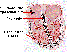
The stimulus that maintains this rhythm is self-contained. Embedded in the wall of the right atrium is a mass of specialized heart tissue called the sino-atrial (S-A) node. The S-A node is also called the pacemaker because it establishes the basic frequency at which the heart beats.
The interior of the fibers of heart muscle, like all cells, is negatively charged with respect to the exterior. In the cells of the pacemaker, this charge breaks down spontaneously about 70 times each minute. This, in turn, initiates a similar discharge of the nearby muscle fibers of the atrium. A tiny wave of current sweeps over the atria, causing them to contract.
When this current reaches the region of insulating connective tissue between the atria and the ventricles, it is picked up by the A-V node (atrio-ventricular node). This leads to a system of branching fibers that carries the current to all parts of the ventricles. The contraction of the heart in response to this electrical activity creates systole. A period of recovery follows called diastole.
- The heart muscle and S-A node become recharged.
- The heart muscle relaxes.
- The atria refill.
The Electrocardiogram
The electrical activity of the heart can be detected by electrodes placed at the surface of the body. Analysis of an electrocardiogram (ECG or EKG) aids in determining, for example, the extent of damage following a heart attack. This is because death of a portion of the heart muscle blocks electrical transmission through that area and alters the appearance of the ECG.
Ventricular Fibrillation
The ventricles can maintain a beat even without a functioning A-V node, although the beat is slower. There is, however, a danger that impulses arising in the ventricles may become disorganized and random. If this happens, they begin to twitch spasmodically, a condition called ventricular fibrillation. Blood flow ceases and unless the heart rhythm is restarted, death follows swiftly. In fact, ventricular fibrillation is the immediate cause of as much as 25% of all deaths.
Hospital emergency rooms, ambulances, commercial air craft and many other public places are now equipped with defibrillators which, by giving the heart a jolt of direct current, may restore its natural rhythm and save the victim's life.
Artificial Pacemakers
These are devices that generate rhythmic impulses that are transmitted to the heart by fine wires. Thanks to miniaturization and long-lived batteries, pacemakers can be implanted just under the skin and reached through a small incision when maintenance is needed.
Auxiliary Control of the Heart
Although the A-V node sets the basic rhythm of the heart, the rate and strength of its beating can be modified by two auxiliary control centers located in the medulla oblongata of the brain.
- One sends nerve impulses down accelerans nerves.
- The other sends nerve impulses down a pair of vagus nerves
The Accelerans Nerve
The accelerans nerve is part of the sympathetic branch of the autonomic nervous system, and like all post-ganglionic sympathetic neurons releases noradrenaline at its endings on the heart. It increases the rate and strength of the heartbeat and thus increase the flow of blood. Its activation usually arises from some stress such as fear or violent exertion. The heartbeat may increase to 180 beats per minute. The strength of contraction increases as well so the amount of blood pumped may increase to as much as 25–30 liters/minute.
The 24 Feb 2000 issue of the New England Journal of Medicine reports on a family some of whose members have inherited a mutant gene for the transporter that is responsible for reuptake of noradrenaline back into the neuron that released it. Those with the mutation are prone to bouts of rapid heartbeat and fainting when they suddenly stand up.
Vigorous exercise accelerates heartbeat in two ways:
- As cellular respiration increases, so does the carbon dioxide level in the blood. This stimulates receptors in the carotid arteries and aorta, and these transmit impulses to the medulla for relay by the accelerans nerve to the heart.
- As muscular activity increases, the muscle pump drives more blood back to the right atrium. The atrium becomes distended with blood, thus stimulating stretch receptors in its wall. These, too, send impulses to the medulla for relay to the heart.
Distention of the wall of the right atrium also triggers the release of atrial natriuretic peptide (ANP) which initiates a set of responses leading to a lowering of blood pressure.
The Vagus Nerves
The vagus nerves are part of the parasympathetic branch of the autonomic nervous system. They, too, run from the medulla oblongata to the heart. Their activity slows the heartbeat. Pressure receptors in the aorta and carotid arteries send impulses to the medulla which relays these by way of the vagus nerves to the heart. Heartbeat and blood pressure diminish.