
24.4: Angiosperm Life Cycle
- Page ID
- 33946
Observe a prepared slide of mature pollen in a Lilium anther cross section. Within the anthers, there are pollen grains. Each pollen grain contains two cells. The tube cell takes up the majority of the pollen grain, engulfing the smaller generative cell. The tube cell will grow a pollen tube down the length of the style and into the ovary. The generative cell divides to produce two sperm by mitosis.
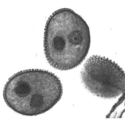
In the image above, you can see three pollen grains. In two of them, you can see the generative cell and the tube cell nucleus.
Observe a prepared slide of a Lilium ovary cross section (pre-fertilization). A Lilium pistil consists of three fused carpels. This is why there are three compartments, called locules, within the ovary.
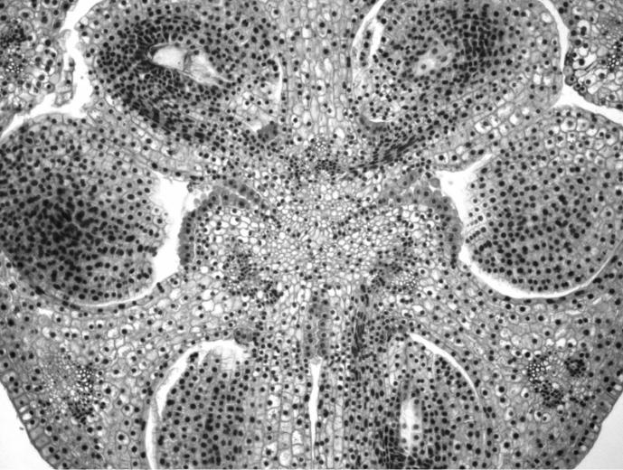
Inside each locule, there are two ovules, each connected to the ovary wall by a funiculus. The funiculus is much like an umbilical cord, providing nutrition to the developing ovule from the sporophyte through the placenta. The tissue surrounding the ovule is called the integument. The small opening in the integument is the micropyle.
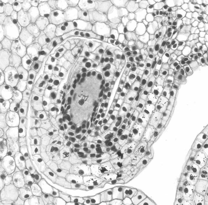
Depending on the pre-fertilization phase you are looking at, the ovule can appear quite differently. In the image above, the ovule is in the earliest stage of development. Currently, all of this tissue is diploid. The large cell in the center, the megasporocyte (2n), will undergo meiosis to produce four haploid cells: the egg and three other cells that will die. This egg will undergo three rounds of mitosis to produce a structure called the embryo sac, which is essentially the megagametophyte (n).
Observe a prepared slide of an embryo sac (this should also be a cross section of a Lilium ovary). The megagametophyte is housed at the center of the ovule and, when it is fully developed, is composed of 7 cells and 8 nuclei.
- The egg sits at the end closest to the micropyle, flanked by two synergids.
- On the other side of the embryo sac, there are three antipodals.
- In the center, there is a large cell with two nuclei. This is alternately called the central cell or the polar nuclei.
Note
Due to the way slides are made (a thin slice of a three dimensional structure), it is difficult to catch all 7 cells in one section unless they happen to be on the same plane.
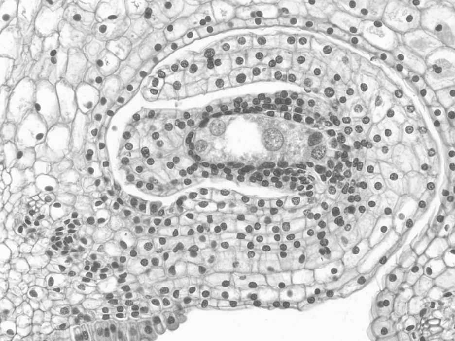
In the image above, you can see what is probably the egg and one synergid on the micropylar (left) side of the embryo sac. The large central cell is just to the right of the blank white area, and two of the three antipodals are visible on the far right.
The egg and the central cell will each be fertilized by one of the two sperm produced by the generative cell. The fertilized egg becomes a diploid zygote. The fertilized central cell becomes the triploid endosperm.
Observe a prepared slide of a Lilium ovary cross section (post-fertilization). Once fertilized by a pollen grain, the ovules become seeds, the megasporangium becomes the nucellus, the micropyle is closed, the integument becomes the seed coat, and the ovary wall begins to develop into the pericarp.
Within the seed, the embryo grows by mitosis and is nourished by the nucellus and endosperm. The seeds develop within the fruit until they are ready for dispersal.
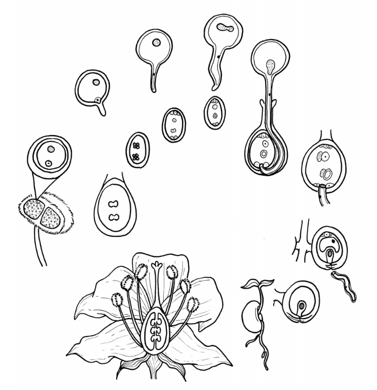
Label all of the bolded components of the angiosperm life cycle in the diagram below. Indicate where meiosis and fertilization occur. Choose a different color to represent haploid, diploid, and triploid tissues and color the tissues accordingly.
Angiosperm Life Cycle Diagram
Seed Germination
In the diagram below, watch a bean seed germinate and develop into a bean plant. Label the seed coat, radical, cotyledons, roop apical meristem, and shoot apical meristem.
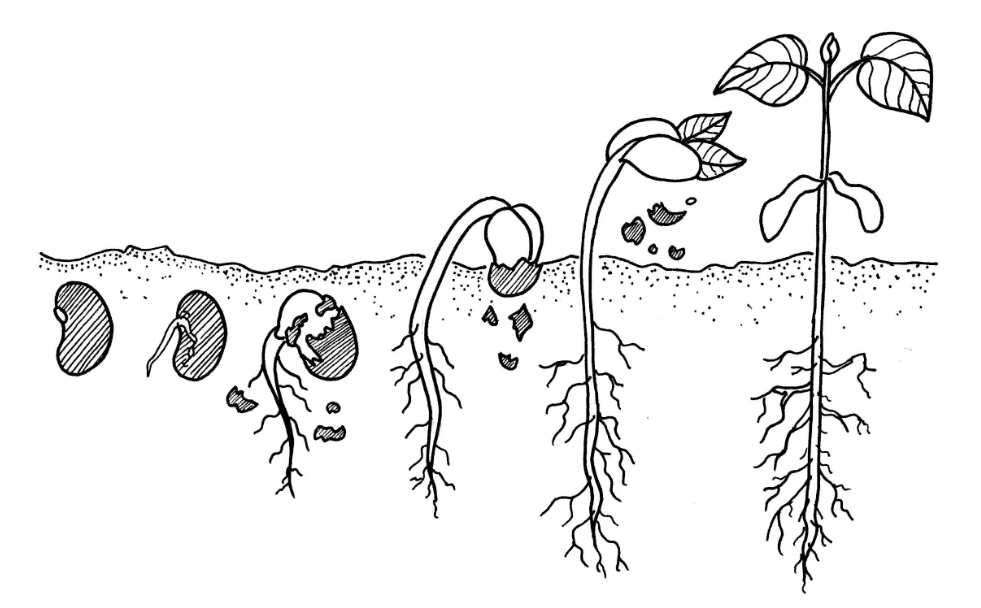
Why would a plant develop a root system before the shoot?