
7.3: Root Structure and Anatomy
- Page ID
- 29548

The root system of a plant serves two primary functions: water absorption and anchorage. Roots with a higher surface area will be better adapted to absorbing water because they have more area to interact with the soil environment. Increased interaction with the soil environment can also contribute to increased anchorage, but there are always trade-offs. If roots become too fine, they will be easily broken and lose the anchorage function. Additionally, finer roots can also lose more water if the soil environment becomes dry.
Root Development
In plants, both roots and shoots grow from the tip or apex of the plant. New cells are produced in these growing tips by meristems, groups of undifferentiated cells whose function is to divide by mitosis to produce new cells. Root growth begins at the root apical meristem (RAM). This meristem divides in two directions, producing a root cap to the outside of the root to protect the growing tip and the primary meristems to the inside: the protoderm, ground meristem, and procambium.
Primary meristems produce the primary tissues in the root:
- Protoderm → Epidermis
- Ground meristem → Cortex (and pith in monocots)
- Procambium → Primary xylem and primary phloem
These primary tissues will then either differentiate into specialized cells or, as is the case in many eudicots, become meristematic and produce secondary tissues. More on this in Lab Secondary Growth.
Observe a long section of Zea mays (corn) root tip. This root tip can be divided into three regions based on what is occurring in that stage of development:
Locate the root cap, RAM, protoderm, ground meristem, and procambium. These can be found at the very tip of the root, where the cells are small and densely clustered. If you trace the columns of cells back to their origin, they will coalesce at the RAM. This area of meristematic activity is called the zone of division.
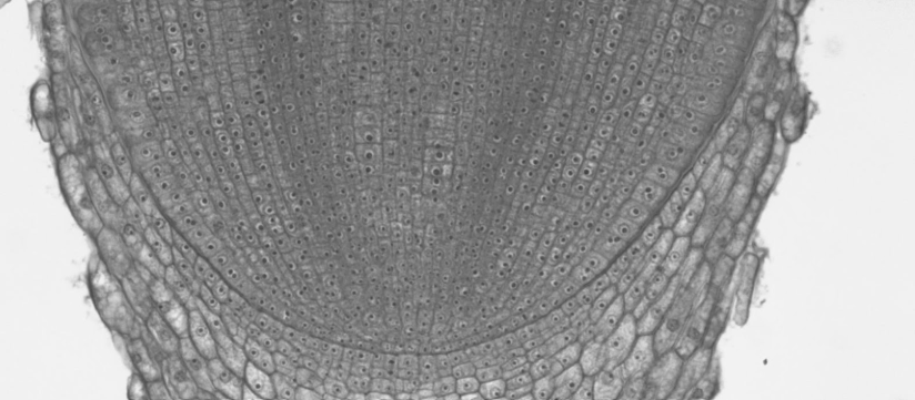
As you move up in the root, the cells begin to get larger, developing into primary tissues. This region is called the zone of elongation.
Further up the root, the larger cells begin to differentiate into specialized cells. In the xylem and phloem, you can find sclerenchyma with secondary walls. In the epidermis, you can see elongated cells called root hairs projecting outward. This region is called the zone of maturation.
Locate each of the zones described above and draw a root tip below. In the zone of division, label the root cap and the following meristems: RAM, protoderm, ground meristem, procambium. In the zone of elongation, label the following primary tissues: epidermis, cortex, pith, primary phloem, primary xylem. In the zone of maturation, label the following specialized cells: root hair, sieve tube element, companion cell, vessel element.
Root Development Flowchart
Below is a flowchart of eudicot root development in primary growth. There are two lines that will later lead to the secondary meristems. With this information and the filled in boxes, you should be able to determine which meristems and tissues go in the empty boxes.
Fill in the diagram below with the following terms: cortex, ground meristem, pericycle, primary phloem, and protoderm. Choose a different color to represent meristems and tissues, then color the boxes accordingly.
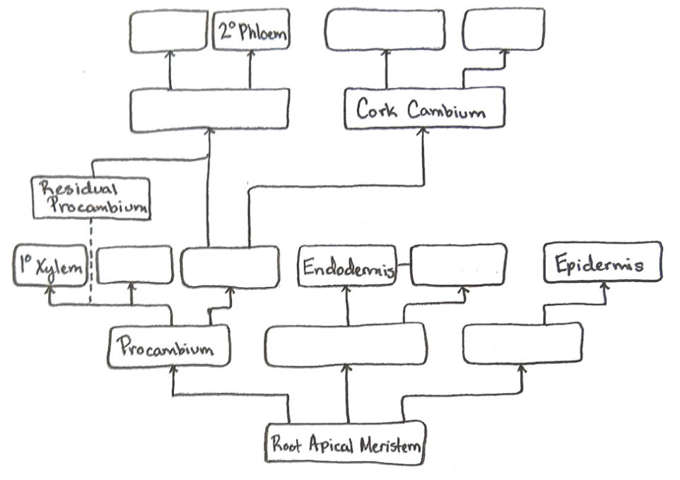
How would this flowchart be different in a monocot? Draw a flowchart of monocot root development in the space below.
Root Anatomy
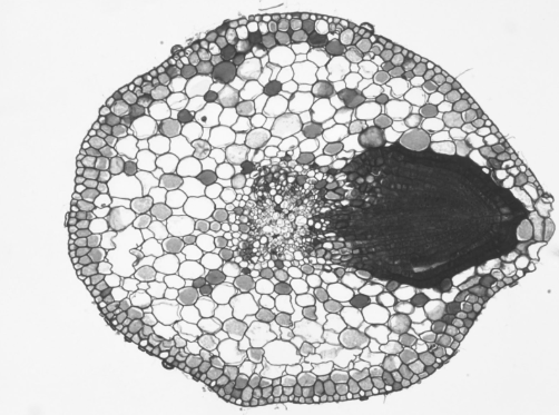
Monocots
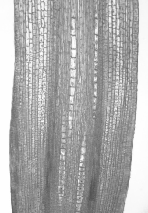
Observe a cross section of the zone of maturation in a Zea mays root. Locate the primary tissues and specialized cells that you found in the long section and label them in the image below.
Eudicots
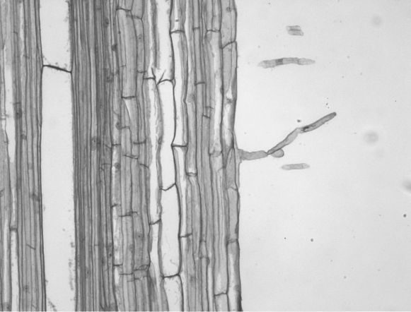
The development of roots in eudicots differs slightly from monocots. Monocots develop a ring of vascular tissue with ground tissue (pith) in the center. In eudicots, the vascular cylinder is closed. The primary xylem develops as an X (or sometimes Y) shape in the center with pockets of primary phloem in the spaces between the arms.
Encircling the conducting tissues is a meristematic layer called the pericycle. This single cell layer is responsible for producing lateral roots. Unlike root hairs which emerge on the outside of the root (exogenous, exo- meaning outside), lateral roots emerge from the internal tissues (endogenous, endo- meaning inside).
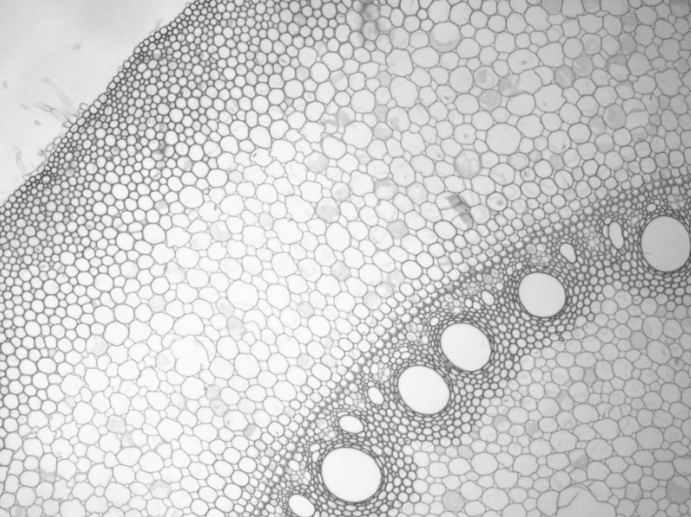
Observe a cross section of a Salix (willow) root producing lateral roots. Locate the primary xylem, primary phloem, pericycle, cortex, and epidermis. Label these features in the image below:.
Just outside the pericycle is the innermost layer of the cortex called the endodermis. The endodermis regulates what enters and exits the vascular cylinder. When the root is young, the epidermis contains many root hairs whose function is to absorb water. This water is transported through the cortex and into the xylem. As the root matures, the root hairs are lost from the epidermis. These areas of the root now function for anchorage and transportation of water. Because these areas are no longer taking up water, the vascular cylinder is sealed off by a waxy layer of suberin that covers the endodermis called the casparian strip. The casparian strip forms in stages with the areas closest to the arms of the xylem forming last. These are the last areas to allow for the passage of water, giving them the name passage cells.
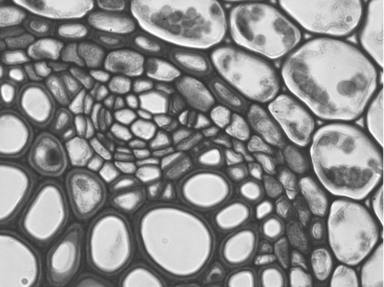
Observe a cross section of a Ranunculus (buttercup) root. Locate the pericycle, endodermis, casparian strip, and passage cells. The suberin in the casparian strip often stains similarly to the secondary walls in the xylem. Draw and label the cross section in the space below.