
3.1.3: Plant Tissues
- Page ID
- 27734

Learning Objectives
- Describe the difference between meristematic and non-meristematic tissues.
- Compare and contrast dermal, ground, and vascular tissue.
Plants are multicellular eukaryotes with tissue systems made of various cell types that carry out specific functions. Plant tissues are composed of cells that are similar and perform a specific function. Together, tissue types combine to form organs. Each organ itself is also specific for a particular function.
Plant tissue systems fall into one of two general types: meristematic tissue, and permanent (or non-meristematic) tissue. Cells of the meristematic tissue are found in meristems, which are plant regions of continuous cell division and growth. Meristematic tissue cells are either undifferentiated or incompletely differentiated, and they continue to divide and contribute to the growth of the plant. In contrast, permanent tissue consists of plant cells that are no longer actively dividing.
Meristematic tissues consist of three types, based on their location in the plant. Apical meristems contain meristematic tissue located at the tips of stems and roots, which enable a plant to extend in length. Lateral meristems facilitate growth in thickness or girth in a maturing plant. Intercalary meristems occur only in monocots, at the bases of leaf blades and at nodes (the areas where leaves attach to a stem). This tissue enables the monocot leaf blade to increase in length from the leaf base; for example, it allows lawn grass leaves to elongate even after repeated mowing.
Meristems produce cells that quickly differentiate, or specialize, and become permanent tissue. Such cells take on specific roles and lose their ability to divide further. They differentiate into three main types: dermal, vascular, and ground tissue. Dermal tissue covers and protects the plant. The ground tissue serves as a site for photosynthesis, provides a supporting matrix for the vascular tissue, and helps to store water and sugars. The vascular tissue transports water, minerals, and sugars to different parts of the plant. Ground tissue is a simple tissue, meaning that each ground tissue consists of only one cell type. Dermal and vascular tissues are complex tissues because they consist of multiple cell types.
Dermal Tissue
Dermal tissue covers the plant and can be found on the outer layer of roots, stems and leaves. Its main functions are transpiration, gas exchange and defense. The epidermis is an example of dermal tissue (Figure \(\PageIndex{1}\)). It is composed of a single layer of epidermis cells. It may contains stomata and guard cells that allow gas exchange. It may contain root hairs that increase surface area or or trichomes used in transpiration or defense. It may contain a waxy cuticle if found on the upper surface of leaves, to aid with lowering transpiration.
In woody plants, the epidermis breaks apart into a thick periderm as secondary growth allows the plant to grow in girth. The cork cambium, which makes cork cells, the cork cells (which are dead at maturity), and the phelloderm (parenchyma cells on the inside of the cork cambium) together make up the periderm (Figure \(\PageIndex{2}\)). The periderm functions as the first line of defense for the plant, protecting it from fire or heat injury, dehydration, freezing conditions, and/or disease.

Ground Tissue
Often times, tissues that are not considered dermal or vascular tissue are noted as ground tissue. These cells store molecules (such as starch), photosynthesize (such as mesophyll cells), or support the plant. There are three types of ground tissue: collenchyma, sclerenchyma, and parenchyma.
Collenchyma (Figures \(\PageIndex{3-4}\)) is living supportive tissue that has elongated cells and an unevenly thickened primary cell wall. Its main function is the mechanical support of young stems and leaves via turgor.
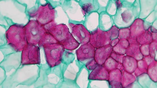
Sclerenchyma is a dead supportive tissue that consists of long sclerenchyma fibers (Figure \(\PageIndex{4}\)) or short, crystal-like cells (sclereids; Figure \(\PageIndex{5}\)). Sclerenchyma fibers occur in groups (bundles). Sclereids may be branched or not and occur individually or in small clusters. Each cell has a uniformly thick secondary wall that is rich in lignin. Its main function is a support of older plant organs, and also hardening different parts of plants (for example, make fruit inedible before ripeness so no one will take the fruit before seeds are ready to be distributed). Without sclerenchyma, if a plant isn’t watered, the leaves will droop because the vacuoles will decrease in size which lowers the turgor. Fibers inside phloem (see below) are sometimes regarded as a separate sclerenchyma.
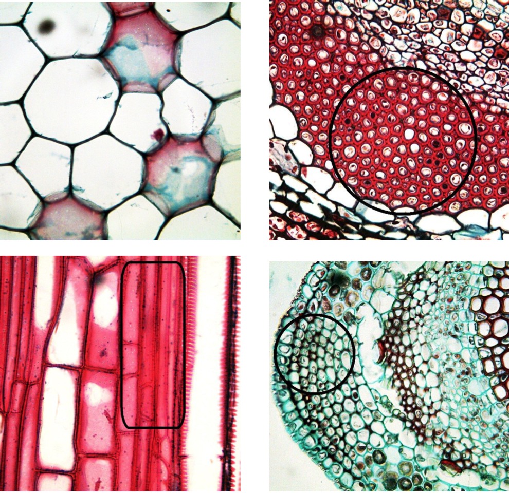
Parenchyma (Figure \(\PageIndex{4}\)) are spherical, elongated cells with a thin primary cell wall. It is a main component of young plant organs. The basic functions of parenchyma are photosynthesis and storage. They are also important in regeneration because they are totipotent (capable of differentiating into any cell type). Parenchyma cells are widespread in plant body. They fill the leaf, frequent in stem cortex and pith and is a component of complex vascular tissues (see below).
Vascular Tissue
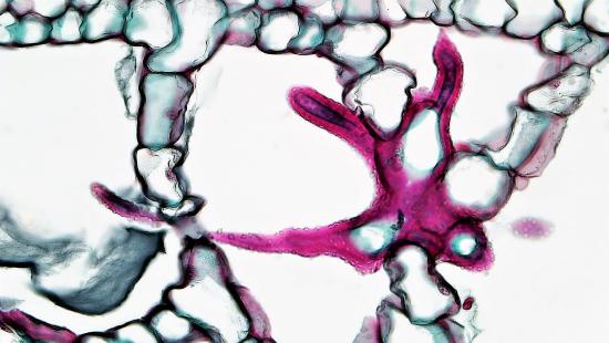
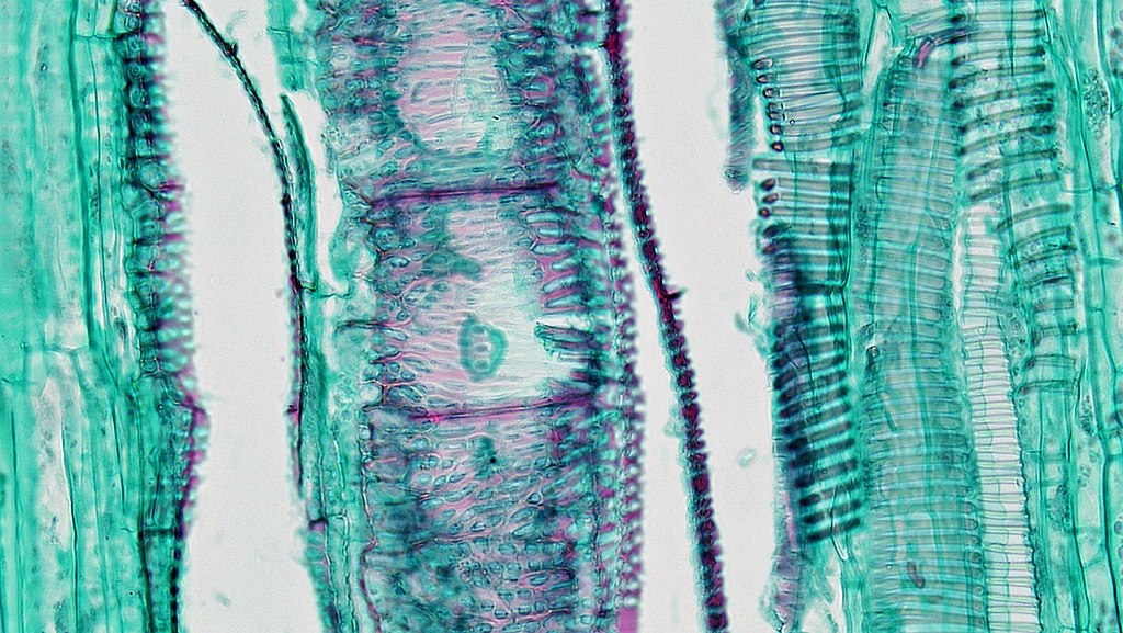
Vascular tissue is the plumbing system of the plant. It allows water, minerals, and dissolved sugars from photosynthesis to pass through roots, stems, leaves, and other parts of the plant. It is primary composed of two types of conducting tissue: xylem and phloem. The veins on leaves are an example of vascular tissue, moving material through the plant in the same manner that our blood vessels carry nutrients through our body. The xylem and phloem always lie adjacent to each other (Figure \(\PageIndex{6}\)). In stems, the xylem and the phloem form a structure called a vascular bundle; in roots, this is termed the vascular stele or vascular cylinder.
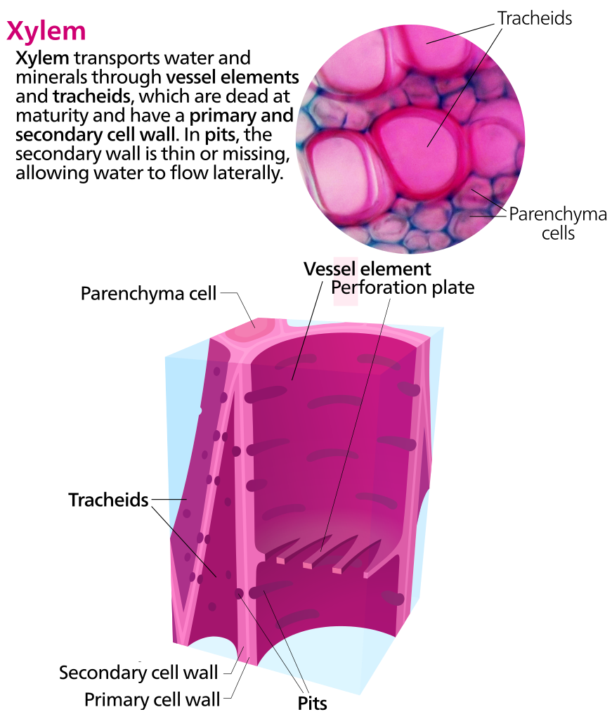
Xylem tissue transports water and minerals from the roots to different parts of the plant. The conducting cells of the xylem are called tracheary elements. Parenchyma cells are also found in the xylem, and sclerenchyma fibers and sclereids are sometimes present.
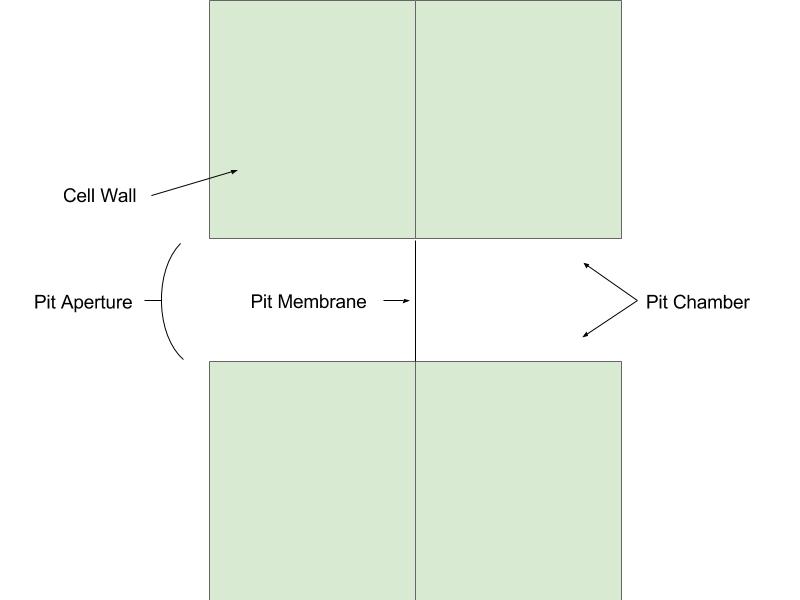
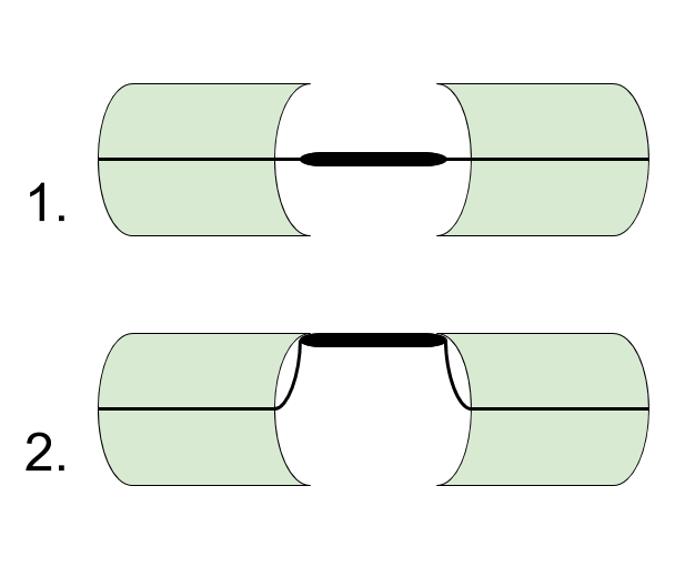
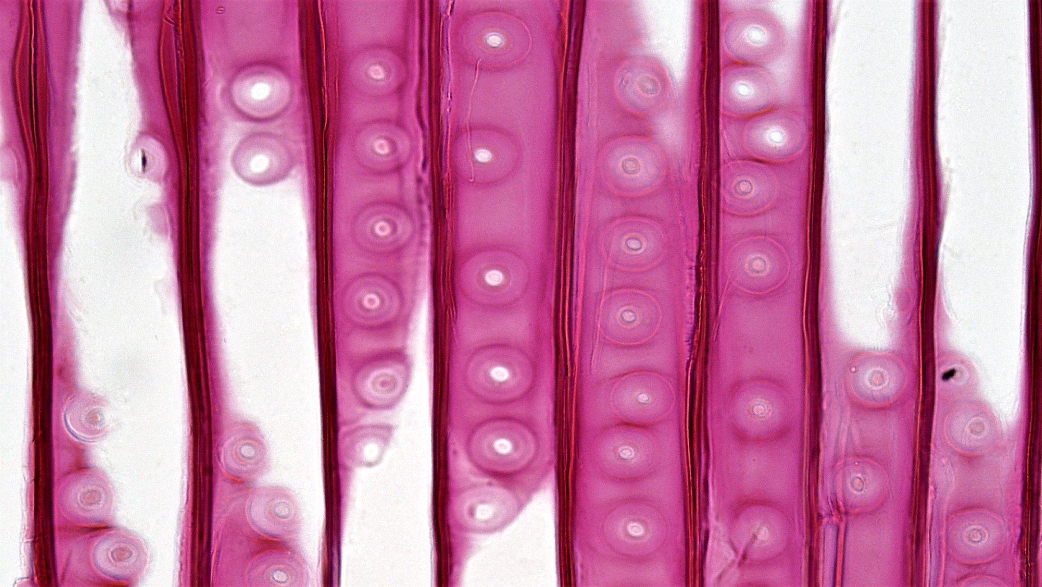
There are two type of tracheary elements: vessel elements and tracheids (Figure \(\PageIndex{7}\)). Both cell types that are dead at maturity and have thickened secondary cell walls. These cells connect to one another and allow water to be transported through them. Structurally, the vessel elements are wider than tracheids and contain perforation plates between adjacent vessel elements (Figure \(\PageIndex{7-8}\)). Wide openings (slits or pores) in perforation plates allow water to flow vertically between vessel elements, forming a continuous tube. Both types of tracheary elements contain pits, gaps in their secondary cell walls. Adjacent cells have pits in the same locations, forming pit pairs, which allow water and minerals to flow between adjacent cells through the pit membrane (the remaining, thin primary cell walls in these regions; Figure \(\PageIndex{9-10}\)). Therefore, water flows through both perforation plates and pit pairs in vessel elements but only through pit pairs in tracheids. While water can move more quickly through vessel elements, they are more susceptible to air bubbles. An air bubble disrupts cohesion in the column of water moving up the tube of vessel elements preventing use of that particular pathway. In tracheids, an air bubble would only decommission a single tracheid rather than an entire column of vessel elements. Vessel elements are found only in angiosperms, but tracheids are found in both angiosperms and gymnosperms.
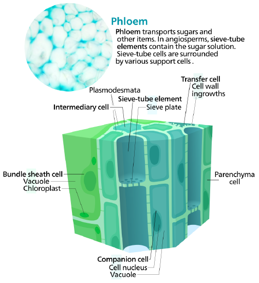
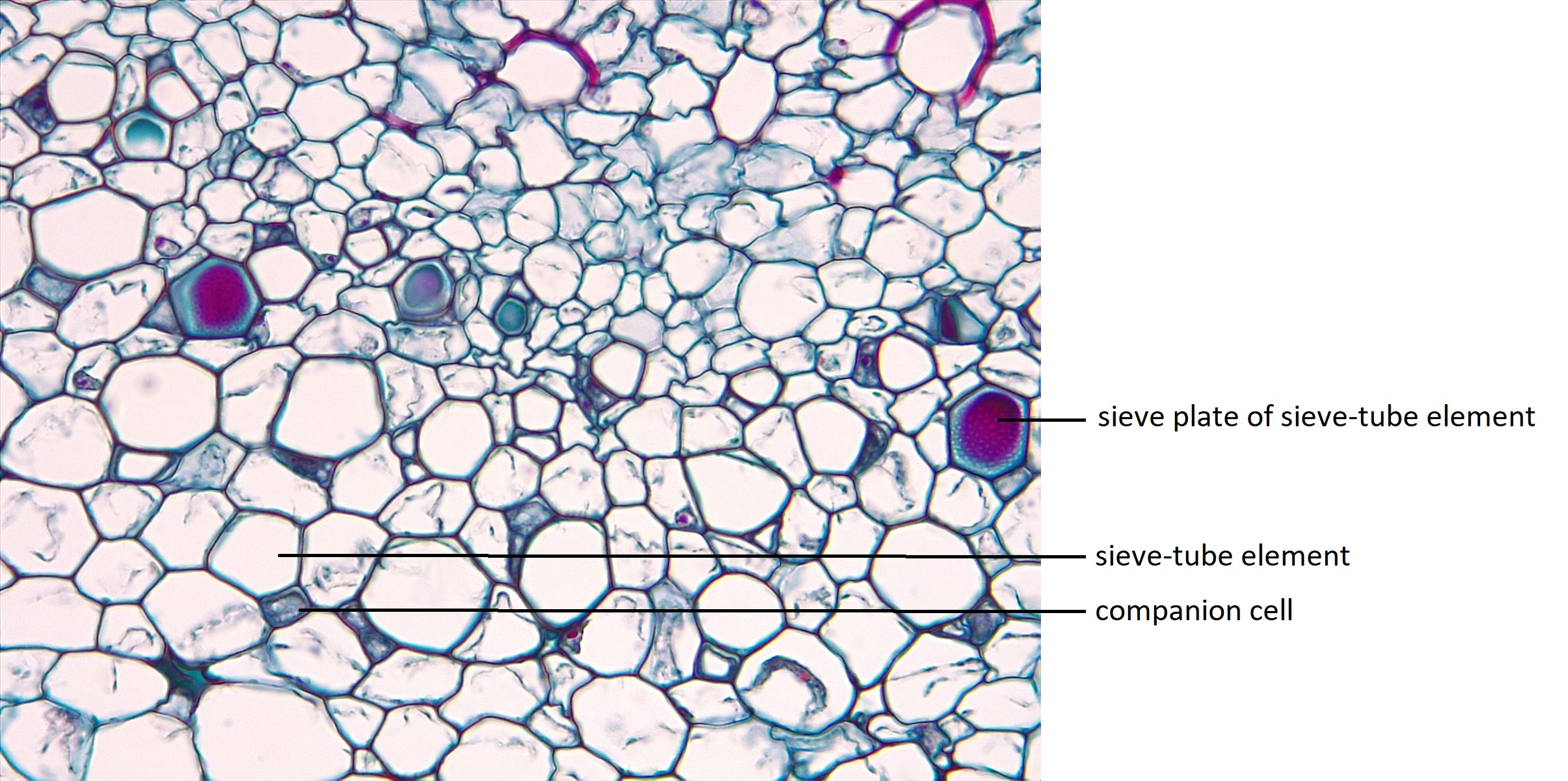
Phloem tissue transports organic compounds such as sugars from the site of photosynthesis to rest of the plant (Figure \(\PageIndex{11-12}\)). The conducting cells of the phloem are called sieve elements. In comparison to tracheary elements, sieve elements have only primary cell walls (and thus thinner cell walls overall) and are alive at maturity; however, they lack certain organelles, including a nucleus. Sieve-tube elements are the sieve elements found only in angiosperms while sieve cells are found only in gymnosperms while. Both types of sieve elements have pores in their cell walls (sieve areas) that allow transfer of materials between adjacent cells, but these are concentrated at sieve plates in sieve-tube elements and evenly distributed in sieve cells. Because they lack essential organelles, sieve elements rely on specialized parenchyma cells to support them. Companion cells support sieve-tube elements in angiosperms, and albuminous cells support sieve cells in gymnosperms. Additionally parenchyma cells and sclerenchyma cells (phloem fibers) are also found in the phloem.
The table below summarizes differences between xylem and phloem:
| Xylem | Phloem | |
|---|---|---|
| Contains mostly | Dead cells | Living cells |
| Transports | Water & Minerals | Sugar |
| Direction | Up | Up and Down |
| Biomass | Big | Small |
Meristematic Tissue
Meristems produce cells that quickly differentiate, or specialize, and become permanent tissue. Such cells take on specific roles and lose their ability to divide further. They differentiate into three main types: dermal, vascular, and ground tissue. Dermal tissue covers and protects the plant, and vascular tissue transports water, minerals, and sugars to different parts of the plant. Ground tissue serves as a site for photosynthesis, provides a supporting matrix for the vascular tissue, and helps to store water and sugars.
Attributions
Curated and authored by Kammy Algiers and Melissa Ha using the following sources:
- 30.1 The Plant Body and 30.2 Stems from Biology 2e by OpenStax (licensed CC-BY). Access for free at openstax.org.
- 5.1 Tissues from Introduction to Botany by Alexey Shipunov (public domain)