
2.4.3.2: Brown Algae and Diatoms
- Page ID
- 31928

Learning Objectives
- Use life history, morphology, and cellular components to identify brown algae.
- Identify the components of a kelp thallus.
- Identify structures and events in the Fucus life cycle and know their ploidy.
- Identify structures and events in the Laminaria life cycle and know their ploidy.
- Use life history, morphology, and cellular components to identify diatoms.
- Classify diatoms based on symmetry and ecology.
- Describe sexual and asexual reproduction in diatoms.
Brown algae (class Phaeophyceae) and diatoms (class Bacillariophyceae) belong to the phylum Ochrophyta. They are the result of a secondary endosymbiosis between a heterokont and a photosynthetic eukaryote. Heterokonts, such as the Oomycota, are united by the presence of a textured, or “hairy,” flagellum and an additional flagellum that lacks hair-like projections (Figure \(\PageIndex{1}\)). Members of this subgroup range in size from single-celled diatoms to the massive and multicellular kelp.
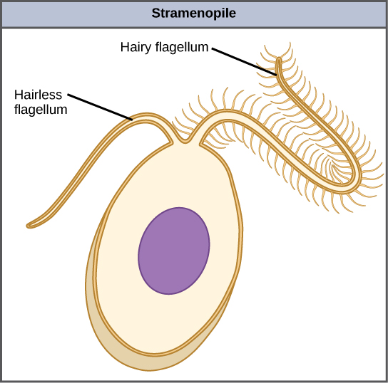
Brown algae, diatoms, and oomycetes belong to a single clade, the stramenopiles (aka heterokonts). The photosynthetic stramenopiles share the following characteristics:
- 4-membraned chloroplasts
- a yellow-brown pigment (which gives them their color). It is a carotenoid called fucoxanthin.
- chlorophylls a and c
- most have a diplontic life cycle (as you'll find below, Laminaria has a haplodiplontic life cycle)
Phaeophyceae
This group is commonly called the brown algae and includes rockweeds and kelps. Kelps are some of the fastest growing organisms on the planet! Brown algae are primarily marine and are often found in the intertidal zone. Members of this phylum are used for food in some coastal areas of the world and harvested in the U. S. for fertilizer and as a source of iodine.
Brown algae are brown due to the large amounts of carotenoids they produce, primarily one called fucoxanthin. These organisms are exclusively multicellular and have filamentous, multinucleate cells (much like oomycetes). They can get so large that they require special conductive cells to transport photosynthates from their blades down to the rest of their tissues. These conductive cells are called trumpet hyphae and have sieve plates and resemble sieve tubes found in flowering plants. Brown algae have cellulose cell walls and store carbohydrates in the form of laminarin. The polymer alginate can also be found in the cell walls of brown algae and is used commercially for a variety of purposes, including the high fidelity molds used in dentistry.
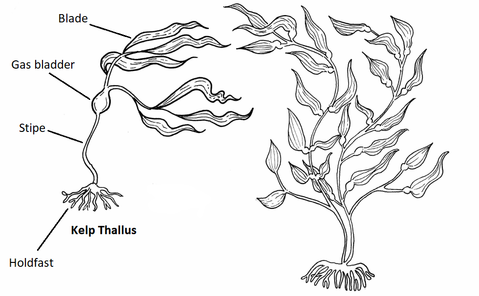
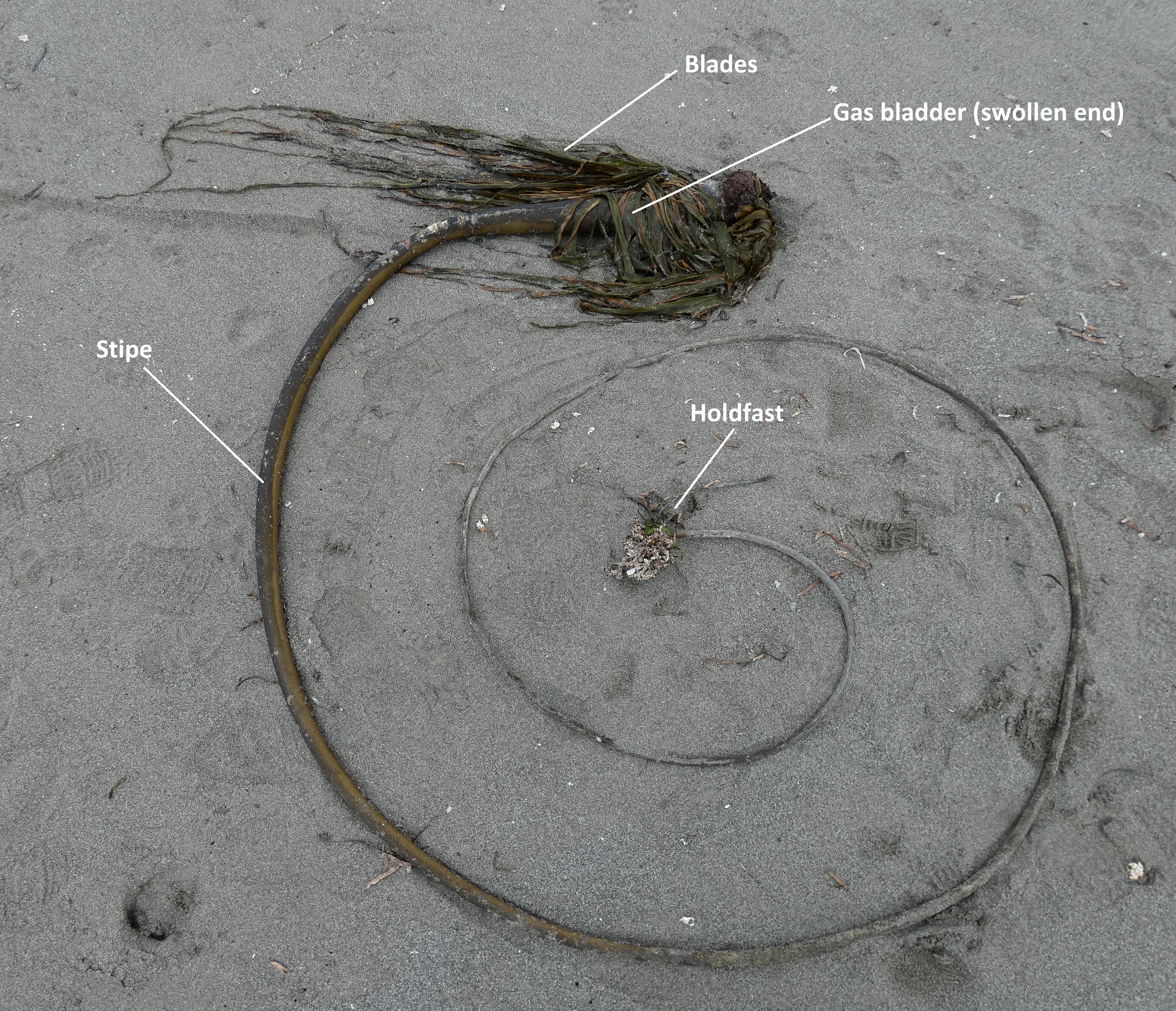
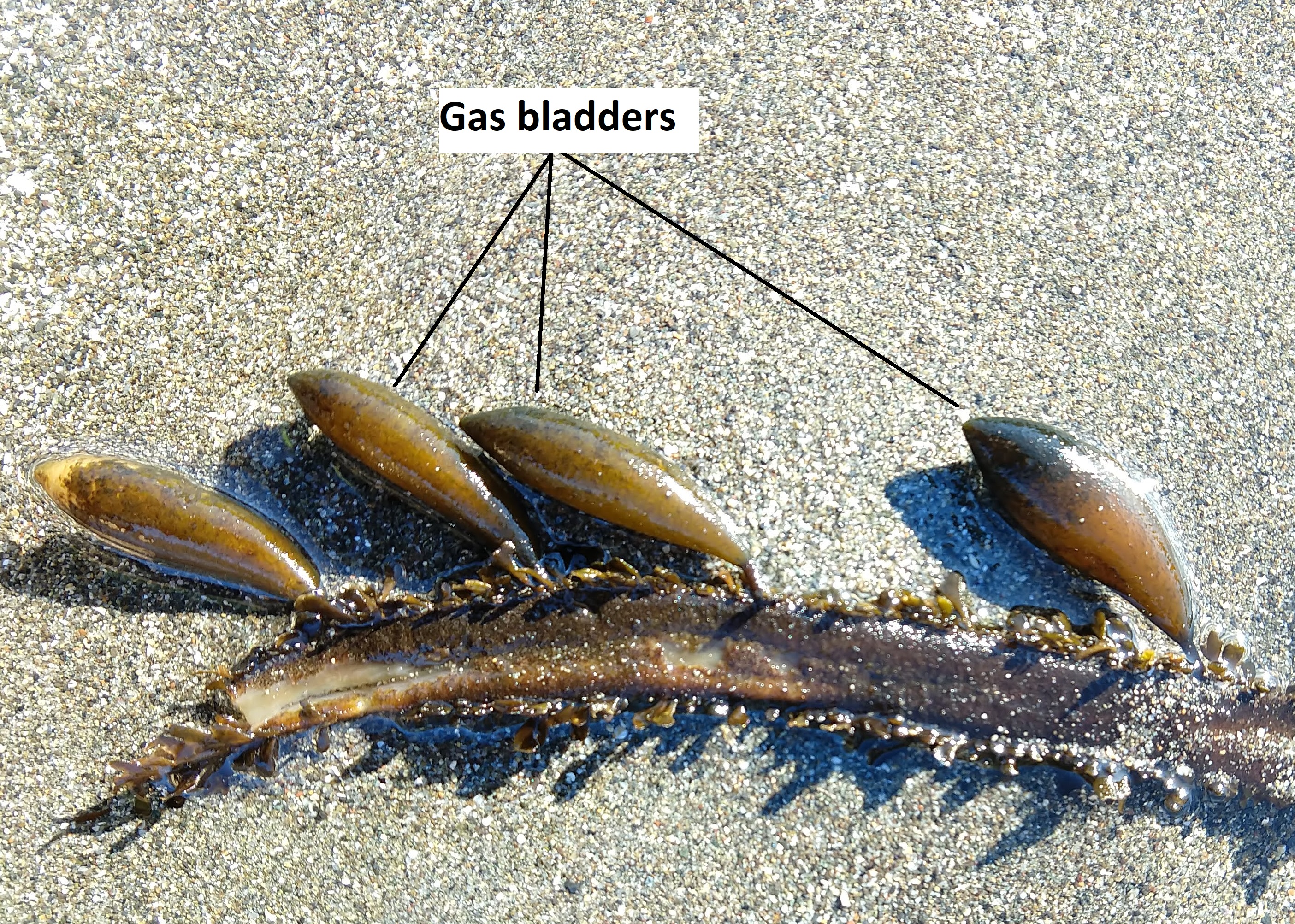
Kelp
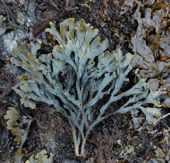
Much like Saprolegnia, the body of an alga is termed a thallus because it is not differentiated into specialized tissues. The general morphology of a brown alga includes a holdfast, stipe, gas bladder(s), and blade(s) (Figures \(\PageIndex{2-4}\)).
Fucus
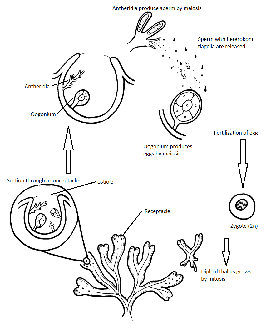
A model organism for the Phaeophyta life cycle is Fucus (rockweed), which, like its relative Saprolegnia, has a diplontic life cycle. The Fucus thallus has dichotomous branching (forking into two equal branches) and swollen, heart-shaped reproductive tips of the branches. These swollen branch tips are called receptacles (Figure \(\PageIndex{5}\)).
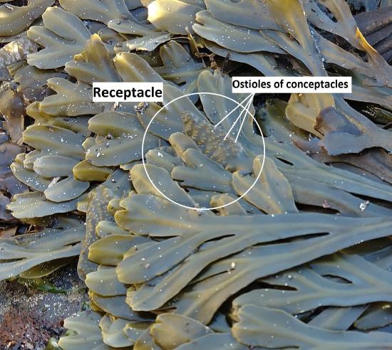
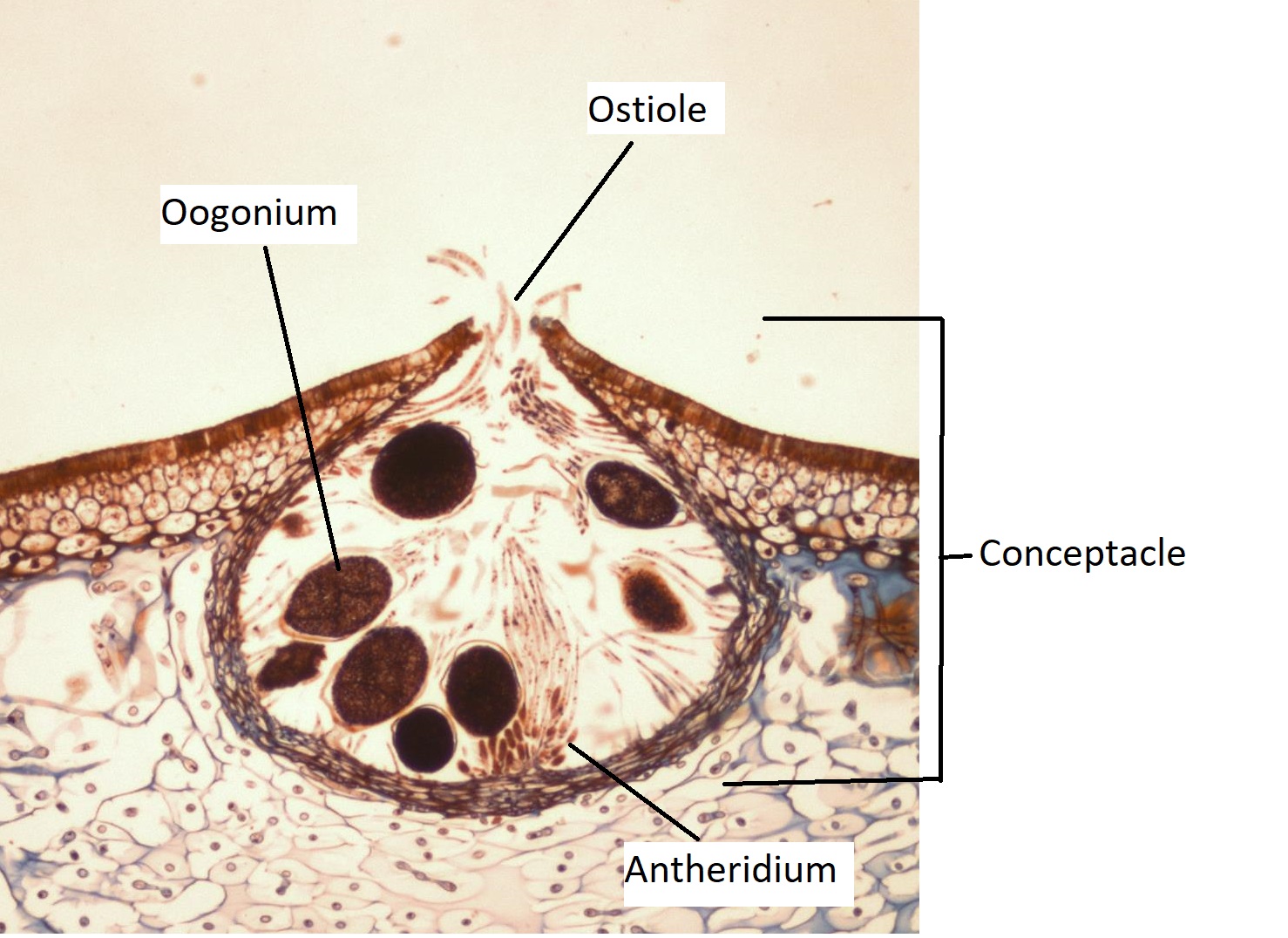
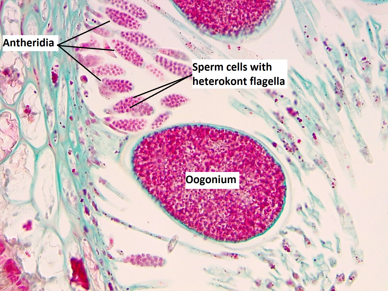
The receptacles are covered in small bumps, each with a pore at the center of the bump called an ostiole. The bumps are conceptacles, chambers that house the gametangia (Figure \(\PageIndex{6}\)). Phaeophyta produce oognia, globose gametangia that undergo meiosis to produce eggs, and antheridia, branched gametangia that undergo meiosis to produce sperm (Figure \(\PageIndex{7}\)).
Fucus Life Cycle
Fucus has a diplontic life cycle (Figure \(\PageIndex{8}\)) where haploid gametes are produced from a diploid thallus. These haploid gametes do not grow, but fuse together to form a zygote. See Figure \(\PageIndex{9}\) for an example of alternation of generations in the Phaeophyta.
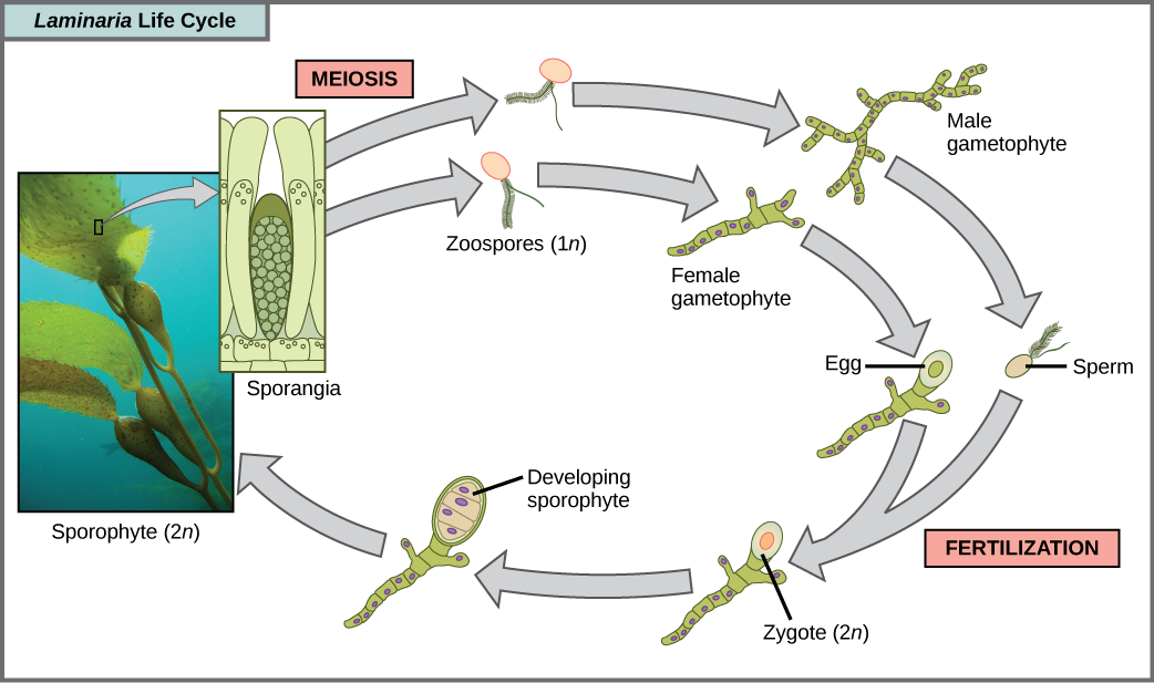
Laminaria Life Cycle
A variety of algal life cycles is represented by the stramenopiles, but the most complex is alternation of generations, in which both haploid and diploid stages involve multicellularity. Compare this life cycle to that of humans, for instance. Haploid gametes produced by meiosis (sperm and egg) combine in fertilization to generate a diploid zygote that undergoes many rounds of mitosis to produce a multicellular embryo and then a fetus. However, the individual sperm and egg themselves never become multicellular beings. Terrestrial plants also have evolved alternation of generations. In the brown algae genus Laminaria, haploid spores develop into multicellular gametophytes, which produce haploid gametes that combine to produce diploid organisms that then become multicellular organisms with a different structure from the haploid form (Figure \(\PageIndex{9}\)). Certain other organisms, such as the red alga Polysiphonia, perform alternation of generations in which both the haploid and diploid forms look the same.
Summary of Characteristics for Brown Algae
- Morphology: Multicellular thallus
- Cell wall composition: Cellulose and calcium alginate
- Chloroplasts: 4 membranes, pigments are chlorophyll a, chlorophyll c, and fucoxanthin
- Storage carbohydrate: Laminarin
- Life cycle: Primarily diplontic (alternation of generations in some species)
- Ecology: Marine
Bacillariophyceae
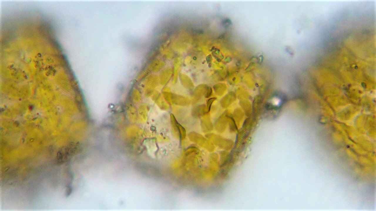
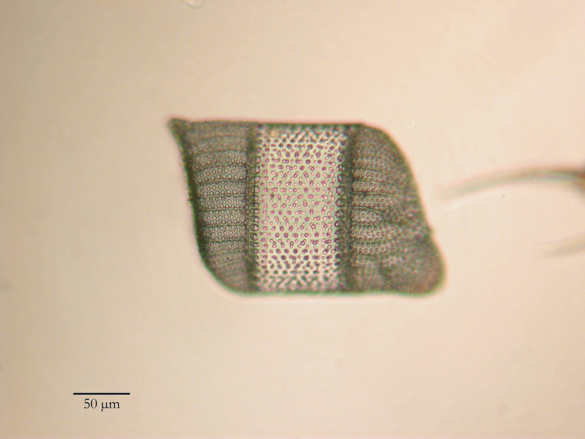
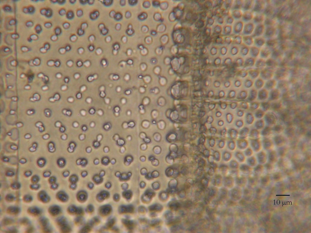
Diatoms are another photosynthetic lineage of photosynthetic heterokonts that was derived from the secondary endosymbiotic event. Diatoms are an incredibly diverse group of unicellular organisms containing anywhere from 20,000 to 2 million species. These organisms are unicellular and surrounded by a frustule, a silica shell made from two distinct valves that enclose the plasma membrane. Frustules are amazingly intricate, covered with small pores in an arrangement specially adapted for capturing sunlight (Figure \(\PageIndex{11}\)). Some diatoms exhibit a slit in their silica shell, called a raphe. By expelling a stream of mucopolysaccharides from the raphe, the diatom can attach to surfaces or propel itself in one direction.
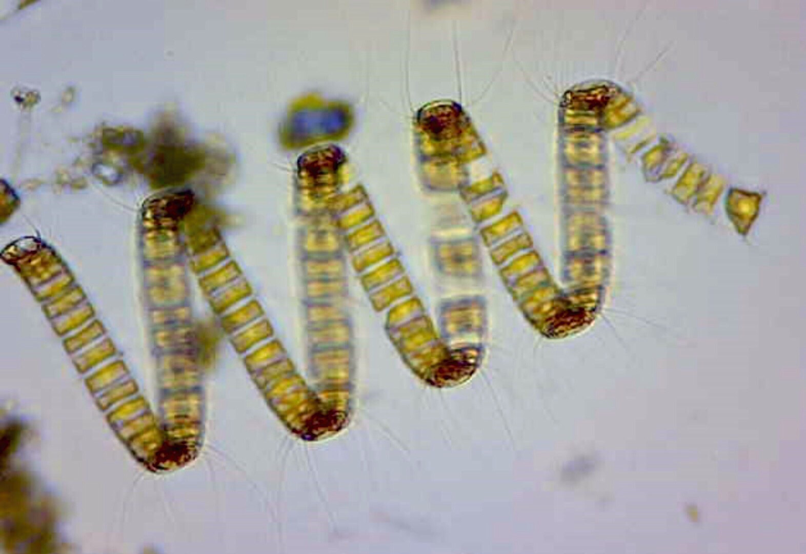
Like the brown algae, they have golden chloroplasts with 4-membranes (Figure \(\PageIndex{10}\)). Diatoms store carbohydrates in the form of chrysolaminarin. The silica frustules of diatoms found in sediments (diatomaceous earth) are used for myriad commercial purposes, including toothpaste additives (as an abrasive), filters, and insulation.
Morphology
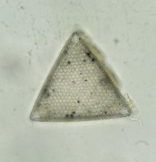
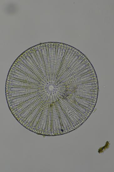
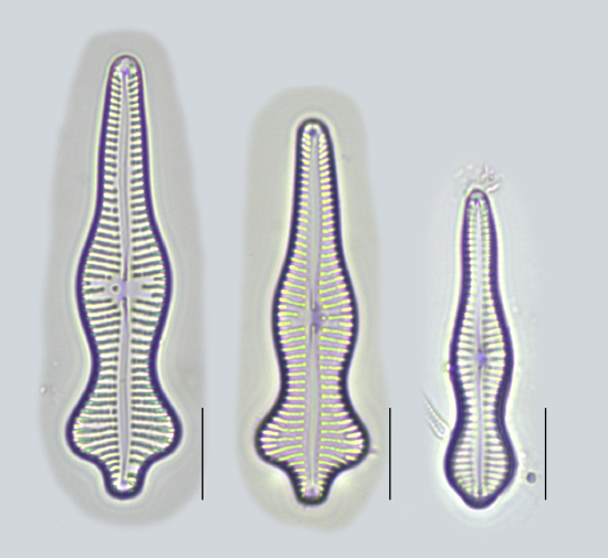
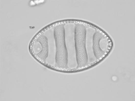
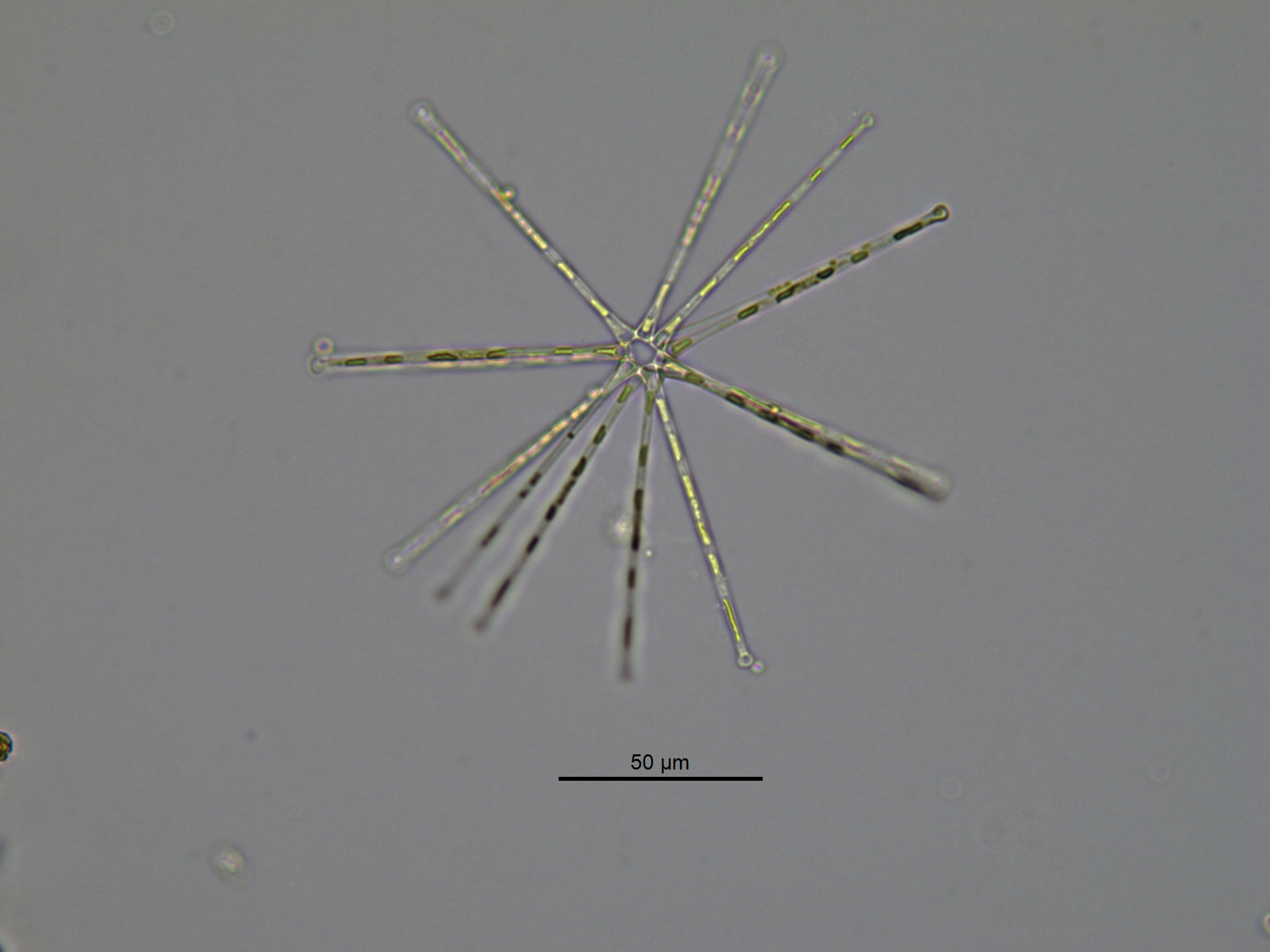
We are still trying to figure out how to determine what a diatom "species" is and, so far, they have been classified based on the morphology of the frustule. Using this classification, historically there were two major groups of diatoms: centric (have radial symmetry, see Figure \(\PageIndex{12}\)) and pennate (have bilateral symmetry, see Figure \(\PageIndex{13}\)). These classifications have improved and increased in complexity, so here we will cover just the broad strokes. For a more in-depth look at current diatom morphological classification and fantastic images, check out this website.
Ecology
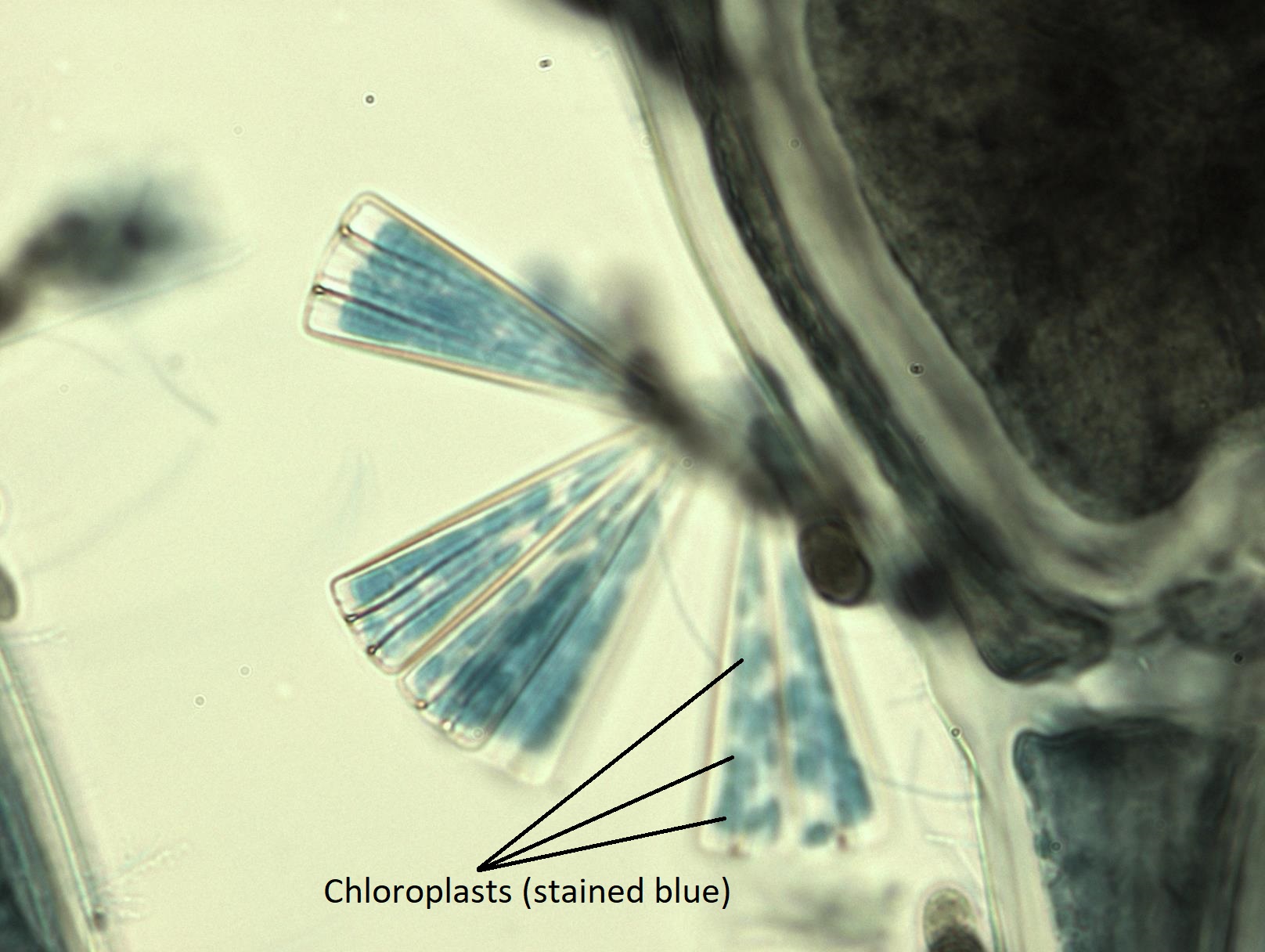
In addition to morphology, diatoms can also be classified by where they occur. Free-floating diatoms are planktonic. Diatoms attached to other organisms (like giant kelp) are epiphytic (Figure \(\PageIndex{15}\)). Epiphytic diatoms can be found in aquatic ecosystems on algae and aquatic angiosperms like eelgrass, as well as terrestrial ecosystems, living in the damp crevices of tree bark. Benthic diatoms tend to dwell toward the bottom of a body of water. In general, these three categorizations refer to aquatic ecosystems. However, diatoms can be found just about anywhere there is water in terrestrial ecosystems. The community composition of diatoms varies depending on location. Because of this, diatoms have been used in forensic investigations to determine where someone drowned (depending on the diatom species present) and how long ago they drown (based on how far the diatoms had migrated into their tissues).
Diatoms are major producers in aquatic environments; that is, they are responsible for as much as 40% of the photosynthesis that occurs in fresh water and in the oceans. They serve as the main base of the food chains in these habitats, supplying calories to heterotrophic protists and small animals. These, in turn, feed larger animals. During periods of nutrient availability, diatom populations bloom to numbers greater than can be consumed by aquatic organisms. The excess diatoms die and sink to the sea floor where they are not easily reached by saprotrophs that feed on dead organisms. As a result, the carbon dioxide that the diatoms had consumed and incorporated into their cells during photosynthesis is not returned to the atmosphere. In general, this process by which carbon is transported deep into the ocean is described as the biological pump, because carbon is “pumped” to the ocean depths where it is inaccessible to the atmosphere as carbon dioxide. The biological carbon pump is a crucial component of the carbon cycle that maintains lower atmospheric carbon dioxide levels.
Reproduction
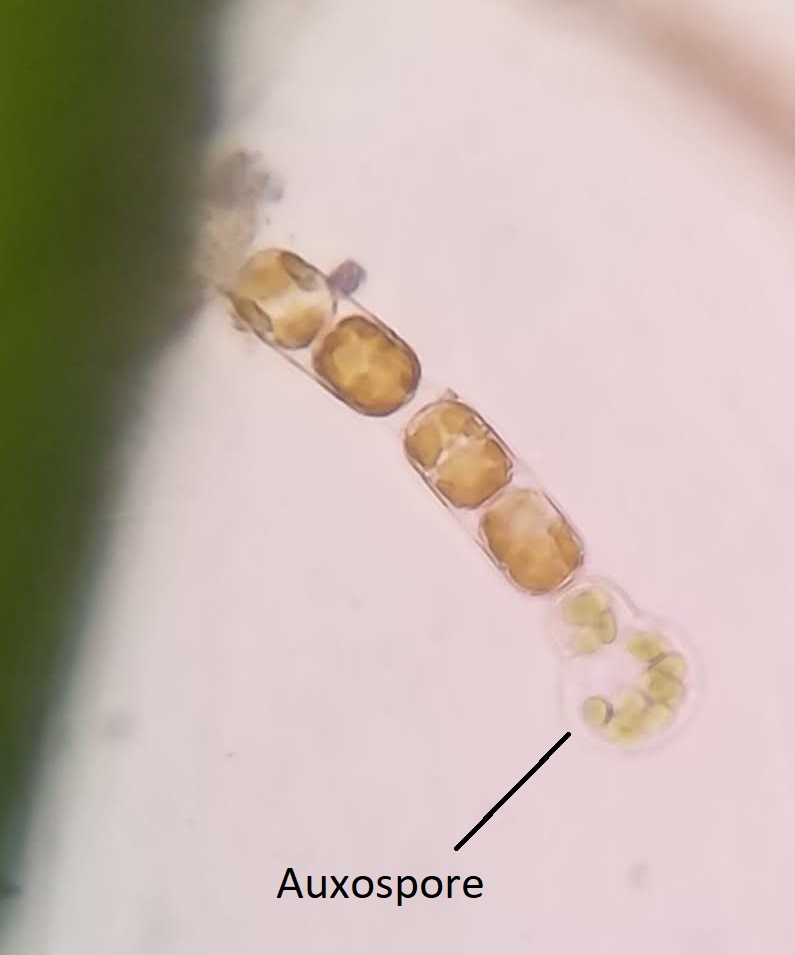
Diatoms primarily reproduce asexually by binary fission, similar to prokaryotes. During binary fission, the two valves of the frustule are separated and each new cell forms a new valve inside the old one. However, the new valve is always smaller. If diatoms only reproduce in this way, it results in a continual decrease in average size. When some minimal size is reached, this can trigger sexual reproduction. When diatoms sexually reproduce, they have a diplontic life cycle and produce a very large auxospore (Figure \(\PageIndex{16}\)).
Diversity
Video \(\PageIndex{1}\): This video shows some of the incredible diversity of diatom shapes and the amazing art Klaus Kemp makes with them. Sourced from YouTube.
Summary of Characteristics for Diatoms
- Morphology: Unicellular
- Cell wall composition: Silica frustule
- Chloroplasts: 4 membranes, pigments are chlorophyll a, chlorophyll c, and fucoxanthin
- Storage carbohydrate: Chrysolaminarin
- Life cycle: Diplontic
- Ecology: Everywhere! Marine, freshwater, and terrestrial.
Summary
Though brown algae and diatoms seem to have very little in common morphologically, they are descended from a common ancestor. Both of these groups have a diplontic life cycle during some stage of which a cell will have heterokont flagella. They have 4-membraned chloroplasts that contain the pigments chlorophyll a, chlorophyll c, and fucoxanthin. This latter pigment gives the chloroplasts in these groups a golden color. This is about where the similarities end.
Brown algae are exclusively multicellular and found in marine habitats, most typically in the intertidal zone. Their cell walls contain cellulose and they store their carbohydrates as laminarin.
Diatoms are exclusively unicellular and found in almost every habitat where there is water. Their single cell is surrounded by a silica frustule composed of two distinct valves. They store their carbohydrates as chrysolaminarin.
Attribution
Curated and authored by Maria Morrow, CC-BY-NC, using the following sources:
- 19.1.2 Protists from Biology by John. W. Kimball (licensed CC-BY)
- 23.3 Groups of Protists from Biology 2e by OpenStax (licensed CC-BY). Access for free at openstax.org.