
2.4.1: Introduction to Protists
- Page ID
- 47625
Learning Objectives
- Explain how organisms were originally classified under Protista.
- Describe the diversity of metabolic strategies and other life history traits within this artificial group.
- Identify evolutionary relationships between "protists" using a phylogenetic tree.
What are Protists?
The term "protist" is a general description for eukaryotic organisms that are not animal, plant, or fungus. These organisms were formerly classified in Kingdom Protista. As we have come to learn about genetics and the evolutionary history of organisms, we have discovered that these organisms come from many distinct evolutionary groups, some of which are closer to fungi and animals than to plants or each other. There are over 100,000 described living species of "protists", and it is unclear how many undescribed species may exist. Since many of these organisms live as commensals or parasites within other organisms, and these relationships are often species-specific, there is a huge potential for diversity that matches the diversity of hosts. As the catchall term for eukaryotic organisms that are not animal, plant, or fungus, it is not surprising that very few characteristics are common to all protists.
- They are eukaryotes because they all have a nucleus.
- Most have mitochondria although some have later lost theirs. Mitochondria were derived from aerobic alpha-proteobacteria that once lived within their cells.
- Many have chloroplasts with which they perform photosynthesis. These chloroplasts were derived from photosynthetic cyanobacteria originally, though have been acquired via secondary endosymbiotic events in several lineages.
- Many are unicellular and all groups (with one exception) contain some unicellular members.
- The name Protista means "the very first", and some of the 80-odd groups of organisms that we once classified as protists may well have had long, independent evolutionary histories stretching as far back as 2 billion years. Genome analysis added to other criteria show that other lineages are derived from more complex ancestors; that is, some are not "primitive" at all.
- Genome analysis also shows that many of the groups placed in the Protista are not at all closely related to one another; that is, the protists do not represent a single clade.
- So we consider them here as a group more for our convenience than as a reflection of close kinship, and a better title for this page would be "Eukaryotes that are neither Animals, Fungi, nor Plants".
Evolutionary Relationships
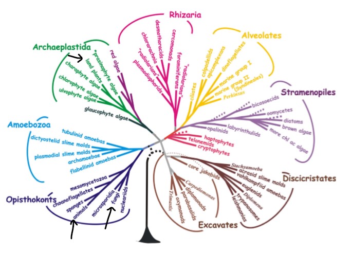
In the phylogenetic tree above (Figure \(\PageIndex{1}\)), protists do not share a common ancestry. Slime molds share a more recent evolutionary history with fungi and animals, while red and green algae are more closely related to land plants than they are to the brown algae (located in the Stramenopiles group). The evolutionary history of protists is not a single story of descent, but rather encompasses the evolutionary history of eukaryotes, in its entirety.
Because groups of protists do not share a common ancestor with each other that is not also shared with plants, fungi, and animals, "protists" represent a polyphyletic group. Only the characteristic of being eukaryotic unites, but is not exclusive to, this group. In the following sections, you will see some of the diversity of life history traits represented by protists.
Cell Structure
The cells of protists are among the most elaborate of all cells. Most protists are microscopic and unicellular, but some true multicellular forms exist (such as in the brown algae, Phaeophyta). A few protists live as colonies that behave in some ways as a group of free-living cells and in other ways as a multicellular organism. Still other protists are composed of enormous, multinucleate, single cells that look like amorphous blobs of slime, or in other cases, like ferns. In fact, many protist cells are multinucleated; in some species, the nuclei are different sizes and have distinct roles in protist cell function.
Single protist cells range in size from less than a micrometer to three meters in length to hectares. Protist cells may be enveloped by animal-like cell membranes or plant-like cell walls. Others are encased in glassy silica-based shells or wound with pellicles of interlocking protein strips. The pellicle functions like a flexible coat of armor, preventing the protist from being torn or pierced without compromising its range of motion.
Metabolism
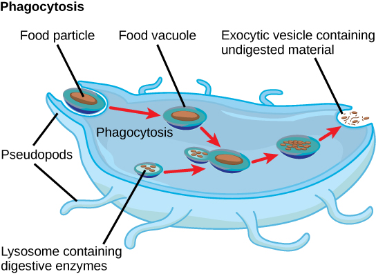
Protists exhibit many forms of nutrition and may be aerobic or anaerobic. Protists that store energy by photosynthesis belong to a group of photoautotrophs and are characterized by the presence of chloroplasts. Other protists are heterotrophic and consume organic materials (such as other organisms) to obtain nutrition. Amoebas and some other heterotrophic protist species ingest particles by a process called phagocytosis, in which the cell membrane engulfs a food particle and brings it inward, pinching off an intracellular membranous sac, or vesicle, called a food vacuole (Figure \(\PageIndex{2}\)). The vesicle containing the ingested particle, the phagosome, then fuses with a lysosome containing hydrolytic enzymes to produce a phagolysosome, and the food particle is broken down into small molecules that can diffuse into the cytoplasm and be used in cellular metabolism. Undigested remains ultimately are expelled from the cell via exocytosis.
Subtypes of heterotrophs, called saprotrophs, absorb nutrients from dead organisms or their organic wastes. Some protists can function as mixotrophs, obtaining nutrition by photoautotrophic or heterotrophic routes, depending on whether sunlight or organic nutrients are available.
Motility
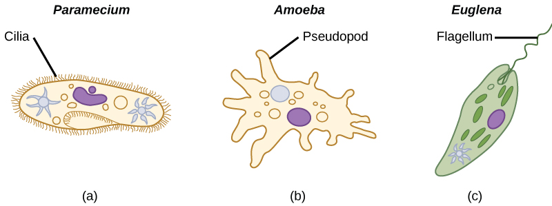
The majority of protists are motile, but different types of protists have evolved varied modes of movement (Figure \(\PageIndex{3}\)). Some protists have one or more flagella, which they rotate or whip. Others are covered in rows or tufts of tiny cilia that they coordinately beat to swim. Still others form cytoplasmic extensions called pseudopodia anywhere on the cell, anchor the pseudopodia to a substrate, and pull themselves forward. Some protists can move toward or away from a stimulus, a movement referred to as taxis. Movement toward light, termed phototaxis, is accomplished by coupling their locomotion strategy with a light-sensing organ.
Life Cycles
Protists reproduce by a variety of mechanisms. Most undergo some form of asexual reproduction, such as binary fission, to produce two daughter cells. In protists, binary fission can be divided into transverse or longitudinal, depending on the axis of orientation; sometimes Paramecium exhibits this method. Some protists such as the true slime molds exhibit multiple fission and simultaneously divide into many daughter cells. Others produce tiny buds that go on to divide and grow to the size of the parental protist. Sexual reproduction, involving meiosis and fertilization, is common among protists, and many protist species can switch from asexual to sexual reproduction when necessary. Sexual reproduction is often associated with periods when nutrients are depleted or environmental changes occur. Sexual reproduction may allow the protist to recombine genes and produce new variations of progeny that may be better suited to surviving in the new environment. However, sexual reproduction is often associated with resistant cysts that are a protective, resting stage. Depending on their habitat, the cysts may be particularly resistant to temperature extremes, desiccation, or low pH. This strategy also allows certain protists to “wait out” stressors until their environment becomes more favorable for survival or until they are carried (such as by wind, water, or transport on a larger organism) to a different environment, because cysts exhibit virtually no cellular metabolism.
Protist life cycles range from simple to extremely elaborate. Certain parasitic protists have complicated life cycles and must infect different host species at different developmental stages to complete their life cycle. Some protists are unicellular in the haploid form and multicellular in the diploid form, a strategy employed by animals. Other protists have multicellular stages in both haploid and diploid forms, a strategy called alternation of generations that is also used by plants.
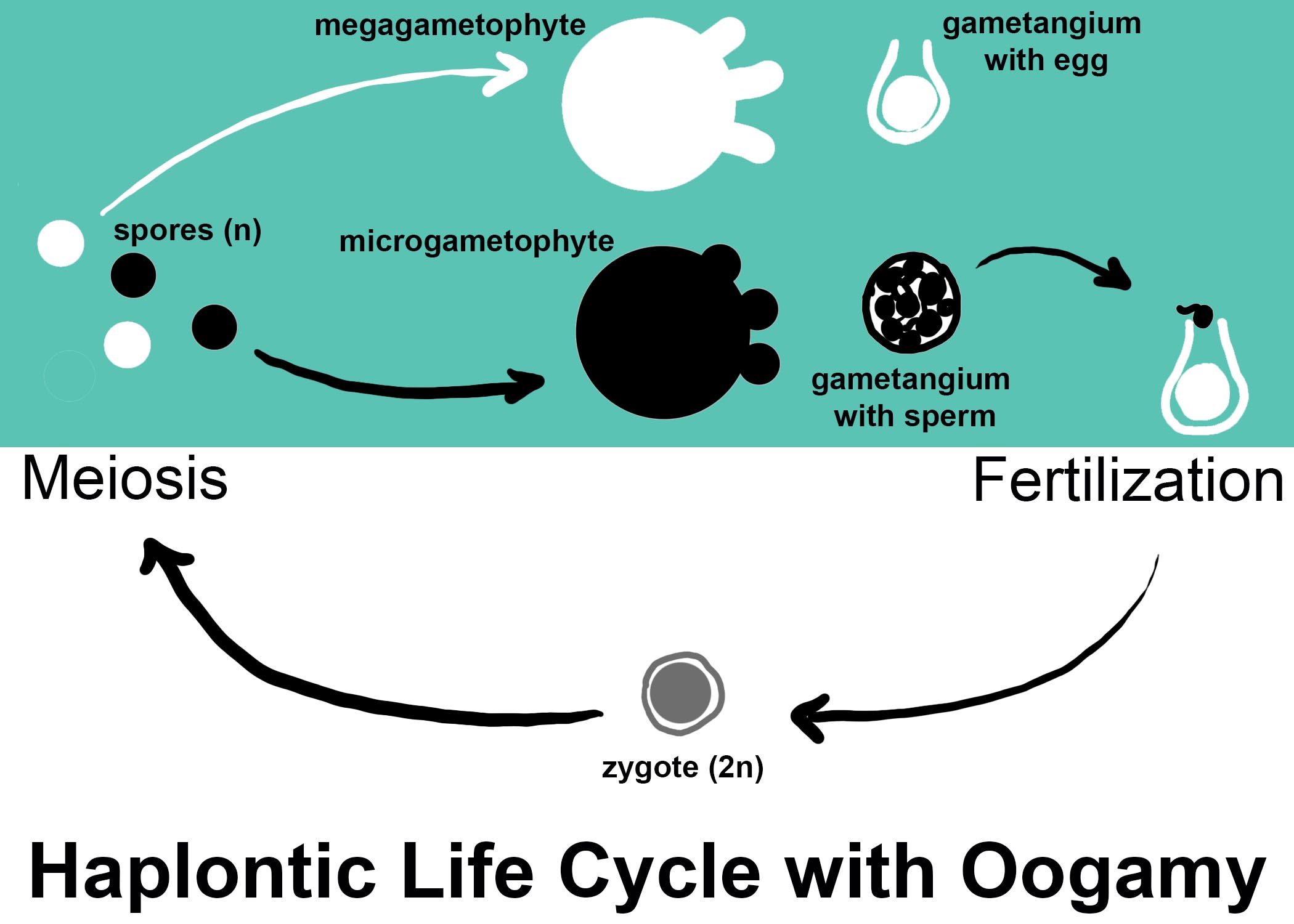
Life cycles can be generally classified as haplontic, diplontic, and haplodiplontic. In a haplontic life cycle, the multicellular stage is haploid and produces gametes from structures called gametangia. In plants and algae, these haploid organisms are sometimes referred to as gametophytes (meaning gamete plants). This life cycle is also called zygotic meiosis, because the zygote does not grow, but instead divides by meiosis to form haploid spores (see Figure \(\PageIndex{4}\)).
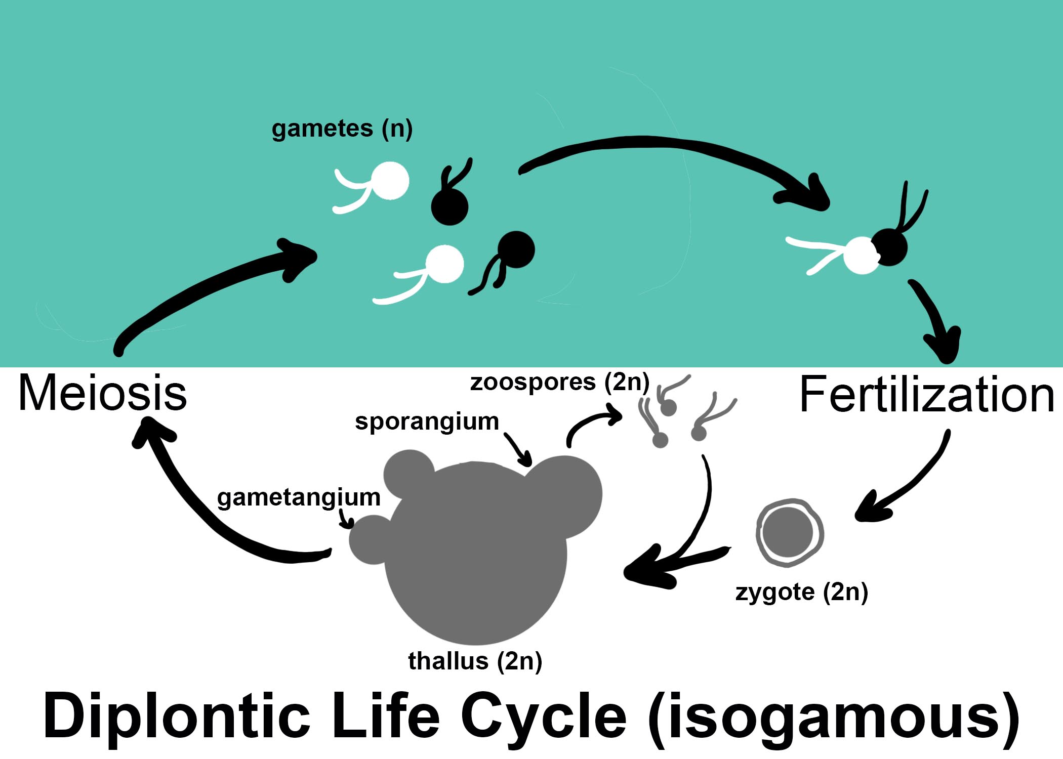
In a diplontic life cycle, the multicellular phase is diploid. The zygote grows by mitosis to form a diploid, multicellular organism. That organism might form sporangia for asexual reproduction. Spores would be diploid and could grow to form a new multicellular diploid organism. Sexual reproduction occurs in gametangia, where cells divide by meiosis to produce gametes. This life cycle is sometimes called gametic meiosis (see Figure \(\PageIndex{5}\)).
The haplodiplontic life cycle, also called alternation of generations, is the most complex. In this life cycle, there are multicelllular haploid and diploid phases. The zygote grows by mitosis to form a diploid sporophyte. The sporophyte, as its name implies, produces haploid spores by meiosis of cells within a sporangium. The spores grow into haploid gametophytes. The gametophytes produce gametes by mitotic division of cells within gametangia. These gametes then fuse to form the diploid zygote.
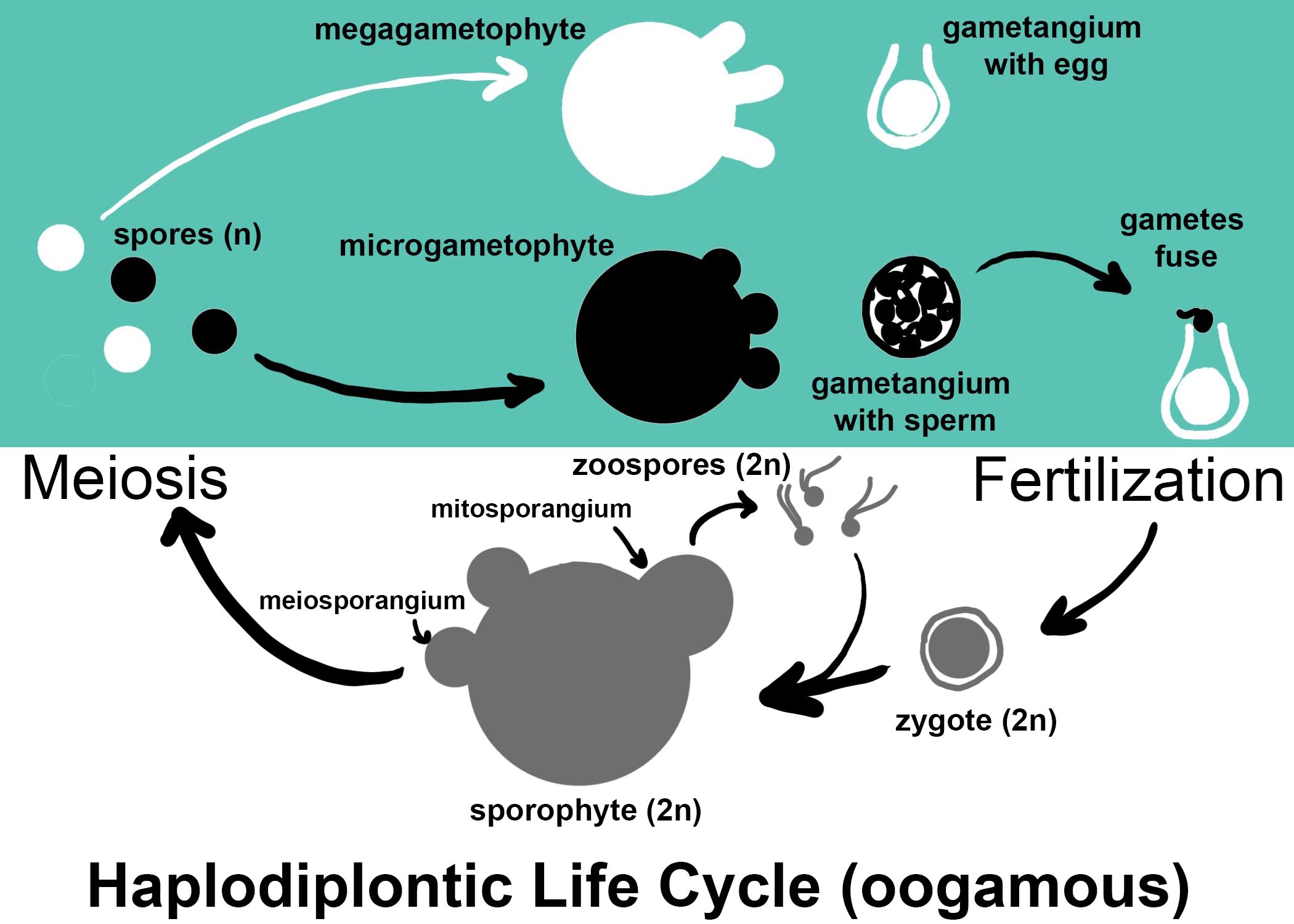
As you saw in Figures \(\PageIndex{d-f}\), there are other distinctions that can be made in life cycles. Isogamous life cycles have gametes that look approximately the same. Oogamous life cycles are heterogamous (meaning the gametes look different, also called anisogamous) in a specific way: the egg is larger and nonmotile, while the sperm are smaller and motile. With all of the complexities in life cycles, it can be helpful to remember this simple rule: spores grow, gametes fuse. Gametes never grow my mitosis, but must fuse together to form a zygote. Spores tend to grow or germinate in some way.
Habitats
Nearly all protists exist in some type of aquatic environment, including freshwater and marine environments, damp soil, and even snow. Several protist species are parasites that infect animals or plants. A few protist species live on dead organisms or their wastes, and contribute to their decay.
Summary
Protists are extremely diverse in terms of their biological and ecological characteristics, partly because they are an artificial assemblage of phylogenetically unrelated groups. Protists display highly varied cell structures, several types of reproductive strategies, virtually every possible type of nutrition, and varied habitats. Most single-celled protists are motile, but these organisms use diverse structures for transportation.
Attributions
Curated and authored by Maria Morrow, CC BY-NC, using the following sources:
- 19.1.2 Protists from Biology by John. W. Kimball (licensed CC-BY)
- 23.2 Characteristics of Protists from Biology 2e by OpenStax (licensed CC-BY). Access for free at openstax.org.