
3.3: Roots
- Page ID
- 47211

Though unseen, the roots of a plant also have specialist anatomical features that enable plants to efficiently obtain nutrients and control the substances entering a plant.
Figure 3.9. Diagram of the structures of and areas of a developing root. (Image from: Marsland, Douglas. (1964) Principles of modern biology. Holt, Rinehart and Winston, New York. Digitized Cornell University Library, No known copyright restrictions. Text Sean Bellairs.)

Figure 3.10. The apical meristem and meristematic tissues developing from the apical meristem of the root. (Image Jen Dixon (CC attribute, share alike). Text and arrows Sean Bellairs.)
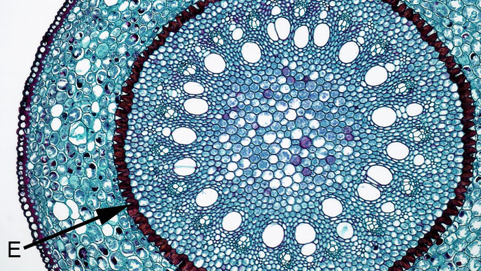
Figure 3.11. The endodermis (E) in the monocot Smilax. Cells making up the endodermis have walls that are heavily impregnated with suberin, forming the Casparian strip. Suberin is a fatty acid and highly hydrophobic, thus creating a barrier to water and solutes. (Berkshire Community College Bioscience Image Library, public domain; text and arrow Sean Bellairs).
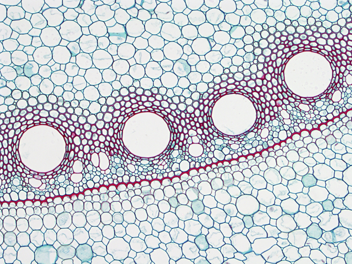
Figure 3.12. The endodermis denoted by the band of the red stained casparian band in Zea mays. The interior of the root is at the top and the casparian band is between the vascular tissue and the cortex. (Image by BlueRidgeKitties (CC attribute, share alike)).
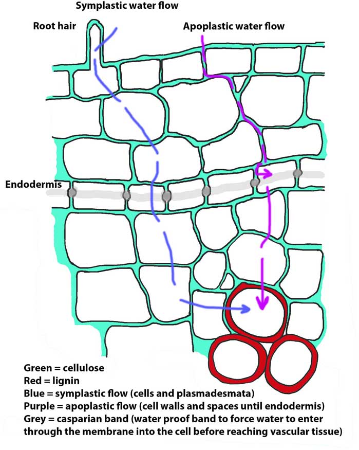
Figure 3.13. Effect of the casparian band on water flow between the cortex and the xylem. In the cortex water and solutes can move symplastically (through the living cells) or apoplastically (through the non-living cellulose cell walls and intercellular spaces. The casparian band forces all water movement into the vascular tissues to move though the cell membranes (Diagram by Sean Bellairs, CC attribute, share alike).