Biology LibreTexts

2.4: Dermal tissues and features

  • Page ID
    47208
  • \( \newcommand{\vecs}[1]{\overset { \scriptstyle \rightharpoonup} {\mathbf{#1}} } \) \( \newcommand{\vecd}[1]{\overset{-\!-\!\rightharpoonup}{\vphantom{a}\smash {#1}}} \)\(\newcommand{\id}{\mathrm{id}}\) \( \newcommand{\Span}{\mathrm{span}}\) \( \newcommand{\kernel}{\mathrm{null}\,}\) \( \newcommand{\range}{\mathrm{range}\,}\) \( \newcommand{\RealPart}{\mathrm{Re}}\) \( \newcommand{\ImaginaryPart}{\mathrm{Im}}\) \( \newcommand{\Argument}{\mathrm{Arg}}\) \( \newcommand{\norm}[1]{\| #1 \|}\) \( \newcommand{\inner}[2]{\langle #1, #2 \rangle}\) \( \newcommand{\Span}{\mathrm{span}}\) \(\newcommand{\id}{\mathrm{id}}\) \( \newcommand{\Span}{\mathrm{span}}\) \( \newcommand{\kernel}{\mathrm{null}\,}\) \( \newcommand{\range}{\mathrm{range}\,}\) \( \newcommand{\RealPart}{\mathrm{Re}}\) \( \newcommand{\ImaginaryPart}{\mathrm{Im}}\) \( \newcommand{\Argument}{\mathrm{Arg}}\) \( \newcommand{\norm}[1]{\| #1 \|}\) \( \newcommand{\inner}[2]{\langle #1, #2 \rangle}\) \( \newcommand{\Span}{\mathrm{span}}\)\(\newcommand{\AA}{\unicode[.8,0]{x212B}}\)

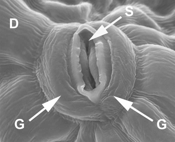
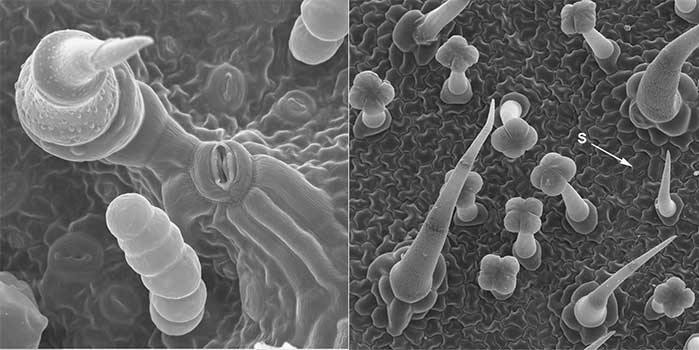
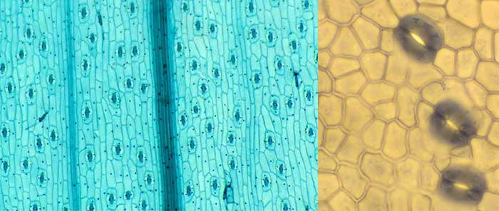
    The dermal tissue is largely composed of squat more or less cubic dermal cells, but it also contains specialist guard cells around the stomata, and various trichomes and root hairs.
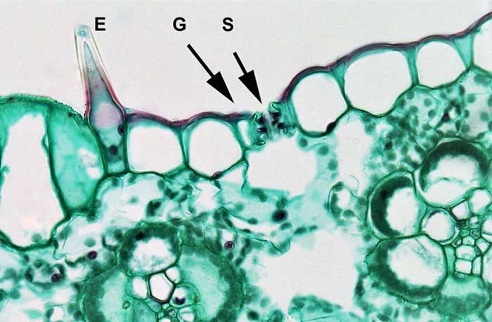
    Cuticle

    The cuticle is a layer of cutin and waxes external to and embedded in the cell wall on the exterior surface of the plant on stems and leaves.

    Cuticle (red stained) and the underlying square dermal cells on the upper surface of a mesophyte leaf.
    Figure 2.14. Cuticle (red stained) and the underlying square dermal cells on the upper surface of a mesophyte leaf. (Berkshire Community College Bioscience Image Library, public domain).

    Stomata, guard cells and trichomes

    Stoma (S), (plural stomata) on the upper surface of a mesophyte leaf, in the middle of two guard cells (G).
    Figure 2.15. Stoma (S), (plural stomata) on the upper surface of a mesophyte leaf, in the middle of two guard cells (G). An epidermal hair or trichome can also be seen (E). (Berkshire Community College Bioscience Image Library, public domain).
    Stomate on the surface of a sunflower leaf
    Figure 2.16. Stomate on the surface of a sunflower leaf (Louisa Howard, Dartmouth College; Public Domain; modified with labels by Sean Bellairs).
    Trichomes and stomata
    Figure 2.17. Trichomes and stomata on the surface of a sunflower leaf (left) and tomato leaf (right) (Louisa Howard, Dartmouth College; Public Domain).
    Stomata across the surface of the epidermis of the
    Figure 2.17. Stomata across the surface of the epidermis of a leaf of two plant species (Left – Tradescantia pallida by BlueRidgeKitties (CC: attribute, share alike); Right – Sean Bellairs (CC: attribute, share alike)).

    This page titled 2.4: Dermal tissues and features is shared under a CC BY-SA license and was authored, remixed, and/or curated by Sean Bellairs (Charles Darwin University) .

    • Was this article helpful?