
15.5: General Morphology
- Page ID
- 33538
Unicellular fungi are called yeasts. Yeasts reproduce asexually by budding, where the cell produces an outgrowth that enlarges and is eventually pinched off, creating an identical copy of the parent cell. The parent cell is left with a small circular scar (called a bud scar) that, at the right angle, refracts the light of the microscope and appears to glow a brighter white.

Place a small drop of the yeast culture on a slide, dilute it with a drop or two of water, and add a cover slip. Observe your slide under the microscope at 400x (or 1000x, if possible) and look for yeast reproducing asexually. Can you see bud scars?
Draw what you see in the space below and label any important structures.
When fungi produce a larger vegetative body, it is called a thallus (a body plan that is not differentiated into tissues). The thallus is composed of microscopic strands called hyphae (pronounced high-fay). As a collective, these hyphae are called a mycelium (pronounced my-seal-ee-um).
Safety Note
Congo Red is carcinogenic, do not contact with skin. Do not wash slides with Phloxine B in the sink, as this compound is harmful to aquatic life. See your institution’s MSDS for safety information on these compounds before use.
Obtain a sample of mycelium, make a wet mount with 5% KOH and stain it with Phloxine B and/or Congo Red. The stains are optional, but usually the fungal mycelium lacks pigment. The stains are picked up by the hyphae and allow them to stand out from the background. Phloxine B stains only the contents of the hyphae, while Congo Red stains the contents and walls. You can combine these two stains together or use separately.
In your prepared slide, can you see divisions of the hyphae into compartments? These dividing walls are called septa (septum singular) and are only present in certain groups of fungi.
Draw a strand of the hyphae (also sometimes called a hyphal thread or filament), label the cell wall, septum (if present), plasma membrane, and any other features you see.
Molds
Molds are the asexual form of a fungus. Before the advent of DNA sequencing, we thought molds were fungi that only reproduced asexually and they were placed in their own group, the Fungi Imperfecti (also sometimes called the Deuteromycota). As we are sequencing different mold species, we are discovering that they are identical to sexually reproducing fungi that we have already identified. This is causing a lot of trouble for taxonomy, as now we have one species with two names.
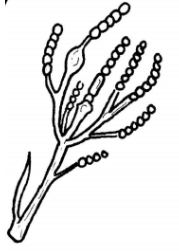
Asexually reproducing fungi produce spores called conidia. Much like the yeasts, many conidia are formed from budding off of another cell. Make a wet mount of a mold by adding a drop of 5% KOH to your slide, scraping a small amount of mold onto a razor blade or other tool, and gently depositing it onto the droplet. GENTLY lay down a cover slip. You may also consider trying to view molds as a dry mount, as they tend to be extremely hydrophobic.
Look for conidia and conidiophores, structures where the conidia are produced. Draw what you find below and label any identifiable structures:
