
4.3: Identifying Cell Types and Tissues
- Page ID
- 29536

Cell Types
Make a thin section of a celery petiole or the main celery stalk. This needs to be very thin to see the features you are looking for, so make a few samples to look at! If you would like to stain your specimen, place the specimen on a slide and add a small drop of Toluidine Blue. Wait a few seconds for the dye to penetrate into the sample, then rinse by adding water to the slide and either soaking up or draining off the excess liquid. When the water is mostly clear, add another drop or two of water and a coverslip.
View your specimen under the compound microscope.
You should be able to see several cell types in your specimen. Most of the cells will be parenchyma. A great place to look for textbook parenchyma cells is the outermost layer of the plant, the epidermis.
You will find collenchyma cells in dense clusters near the epidermis in a region called the cortex, forming the strings that you would find in your celery. They appear to have an almost checkerboard-like pattern, due to the unevenly thickened primary walls. In Toluidine Blue, primary walls stain purple.
Some specialized cells can be found in the vascular tissue, organized regions of cells that are transporting water, sugars, and other chemicals throughout the plant body. The xylem is the tissue responsible for conducting water. Specialized cells in the xylem tissue called tracheids and vessel elements have evolved specifically for this ability by forming hollow tubes with lignified secondary walls. Tracheids evolved first and are narrow with tapered ends. Vessel elements evolved in the most recent group of plants, the Angiosperms, and are usually much wider than tracheids. In Toluidine Blue, the lignin in the secondary wall stains bright aqua blue.
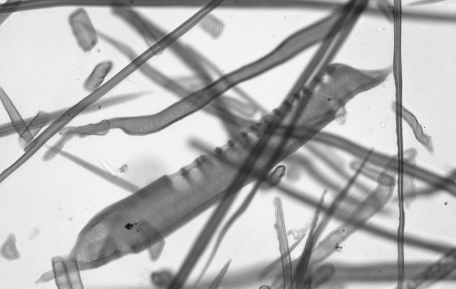
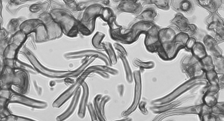
The image above shows three different types of cells with secondary walls found in wood pulp. A vessel element is shown in the center with a tracheid running parallel just above it. Note the pits in the walls of both of these cells and the large holes (perforation plates) on the ends of the vessel element only. Criss-crossing the rest of the slide are many thin fibers. All of these cells are dead at maturity and provide structural support due to the lignin in the cell walls.
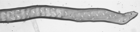
In the image above, you can see the pits in the walls of a tracheid.
Unlike the xylem, conducting cells in the phloem tissue are alive so they may transport sugars and communication signals in any direction. These cells, sieve tube elements and companion cells, are more similar to parenchyma. The sieve tube elements conduct sugars and have specialized to do this by having reduced cytoplasm contents: sieve tube elements have no nucleus (or vacuole)! These cells are controlled by small, adjacent cells called companion cells. If you look closely, you can also see some sclerenchyma bunched together in the phloem. These are the phloem fibers. Their thick secondary walls should stain the same color as the tracheids and vessel elements.
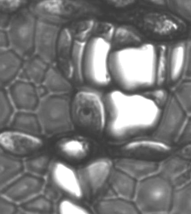
In the image above, you can see clusters of thick walled fibers, large open sieve tube elements, and small companion cells containing nuclei.
Draw a cross section of the celery petiole, labeling parenchyma in the epidermis, collenchyma in the cortex, and sclerenchyma in the vascular tissue. Make notes about the differences in the cell wall for your future study.
The central region of the celery petiole is called the pith. What type of cells are present in this region?
While collenchyma tissue tends to have one job--flexible support--parenchyma and sclerenchyma can fill a diverse set of roles. In a developing pear, there is a high density of a second type of sclerenchyma cells called sclereids (the first type of sclerenchyma cells were fibers). These cells cause young pears to be tough and unpalatable, as the seeds inside are still developing. As the seeds mature, the pear ripens, making more parenchyma cells for storing large amounts of sugar, while the tough sclereids are slowly outnumbered by the larger, juicier cells. The grit that you feel when eating a pear are these remaining sclereids.
What other cellular changes might occur to signal that a pear is ripe?
Make a squash mount of the flesh of a pear (not the skin) by scraping off a small amount with a razorblade. Do not take a slice or a chunk, just a tiny bit of pulp (consider chopping it up on the slide). Again, I recommend staining with Toluidine blue, as this should make the thick secondary walls of the sclereids appear a bright aqua blue. Sclereids tend to occur in clusters, surrounded by large parenchyma cells. You may need to gently squish your coverslip down a bit to help disperse these clumps. Coverslips are fragile, so ask your instructor what they recommend before doing anything that might result with glass in your fingers.
When you find a sclereid, you should see lines running through the secondary wall. These are channels where the plasmodesmata extended through to connect to other cells. After the cell dies, only the empty channels (called pits) remain.
Draw a sclereid, located in the ground tissue of a pear. Label the secondary wall, pits, an adjacent parenchyma cell, and the primary wall of that parenchyma cell.
Is this sclereid alive or dead? What about the parenchyma cells around it?
What is the compound in the secondary wall that stains differently from the primary wall?
Tissues in the Leaf
When cells of the same type work together to perform a collective function, the collection of cells is called a tissue. For example, the epidermis is a collection of parenchyma-like cells working together to separate the internal environment of the plant from the exterior. The epidermis also contains specialized cells. Trichomes are outgrowths from the epidermis that look like hairs. These can protect the plant from sun damage by being white and reflective, trap evaporating moisture on the plant’s surface, secrete sticky substances, and be unpleasant for herbivores.
A second type of specialized cell in the epidermis is the guard cell. Guard cells are shaped like parentheses and flank small pores in the epidermis called stomata (sing. stoma). When the plant has adequate water, the guard cells inflate and the stoma is open, allowing water vapor to escape through transpiration. When the plant is low on water, the guard cells collapse, closing the stoma and trapping water inside. However, for the plant to perform photosynthesis, it must have access to carbon dioxide and be able to release oxygen. Both of these gases are exchanged through the stomata.
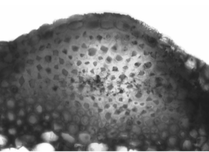
The image above is from the lower epidermis of a Nerium leaf. These plants live in harsh, dry environments and have many adaptations to prevent water loss. This is a pocket on the lower side of the leaf where stomata are located. You can see three different sets of guard cells, currently closed, appearing slightly darker than the other epidermal cells. Surrounding these stomata and filling the pocket are trichomes.
How does the location of the trichomes relate to prevention of water loss?
View a leaf under the dissecting scope. Can you find trichomes, guard cells, or other specialized epidermal cells?
Peel off the lower epidermis of the leaf, similar to how you removed it from the onion. It may help to break the leaf slowly, hopefully getting a piece of the epidermis that you can peel off. It will look like a transparent layer of skin. Make a wet mount of the epidermis and view it under the compound microscope. Draw what you see below, labeling any specialized epidermal cells.
What cell type (-enchyma) are these cells most similar to?
When multiple tissues work together to perform a collective function, this collection of tissues is called an organ. While we are familiar with the concept of organs in animals, it can sometimes be surprising to consider this aspect of plants.
An example of an organ in a plant is the leaf. A leaf is surrounded by epidermal tissue, protecting the interior environment, and allowing for the exchange of gases with the environment. The xylem tissue, found in the veins of the leaf, provides the water needed for specialized parenchyma, mesophyll cells, to carry out photosynthesis. Phloem tissue runs alongside the xylem tissue, transporting sugars made during photosynthesis to other areas of the plant for either immediate use or storage. Together, these tissues allow the leaf to function as an organ specialized for photosynthesis.
View a prepared slide of a leaf cross section. Draw what you see below. Identify and label as many tissues, cell types, and specialized cells as you can.
Summary Table of Cells and Tissues in the Leaf Organ
A simple tissue contains only a single cell type, while a complex tissue contains multiple cell types. The leaf organ is composed of both simple and complex tissues. In the table below under Tissue Type, try to identify whether it is a simple or complex tissue. If it is a simple tissue, identify which cell type it is composed of.
|
Tissue |
Specialized Cells |
Tissue Type |
Function |
|---|---|---|---|
|
Epidermis |
1. Guard cell 2. Trichome |
||
|
Mesophyll |
Mesophyll cells |
||
|
Xylem |
Tracheids & vessel elements |
||
|
Phloem |
1. Sieve tube elements 2. Companion cells 3. Phloem fibers |
Why didn’t I include a stoma among the specialized cells in the epidermis?