
11.5: Secondary Tissues in the Shoot
- Page ID
- 33484

Secondary Vascular Tissue
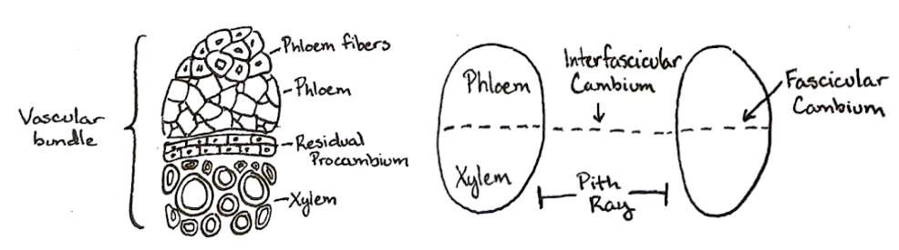
Though the secondary tissues are all the same, the sequence of secondary meristem development in shoots is a bit different than in roots. Shoots have no pericycle, so the secondary meristems must be formed from different tissues. In shoots, the vascular cambium is formed from residual procambium within the vascular bundles (fascicular cambium) joined by tissue in the pith rays (interfascicular cambium).
This layer of cells will divide to produce secondary xylem toward the inside of the stem and secondary phloem toward the outside. The conducting cells in the secondary xylem are dead at maturity. As the stem matures, these dead cells become separated from the living cells and the plant would be unable to direct the transport of materials into and out of the center of the plant. To solve this, plants have pathways of living cells that traverse the xylem and phloem called rays. Xylem rays radiate outward from the center of the stem until they reach the vascular cambium. At this point, they become phloem rays. Some of these will balloon out into large deltas of cells in the phloem tissue, while others will remain narrow strips of cells.
The secondary phloem becomes densely packed with layers secondary phloem fibers that provide structural support for the stem to grow tall.

Vessel elements in secondary xylem
In the secondary xylem, different diameter conducting cells form in wet and dry conditions. Larger diameter cells form in the wet season when water is plentiful. As the seasons transition to drier whether or approach freezing conditions, cells are smaller. Because most climates have a wet and dry season each year, you can track years of growth by the formation of these annual growth rings in the secondary xylem.
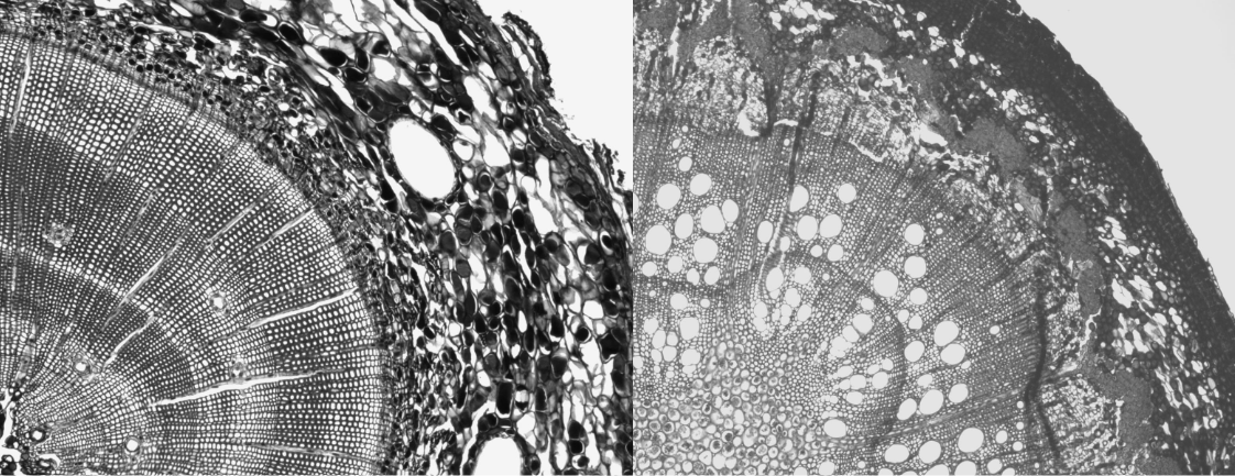
On the left is a pine stem, on the right is an oak. In the pine stem, the cells are a more similar diameter because they are all tracheids. On the right, The largest diameter cells are vessel elements. How old are each of these stems?
Resin canals
In answer to the previous question about the roots, those giant holes in the pine stem (as in the roots) are resin canals. These canals transport a sticky substance called resin. Much like the latex produced in some flowering plants, resin acts as a defense compound and a wound sealant.
Resin canals are also present in some groups of angiosperms, particularly tropical trees. Why might these structures occur more commonly in tropical plants?
The cork cambium is formed directly from the cortex. Layers of periderm are continually formed and shed until the cortex runs out. At this point, secondary phloem de-differentiates and forms the new layers of cork cambium.
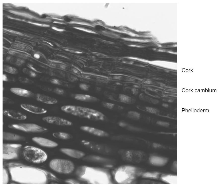
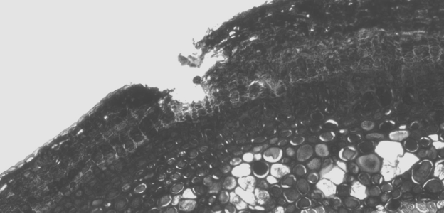
Because the periderm is suberized and dead, it cannot have living guard cells to regulate pores and control gas exchange with the environment. Because the cells on the inside are still alive and respiring, gas exchange is still necessary. This is accomplished through tears in the periderm called lenticels (shown below).
In a mature woody stem, the layers of periderm are referred to as the outer bark and the secondary phloem is the inner bark. The secondary xylem is called wood. Secondary xylem that is still actively involved in conducting water is called sapwood, while inactive xylem is called heartwood. The cross section below is too young to have these last two features.
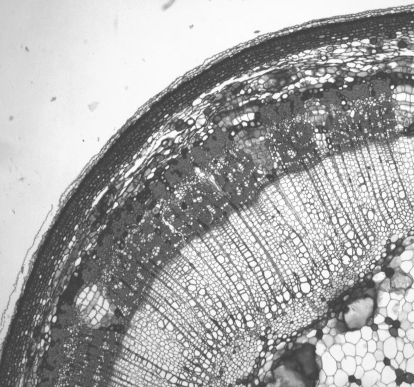
In the stem cross section above, label the following: pith, secondary xylem, xylem ray, vascular cambium, secondary phloem, phloem ray, cortex, phelloderm, cork cambium, cork, and epidermis. Next, indicate where the oldest and youngest xylem cells are located.
Note: the newest cells will be located closest to the meristem that produced them!
Where is the vascular cambium?