Biology LibreTexts

8.5: Eudicots

  • Page ID
    33462
  • \( \newcommand{\vecs}[1]{\overset { \scriptstyle \rightharpoonup} {\mathbf{#1}} } \) \( \newcommand{\vecd}[1]{\overset{-\!-\!\rightharpoonup}{\vphantom{a}\smash {#1}}} \)\(\newcommand{\id}{\mathrm{id}}\) \( \newcommand{\Span}{\mathrm{span}}\) \( \newcommand{\kernel}{\mathrm{null}\,}\) \( \newcommand{\range}{\mathrm{range}\,}\) \( \newcommand{\RealPart}{\mathrm{Re}}\) \( \newcommand{\ImaginaryPart}{\mathrm{Im}}\) \( \newcommand{\Argument}{\mathrm{Arg}}\) \( \newcommand{\norm}[1]{\| #1 \|}\) \( \newcommand{\inner}[2]{\langle #1, #2 \rangle}\) \( \newcommand{\Span}{\mathrm{span}}\) \(\newcommand{\id}{\mathrm{id}}\) \( \newcommand{\Span}{\mathrm{span}}\) \( \newcommand{\kernel}{\mathrm{null}\,}\) \( \newcommand{\range}{\mathrm{range}\,}\) \( \newcommand{\RealPart}{\mathrm{Re}}\) \( \newcommand{\ImaginaryPart}{\mathrm{Im}}\) \( \newcommand{\Argument}{\mathrm{Arg}}\) \( \newcommand{\norm}[1]{\| #1 \|}\) \( \newcommand{\inner}[2]{\langle #1, #2 \rangle}\) \( \newcommand{\Span}{\mathrm{span}}\)\(\newcommand{\AA}{\unicode[.8,0]{x212B}}\)

    In eudicot stems, the vascular tissue is arranged into a ring (the vascular cylinder) that separates the ground tissue into two distinct regions. The region of ground tissue contained within the vascular cylinder is called the pith. The region of ground tissue that is between the vascular cylinder and the epidermis is called the cortex. Similar to the monocots, the primary xylem is located in the region of the vascular tissue facing the center of the stem, while the primary phloem is produced toward the exterior of the stem.

    clipboard_e9f31f0fab07de3b60e339bfebaefd060.png
    Figure \(\PageIndex{1}\): Helianthus stem cross section

    Observe a cross section of a young Helianthus (or other eudicot) stem. Locate the epidermis, cortex, primary phloem, primary xylem, pith, and pith rays. Label these tissues in the image above.

    What cell types (-enchymas) can you identify in the Tilia cross section? Which tissue(s) do you find each cell type in?

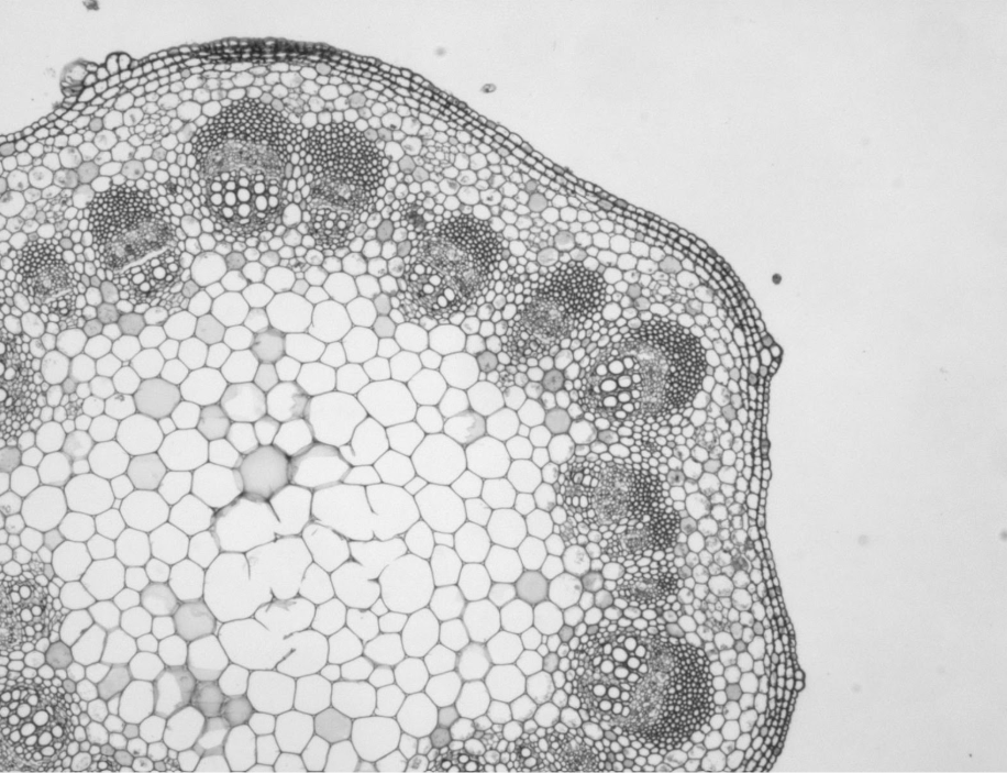
    This image shows the epidermis and cortex of a young Tilia stem in primary growth. The epidermis is the outermost layer of cells and it is covered in a thin layer of wax, the cuticle. Just inside the epidermis is the cortex. Because this is from a young, growing stem, the outer cortex is full of collenchyma cells.

    clipboard_e5310d081624eb5d706599dead16fb447.png
    Figure \(\PageIndex{2}\): Close up of Tilia cortex and epidermis

    In the image on the right, label the cuticle, epidermis, cortex, a collenchyma cell, and a parenchyma cell.

    Why would the stem be covered in wax?

    Contributors and Attributions


    This page titled 8.5: Eudicots is shared under a CC BY-NC license and was authored, remixed, and/or curated by Maria Morrow (ASCCC Open Educational Resources Initiative) .

    • Was this article helpful?