5.4: Mitosis
- Page ID
- 33232

Mitosis is the division of the nucleus in eukaryotic cells to make two identical nuclei. This is accompanied by cytokinesis (cyto- meaning cell, kinesis meaning movement), division of the cytoplasm, to result in division of the entire cell into two identical daughter cells.
A unicellular eukaryote might do mitosis to reproduce asexually, making an identical copy of itself. This is common practice in many groups of organisms. For example, molds are fungi that are reproducing asexually--each mold spore is genetically identical to the others. Many unicellular algae will also reproduce this way.
Multicellular eukaryotes also do mitosis -- that is how they became multicellular! Multicellular eukaryotes primarily use mitosis for growth and repair of damage, though some also reproduce asexually (such as strawberries and mint). To see mitosis occurring in a plant, the best place to look are the growing tips, as most plants experience apical growth (growth from the tips).
Obtain a prepared slide of an onion root tip (Allium cepa). Cells toward the apex (pointed end) are likely to have been caught in a stage of active division.
Prophase
Mitosis begins with prophase. During this portion of mitosis, the chromatin condenses into a more organized state -- the chromosome. The nuclear envelope begins to break down and any nucleoli dissolve. The cell begins to produce a network of microtubules called the spindle (in animal cells, the production and organization of the spindle is organized by a set of organelles called centrioles that assemble together into a centrosome). These networks begin to form on opposite sides of the cell.
You can identify cells in prophase by looking for the following:
- The nuclear region has expanded and moved to the center of the cell
- DNA appears as distinct structures (sausage-like), still contained in the center of the cell
- At the end of prophase, no nucleolus is present
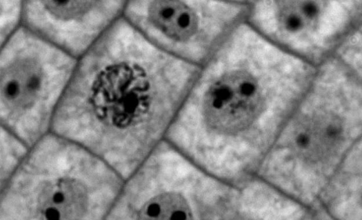
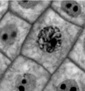
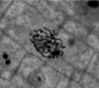
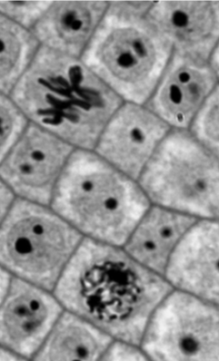
The image shows a cell in early prophase, surrounded by cells in interphase. The nucleoli are still visible in all of these cells as dark dots, but the cell in early prophase has condensed its chromatin into darkly stained chromosomes, whereas the cells in interphase still have the lightly staining grey chromatin. The circular organization of the chromosomes indicates that the nuclear envelope has not finished breaking down yet.
Compare the cell in early prophase (left) to a cell in late prophase (right). As the nuclear envelope finishes breaking down, the chromosomes become more spread out. Additionally, no nucleolus is visible. During this time, the spindle fibers attach to the kinetochores and are about to start pushing and pulling the chromosomes into the middle of the cell.
Draw an onion cell in prophase. Label the cell wall, plasma membrane, nuclear envelope (or where it would be), and chromosomes. If a nucleolus is still present or you can distinguish a forming spindle, label these as well.
Metaphase
As the cell transitions into metaphase, referred to as prometaphase, the spindle fibers attach to sticky regions called kinetochores that flank the central region of the chromosome, the centromere. The centromere holds the two identical pieces of the chromosome, the sister chromatids, together. The spindle fibers contract and expand to push and pull the chromosomes into a line in the center of the cell, the metaphase plate. This is where the new cell wall will form
You can identify cells in metaphase by looking for the following: Chromosomes are found in the center of the cell, they appear to be connected, often forming a straight line.
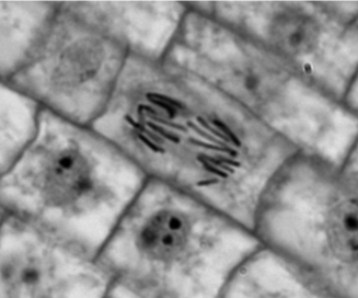
There are two cells in this image not currently in interphase. The cell on the top is in metaphase.
Which stage of mitosis is the cell on the bottom in? How can you tell?
Draw an onion cell in metaphase. Label the cell wall, plasma membrane, chromosomes, spindle, and metaphase plate.
Anaphase
Anaphase begins when the two networks of spindle fibers start to contract. During this process, sister chromatids of each chromosome are pulled apart, each becoming an individual chromosome the moment they are separated. These chromosomes are pulled to either side of the cell in preparation to form two new nuclei.
You can identify cells in anaphase by looking for the following:
- There should be two distinct groups of chromosomes
- individual chromosomes can still be distinguished.
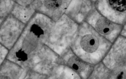
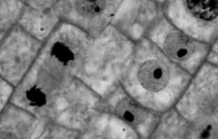
This image depicts a cell in anaphase. Are the dark lines in this cell chromatids or chromosomes? How can you tell?
Draw an onion cell in anaphase. Label the cell wall, plasma membrane, chromosomes, spindle, and metaphase plate.
Telophase
The final stage of mitosis is telophase. During this stage, a nuclear envelope begins to reform around each group of chromosomes and the chromosomes decondense into chromatin. The spindle fibers begin to break down and any nucleoli reform.
You can identify cells in telophase by looking for the following:
- There should be two distinct darkened regions, but individual chromosomes cannot be distinguished.
- The formation of the new cell wall, which occurs during cytokinesis, is still incomplete and the area between the two forming nuclei appears blurry.
This image shows a cell on the left that is currently in telophase, forming two new nuclei. On the right side of the image there are three cells that have probably recently finished dividing and have entered interphase.
Draw an onion cell in telophase. Label the cell walls, plasma membranes, forming nuclear envelope, and chromatin. (It is likely that the DNA is in a transitional state between chromosomes and chromatin)
Cytokinesis
The formation of the new cell wall and division of the cytoplasm is called cytokinesis. This stage of cell division happens concurrently with telophase, but is not part of mitosis, as that refers strictly to the division of the nucleus.
In the image below, label the new cell wall that is forming during cytokinesis.
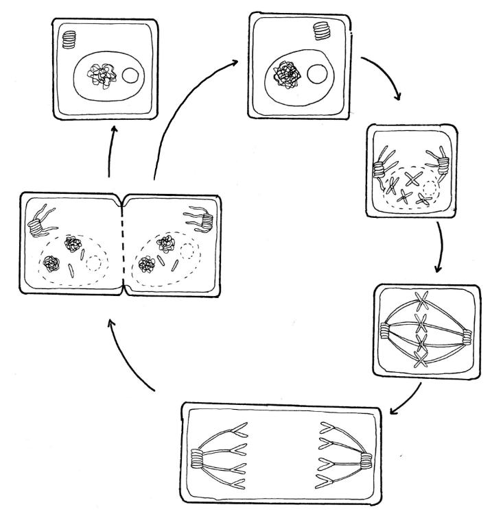
Below is a diagram of mitosis. Label each stage and the relevant structures. Make note of important events.