
3.8: Cell Structures and Organelles
- Page ID
- 33220

Because Azolla is a plant, its cells are surrounded by a rigid cell wall containing cellulose. Like the bacteria, the plasma membrane is located just inside the cell wall and contains the cytoplasm. The cell wall tends to give plant cells a boxy, rigid structure.
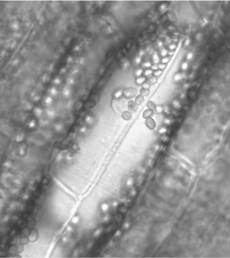
The most obvious of the membrane-bound organelles you will see are the chloroplasts. These numerous, green, disc-like structures are responsible for doing photosynthesis, making food for the plant. Their color is derived from the presence of chlorophylls, green-pigmented molecules involved in harvesting light for photosynthesis. You might also see that these chloroplasts are pushed toward the edges of the cell. This is because the majority of the cell is taken up by a large, liquid-filled structure called the central vacuole, surrounded by a membrane called the tonoplast. The central vacuole contributes to the plant’s overall structure by drawing in water, inflating the cell to a turgid state (more on this in lab 5).
Above is a cell from the aquatic plant Elodea. The cell wall, nucleus, and chloroplasts are visible.
Draw a cell from the Azolla in the space below. Label the cell wall, plasma membrane, cytoplasm, chloroplasts, nucleus (if you see it), central vacuole, and tonoplast.
The layers in an onion bulb are fleshy leaves that have been modified to store starches for the plant. At the base of the bulb, you will see a short stem with roots emerging. The part of the onion that you will be observing is the epidermis (outermost layer) of the leaf.
Cut a small section of an onion leaf. Carefully peel off a section of the epidermis--not the pigmented side, but the paler side that faces the interior of the onion. It should come off as a translucent sheet. Prepare a wet mount of onion epidermis using a drop or two of IKI solution.
This iodine solution increases the refractivity of the light and makes the nucleus visible. You should also be able to see at least one dark dot in the nucleus. This is the nucleolus (plural, nucleoli), a region of the nucleus where ribosomes are assembled.
View your specimen with the compound microscope. Draw an onion cell and label the cell wall, plasma membrane, cytoplasm, nucleus, nucleolus, central vacuole, and tonoplast.
Why are there no chloroplasts in this plant cell? Consider where an onion bulb would be located in a garden setting.
Plant cells are static within the plant (they do not move from place to place) and are surrounded by a cell wall. Adjacent cells are “glued” together by a pectin-rich layer called the middle lamella. As you saw above, the onion cells did not contain chloroplasts, meaning they are not producing their own food, yet they are used to store large amounts of sugars in the form of starch. How then do these cells communicate with each other and exchange nutrients? When plant cells divide, the division is incomplete, leaving small channels through the cell wall and middle lamella. These channels are called plasmodesmata (plasmodesma, singular). You can look for these in the epidermal cells of a red pepper (or similar fruit).
Peppers are fruits whose function is to attract animals to disperse the seeds of the pepper plant. To do this, the pepper goes through a ripening process. Pectins (a form of starch) in the middle lamella begin to break down into simple sugars, making the pepper both sweeter and softer. The color of the pepper also begins to change as chloroplasts are converted to chromoplasts. These organelles contain pigments called carotenoids that produce yellows, oranges, and reds, the last of which is highly attractive to animals like us.
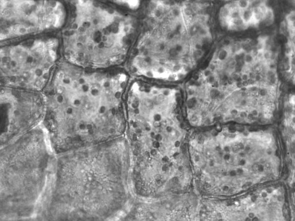
The image above shows epidermal cells in a red pepper. They are filled with small, red, disc-like plastids. These are chromoplasts filled with carotenoids.
In the cell walls, you can see tiny gaps that look more like constrictions. These are the plasmodesmata.
Peel off a small piece of red pepper epidermis and make a wet mount with water. Using the compound microscope, look for small, red chromoplasts within the cells. Next, look for breaks in the regions between the cells. These are the plasmodesmata.
Draw two adjacent pepper epidermal cells. Label the cell wall, middle lamella, plasmodesmata, and chromoplasts. You are encouraged to identify and label other cell components, such as the nucleus and nucleolus, if they are visible.
A potato is a modified part of the plant called a tuber. Much like an onion, a tuber is a part of the plant--this time the stem--adapted for storing starch. Potatoes synthesize and store these starches in organelles called amyloplasts (named for the starch amylose). In other cases, similar organelles are used to store lipids or other complex molecules. In general, these organelles are called leucoplasts (leuco- meaning white), because they lack pigments.
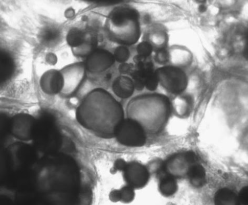
Using a razor blade or sharp probe, scrape a small amount of the interior of a potato (not the skin) onto a slide. Make a wet mount using one drop of IKI followed by one drop of water. The iodine in the IKI solution will surround the starch molecules, causing starches to appear to stain blue to purple. This will help you locate the amyloplasts within the potato cells, as they will absorb the IKI and the entire organelle is colored purple.
Draw a potato cell and label the cell wall, plasma membrane, cytoplasm, and amyloplasts.