The morphology and anatomy of a leaf can allow you to predict the conditions that the plant is adapted to. In particular, what is the water availability in that plant's environment?
Mesophytic Leaves
A leaf in "normal" conditions is called mesophytic (meso- means middle), meaning it is not particularly adapted for either high or low water conditions.
Figure \(\PageIndex{1}\): A cross section through a mesophytic leaf. The arrangement of tissues in a mesophytic leaf is as described in Fig. 13.3.2. As you look at adaptations to water availability, the arrangement of tissues in the leaf above will serve as the "standard" condition for leaves. Photo by Maria Morrow, CC BY-NC.
Hydrophytic Leaves
Hydrophytes (literally "water plants") are adapted to living in aquatic conditions.
Figure \(\PageIndex{2}\): A cross section through a dichotyledonous hydrophyte, Nymphaea (a water lily). The organization of tissues in this leaf is described in depth in Fig. 13.3.1.4. The important point here is to note the similarities between this eudicot leaf and the monocot leaf shown in Fig. 13.3.1.3. The similarities in tissue arrangement are due to convergent evolution to an aquatic lifestyle, as opposed to relatedness. Photo by Maria Morrow, CC BY-NC.
Below is an image of another hydrophytic leaf. This one is from a monocot, Potamogeton. Note the similarity to the Nymphaea leaf and the distinct differentiation between regions of mesophyll. A good example of convergent evolution to similar environmental pressures!
Figure \(\PageIndex{3}\): A cross section through a hydrophytic monocot leaf (Potamogeton). Note the similarity in internal anatomy to the hydrophytic eudicot Nymphaea shown in Fig. 13.3.1.2. Image is in the Public Domain, sourced from Berkshire Community College Bioscience Image Library.
Tissue Organization
Figure \(\PageIndex{4}\): A cross section through a dichotyledonous hydrophyte, Nymphaea (a water lily). The upper epidermis is a thin layer of parenchyma with many stomata. Below each stoma, there is a chamber of air located within the palisade mesophyll (this makes them easier to find). Under the palisade mesophyll is a much larger region of spongy mesophyll than we would find in a mesophytic plant leaf. Most of the space is taken up by large air pockets, making this tissue aerenchyma. The lower epidermis has no stomata. Within the mesophyll, there are spiky, pink-stained astrosclereids that have been caught in strange views during the sectioning process. Photo by Maria Morrow, CC BY-NC.
Location of Stomata
Because Nymphaea is aquatic and sits on top of the water, the stomata are located only in the upper epidermis. You can locate them in the cross section by finding the gaps (stomatal pits) in the palisade mesophyll. Why wouldn't there be stomata in the lower epidermis?
Figure \(\PageIndex{5}\): A close up of the stomata and stomatal chambers in Nymphaea. In the center of the image, there is an elongated empty space within the palisade mesophyll. At the top of this space, there are two guard cells flanking a closed stoma. At least three other stoma are present in the upper epidermis captured in this image, though the stomatal chambers haven't been captured as clearly in this section. Can you find them? Image is in the Public Domain, sourced from Berkshire Community College Bioscience Image Library.
Other Features
If you see strange branching structures within your Nymphaea leaf cross section, you may be looking at an astrosclereid (astro- meaning star). This is a branching sclerenchyma cell with a thick secondary wall. What function might these cells have?
Figure \(\PageIndex{6}\): A close up of an astrosclereid found in a Nymphaea leaf, caught in partial section. These sclerenchyma cells are covered in pits and have long, sharp arms. Some span the entire width of the leaf. Image is in the Public Domain, sourced from Berkshire Community College Bioscience Image Library.
Xerophytic Leaves
Xerophytes (literally "dry plants") are adapted to living in dry conditions with low water availability.
Figure \(\PageIndex{7}\): A cross section through a xerophytic leaf (Nerium). At first glance, the organization looks similar to a mesophytic leaf. However, there are several adaptations that allow this plant to lose less water (see Figure \(\PageIndex{8}\)). Photo by Maria Morrow, CC BY-NC.
Tissue Organization
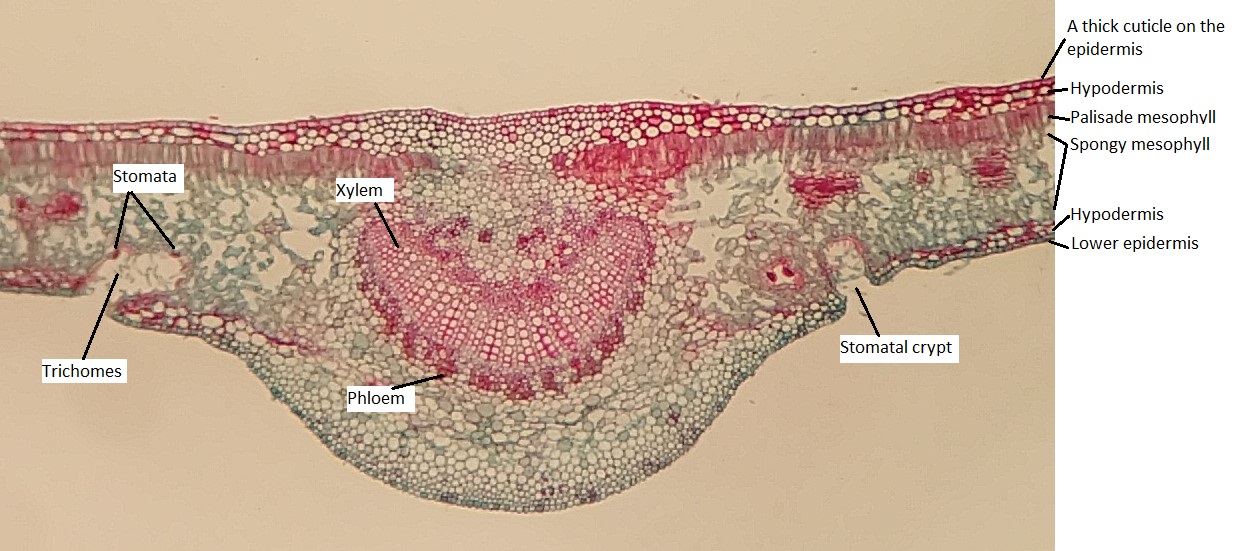
Figure \(\PageIndex{8}\): Tissue organization through a xerophytic leaf (Nerium). On the right side of the image, the layers of tissue are labeled from the upper surface of the leaf to the lower. The upper epidermis of the leaf is sealed by a thick, waxy cuticle. There are no stomata present in the upper epidermis. Just below the epidermis are several layers of tightly packed cells called the hypodermis. Beneath the hypodermis, the palisade and spongy mesophylls are arranged as in a mesophytic leaf. There are more layers of hypodermis between the spongy mesophyll and the lower epidermis. There are invaginations in the lower epidermis called stomatal crypts (see Figure \(\PageIndex{9}\)). Stomata are located within these, surrounded by trichomes. Photo by Maria Morrow, CC BY-NC.
Cuticle Thickness
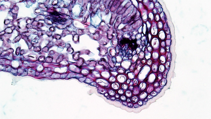
The image below shows the cuticle of the Nerium leaf. Notice how thick it is on the upper (adaxial) surface of the leaf and how it changes in thickness as it transitions to the lower (abaxial) surface.
Figure \(\PageIndex{9}\): The margin of a xerophytic leaf. There is a thick cuticle on the upper epidermis (it looks like a transparent skin). The cuticle gets thinner as it transitions to the lower epidermis. Image is in the Public Domain, sourced from Berkshire Community College Bioscience Image Library.
Location of Stomata
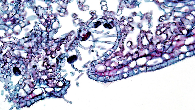
In xerophytic leaves, stomata tend to be located on the lower (abaxial) surface. This side of the leaf is usually cooler, as the upper (adaxial) surface is facing the sun. In extremely dry conditions, stomata might be further protected from the desiccating outer air by being located in stomatal crypts.
Figure \(\PageIndex{10}\): A stomatal crypt in the lower epidermis of a Nerium leaf. The epidermis folds inward, creating a small cave-like structure called a crypt. The stomata are located on the epidermis inside the crypt, surrounded by trichomes. Image is in the Public Domain, sourced from Berkshire Community College Bioscience Image Library.
Gymnosperms: The Original Xerophytes
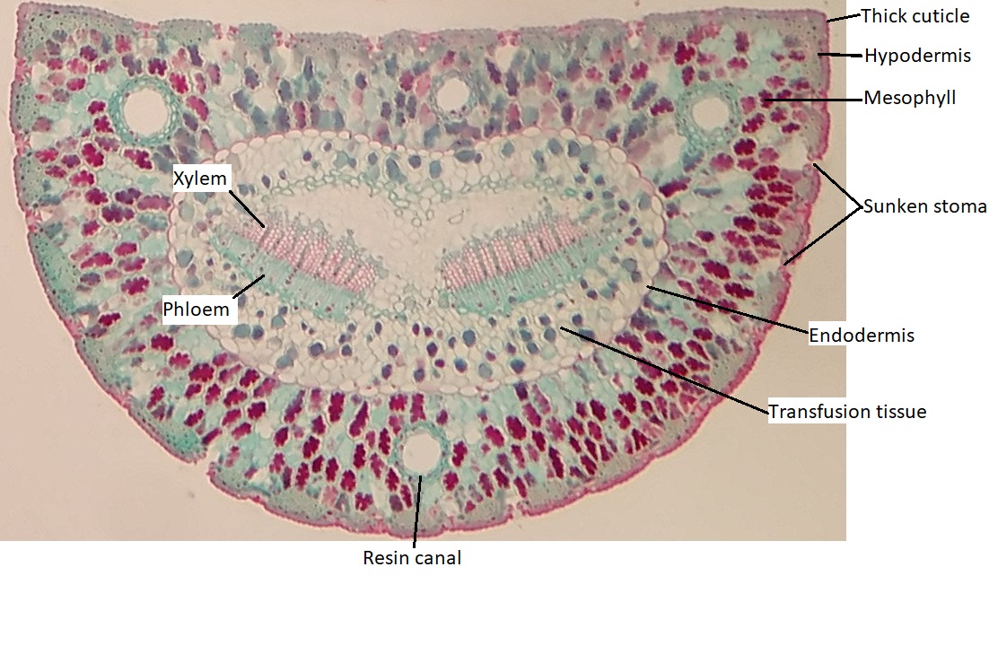
Though pines are not angiosperms, they have xerophytic leaves (needles). Note the features this pine needle has in common with the Nerium leaf.
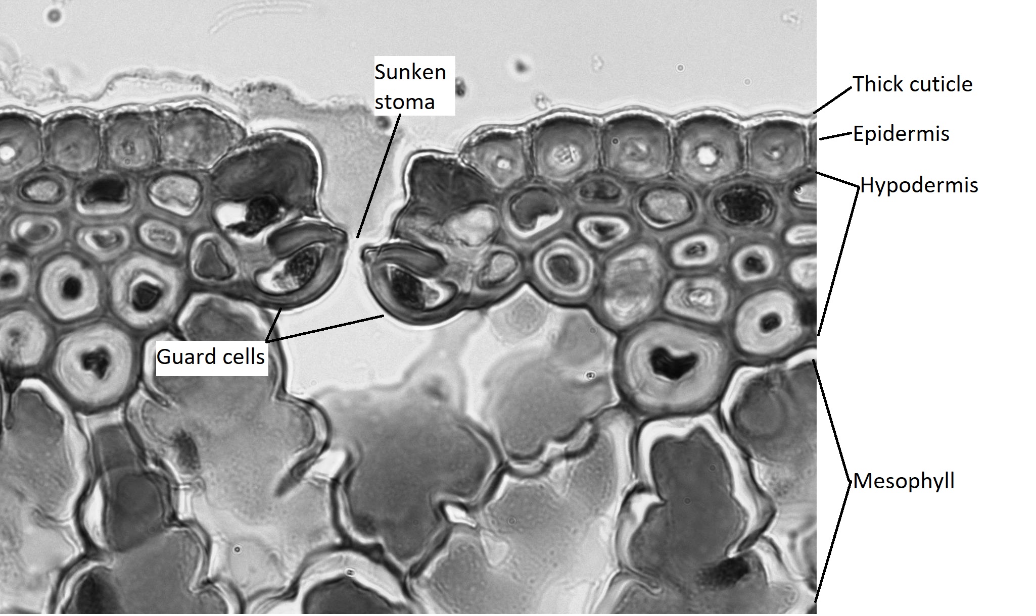
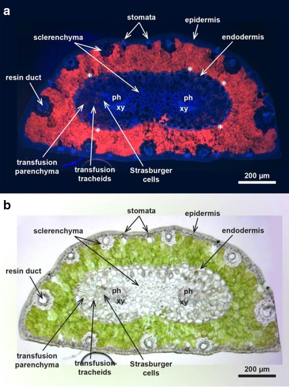
Figure \(\PageIndex{11}\): A cross section through a pine needle. Much like the Nerium leaf, this leaf is coated in a thick cuticle and there is a hypodermis below the epidermis (because this leaf is so round, there is not really a distinct 'upper' and 'lower'). There are no stomatal crypts, but the stomata are sunken, located in the hypodermis (see Figure \(\PageIndex{12}\)). The leaf has a low surface area to volume ratio (more volume, less surface area), which decreases water loss. In the center of the leaf, There is a large region surrounded by an suberized endodermis (much look in a root). There are two vascular bundles within this region, surrounded by transfusion tissue. Photo by Maria Morrow, CC BY-NC.Figure \(\PageIndex{12}\): A closer view of a sunken stoma and the outermost layers of the pine needle. The thick cuticle is visible as a transparent layer coating the small epidermal cells. Each of the epidermal cells has a thick cell wall. The hypodermis is composed of 3-4 layers of small, tightly packed cells that also have thick walls (sclerenchyma). The guard cells of the stoma are located about 3 layers below the epidermis and the cuticle can be seen extending down over them. The stoma is open in this image. Below the stoma, there is a gap of air space, then highly invaginated mesophyll cells. Photo by Maria Morrow, CC BY-NC.Figure \(\PageIndex{13}\): Two pine needle cross sections. In a) the mesophyll cells glow red (autofluoresce), in b) the mesophyll cells are green due to chlorophyll. Photosynthesis takes place in the mesophyll cells. "Scots pine needle cross-section photographed using a light microscope with UV excitation at 365 nm resulting in tissue autofluorescence (a), the same section photographed in the bright field (b). x xylem, ph phloem, asterisks indicate the radial thickening of the endodermal cells resembling the Casparian strip." Image and quoted caption text from the open-access paper Tissue-specific element profiles in Scots pine (Pinus sylvestris L.) needles. Pongrac, P., Baltrenaite, E., Vavpetič, P. et al.Tissue-specific element profiles in Scots pine (Pinus sylvestris L.) needles. Trees33, 91–101 (2019).